Abstract
Purpose
Odontomas represent a common clinical entity among odontogenic tumors, but are not well-addressed in the Vietnamese population. The present study aimed to determine the clinical and preclinical characteristics of odontomas and associated factors in the Vietnamese population.
Materials and Methods
This retrospective study retrieved data from histopathological diagnoses from 2 central hospitals of Odonto-Stomatology in Ho Chi Minh City, Vietnam during 2004-2017. The odontomas were classified as complex (CxOD) or compound (CpOD) subtypes. The epidemiological, clinical, and radiological characteristics of the odontomas, stratified by subtype and sex, were obtained and analyzed.
Results
Ninety cases, consisting of 46 CxODs and 44 CpODs, were included. The average age of patients was 32.4 (±20.2) years. The patients with CxOD were older than those with CpOD (P<0.05). Clinically, 67% of patients showed an intraoral bone expansion. Approximately 60% of patients with CxOD exhibited a painful symptom, about 3-fold more than those with CpOD (P<0.05), whereas almost all patients with CpOD exhibited perturbations of dentition, unlike those with CxOD (P<0.05). Radiologically, CxOD was characterized by a larger dimension than CpOD in both sexes (P<0.05), and CpOD induced complications in adjacent teeth more often than CxOD (P<0.05). The development of odontoma with advancing age differed significantly in odontoma subtypes related to their pathological origins, and between the sexes, resulting from different physiological states.
Conclusion
The findings of this study highlight the value of clinical and radiological features of odontomas and their associated factors for the early diagnosis and adequate treatment of younger patients.
Keywords: Odontogenic Tumors; Odontoma; Radiography, Panoramic; Tooth, Impacted
Introduction
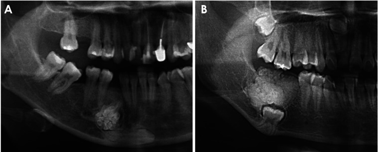
Odontomas represent a large percentage (23%-77%) of all odontogenic tumors.1,2,3 Several studies have examined large series of these tumors and identified odontomas as the most common odontogenic tumors.3,4 The latest fourth edition of the World Health Organization (WHO) Classification of Head and Neck Tumors5 classifies odontomas as compound odontomas (CpODs) (Fig. 1A), characterized by numerous tooth-like structures, and complex odontomas (CxODs) (Fig. 1B), manifesting as disorganized masses of calcified tissue. According to the literature, the incidence of CpODs and CxODs is 32%-60% and 40%-68%, respectively. No sex predilection has been indicated.2,6,7
Fig. 1. Representative panoramic radiographs of compound odontoma (A) and complex odontoma (B).
Odontomas are typically characterized by benign and asymptomatic lesions that may lead to disturbances in the eruption of teeth, such as delayed eruption of primary or permanent teeth or retention of primary teeth. The majority of patients are diagnosed with odontoma during a routine radiographic examination. Odontomas are considered to be developmental hamartomas rather than true neoplasms of odontogenic origin8,9 because a neoplasm has the distinguishing feature of persistent, uncoordinated growth. However, many diagnosed odontomas become sufficiently large to show almost neoplastic potential and cause various complications, such as the expansion of the jaw bone, tooth displacement, or missing teeth,10,11 and even signs of infection, pain, and suppuration.11,12 Indeed, whether these odontogenic lesions represent a true neoplasm or only a hamartoma is still not well established and requires additional studies.
Radiographically, an odontoma appears as a clearly outlined, dense, radiopaque lesion surrounded by a thin radiolucent zone corresponding to a thin soft tissue capsule, in which CpODs present numerous tooth-like structures, while CxODs appear as disorganized masses of calcified tissue.5 However, various highly calcified bone lesions that appear as mixed radiopaque and radiolucent images confuse clinicians in the differential diagnosis of odontomas, in particular, a diagnosis involving CxOD. The definitive odontoma diagnosis is usually based on the pathological examination after the surgical removal of the tumor, while the primary diagnosis plays an important role in determining the intervention plan at the early stage of management.
Several retrospective studies on odontogenic tumors have reported geographic or racial variations in the incidence of odontoma from almost all continents: Asia,6,13 Africa,14 South America,1,15 and Europe.2,16 However, studies focused on odontomas have been poorly documented,7,12,16 except for the published case reports.10,17,18 In addition, most studies have not adequately investigated the characteristics of odontomas in terms of their clinical and radiographic aspects and have even failed to consider these features in the context of the distinct complex and compound subtypes.14,15 Thus, it would be inaccurate to compare the outcomes of these various studies without accounting for subtype or sex.
Vietnam is the 15th most populous country in the world, with a population of more than 99 million in 2023, of which the median age is 32 years.19 Surprisingly, however, there has been no systematic documentation of the incidence of odontomas in Vietnam over the past 15 years.
In evidence-based medicine, knowledge concerned with clinical and preclinical features, and even more so the relationships between them, has contributed to the diagnosis and prognosis of disease. Hence, studying the associations between disease behaviors in a general manner has emerged as a novel strategy in research.
To contribute to the existing literature and to complement previous studies on odontomas, the purposes of this study were as follows: 1) to analyze the incidence, diagnostic findings, anatomic location, tumor size, and adverse effects on adjacent structures of CxOD and CpOD; 2) to perform multivariable regression analysis to establish the possible association between age at diagnosis and tumor size stratified by sex; and 3) to compare this study’s findings on odontoma characteristics in the Vietnamese population to those documented in other populations around the world.
Materials and Methods
Study design
In this retrospective study, demographic and diagnostic data of patients were extracted from the numeric registries in the computer systems of the Hospital of Odonto-Stomatology of Ho Chi Minh City and the National Hospital of Odonto-Stomatology. Any patients admitted and treated for odontomas at these 2 hospitals were considered for this study, without accounting for patients’ provinces of residence in their records. The coverage period was from January 1, 2004, to December 31, 2017. The study was performed in Ho Chi Minh City because 1) it is one of the largest centers in the country (along with Hanoi), in which the national hospitals are responsible for the treatment of maxillofacial pathologies in the southern region are located; and 2) the patients’ records at hospitals in Ho Chi Minh City are more complete than in other provinces of Vietnam. This study was approved by the Ethics Committee of the University of Medicine and Pharmacy of Ho Chi Minh City, approval number: 1547-DHYD. Because all data were anonymized, no individual patient consent was required.
Data collection
The patient data considered for this study were age, sex, reasons for consultation, clinical symptoms, radiographic imaging, histological diagnosis, and adverse effects on adjacent teeth. The 2017 WHO classification was used for the final classification of the lesions as either CxOD or CpOD.5 The cases with poor radiographs, insufficient records, or a combined diagnosis of the odontoma with other lesions in the oral cavity (e.g., ameloblastic fibroma, non-odontogenic cyst, maxillofacial trauma) were excluded from the study. All included cases were definitively diagnosed as odontomas through the histologic examination of removed samples performed by pathologists at the Department of Histopathology in Cho Ray Hospital, Ho Chi Minh City, Vietnam.
Data filtered from the numeric registry was further validated through patient identification and patients’ physical records. Due to the limited availability of digital radiographs in the panoramic radiologic system (i.e., within the past 5 years), radiographic films were used for all cases. The same panoramic machine (Orthophos XG 5 and SIDEXIS XG software, version 2.63, Sirona Dental System GmbH, Bensheim, Germany) was used at the 2 hospitals from which the data were collected. A calibration procedure was performed twice per year to ensure that the magnification factor was 1 : 1. The collected data variables were then coded, including tumor location, odontoma subtype, reasons for consultation, clinical symptoms, and complications in adjacent teeth and surrounding anatomic structures.
The locations of the odontomas were categorized by anatomic site (maxilla or mandible) and region (anterior, defined as extending from the distal surface of the right canine to the distal surface of the left canine, or posterior, defined as extending from the mesial aspect of the first premolar to the distal aspect of the third molar).
The size of each tumor mass was measured using the major axis of the mass in the panoramic radiograph and the scale bar printed in the films as a standard rule. Both authors of this study participated in the data collection. To evaluate the accordance between these authors, 20 cases were randomly chosen, and the mass sizes were independently measured from the radiograph films. The coefficient of variation was then calculated at approximately 2%.
The mass sizes were classified into 5 categories for analysis: <10 mm, 10 to <20 mm, 20 to <30 mm, 30 to <40 mm, and ≥40 mm. Size was also used as a continuous variable depending on the required analysis.
The age variable was categorized as 10-year intervals, in which the minimum age was 8 years and the maximum age was 79 years: 8-10, 11-20, 21-30, 31-40, 41-50, 51-60, 61-70, 71-79. The age variable was aggregated as 10-year intervals to facilitate comparison to other studies.
Data analysis
Both descriptive and inferential analyses were conducted to address the main aims of the study. In the descriptive analysis, the normal distribution of a continuous variable (i.e., age, mass size) was checked by the Shapiro-Wilk test and Quantile-Quantile plots. The differences between subgroups of sex and odontoma subtypes were analyzed using the t-test for normally-distributed data or the bootstrap method20 for non-normally distributed data to report a 95% confidence interval (CI), and the chi-square test for categorical variables. Specifically, the bootstrap method was conducted in the packages “boot” and “simple boot.” Data were randomly resampled 1,000 times to generate the characteristics of the data because the high number of replicates maximized accuracy in the prediction of median differences and power.21 Bias-corrected and accelerated CIs were used.20
A multivariable linear regression analysis was performed to assess the relationship between age and mass size. In this analysis, mass size was considered the dependent variable, age was treated as an independent variable; odontoma subtype and interaction between age and odontoma subtype were covariates. The strength of the association between age or covariates and mass size was estimated by the regression coefficient and standard error. For the association between the presence of CxOD and mass size stratified by sex, a multivariable logistic regression analysis was conducted.
R software environment version 4.1.2 was used to perform all statistical analyses (R Foundation for Statistical Computing).22 A nominal P-value<0.05 was considered to indicate statistical significance.
Results
Patients
Between January 1, 2004, and December 31, 2017, 90 cases of odontoma (63 female patients; 70%) were registered. Female patients accounted for 76% of CxOD cases (35/46) and 63% of CpOD cases (28/44). The mean age (±standard deviation [SD]) at diagnosis was 32.4±20.2 years (range: 8-79 years). The median age was 28 years (interquartile range [IQR]: 18.5-54.0 years) and 22 years (IQR: 15.5-32.0 years) for female and male patients, respectively (difference in medians, -22.0 years; 95% CI: -23.0 to 1.0 years; P<0.05). It was also shown that the patients with CxOD tended to be older than patients with CpOD (difference in medians, +15.8 years, 95% CI: 4.0-35.5 years; P<0.05).
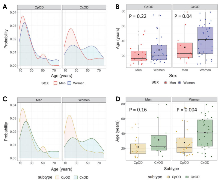
Among the 90 cases of odontoma included in this study, 49% (44/90) were CpODs and 51% (46/90) were CxODs. In general, tumors were more frequently found in the second and third decades of life (51%). This percentage was 56% in male patients (15/27) and 49% in female patients (31/63). Stratifying by sex or odontoma subtype (Fig. 2) revealed that in male patients, the majority of CpOD and CxOD diagnoses were made between the ages of 10-30 years. In contrast, this age distribution was only observed in female patients with CpOD, whereas the ages were scattered throughout the lifespan in female patients with CxOD. The age of female patients with CxOD was significantly greater than that of male patients (median [IQR], 42.0 years [9.0-79.0 years] vs. 23.0 years [10.0-79.0 years]; P<0.05), and female patients with CxOD were also older than those with CpOD (median [IQR], 42.0 years [9.0-79.0 years] vs. 21.0 years [10.0-75.0 years]; P<0.05), whereas these differences did not reach statistical significance in CpOD or male patients, respectively.
Fig. 2. A and B. Distribution of patients’ age according to sex, stratified by odontoma subtypes. C and D. Distribution of patients’ age according to odontoma subtype, stratified by sex. The horizontal line and the black square (▮) in the box plots correspond to the median and mean values, respectively. CpOD: compound odontoma; CxOD: complex odontoma.
Clinical characteristics of odontomas
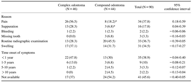
Table 1 displays the patients’ main reasons for the consultation. The most common reason was pain (38%), followed by a routine radiographic examination (37%) and swelling (35%). Those were also the most common reasons for patients with CpOD, among whom about half of the cases were detected on routine radiographic examinations (20/44 cases). By contrast, the majority of patients with CxOD presented with pain (57%), followed by swelling (37%) and suppuration (28%). In particular, pain and suppuration were reported by 38% (95% CI: 18%-59%; P<0.05) and 22% (95% CI: 4%-39%; P<0.05) in patients with CpOD less than in those with CxOD. The time onset of symptoms until treatment was mostly within 1 year in both subtypes (P<0.05), and no significant difference between the subtypes was detected in either period defined as a categorical variable.
Table 1. Main reasons for consultation and time onset of symptoms stratified by odontoma subtype (number [%]).
*: P<0.05 compared with complex odontoma
Table 2 shows significant differences in clinical characteristics in relation to odontoma subtype. As expected, about 59% of patients with CxOD reported pain as a symptom, while this percentage was only 21% in those with CpOD (95% CI: 17%-59%; P<0.05). In contrast, CpOD was characterized by the retention of a primary tooth (30%) and the lack of a permanent tooth (64%) at a significantly higher rate than CxOD (P<0.05). Bone expansion was frequently detected in both subtypes (i.e., 76% of CxODs and 57% of CpODs), and no significant difference was observed between them.
Table 2. Distribution of clinical findings stratified by odontoma subtype (number [%]).
*: P<0.05 compared with complex odontoma
Radiological characteristics of odontomas
Regarding the tumor location on radiographs, CxOD was most frequently located in the mandible (34/46, 74%, P<0.05) and the posterior region (39/46, 85%, P<0.05) than CpOD (21/44, 48% and 11/44, 25%, respectively). CpOD was commonly found in the anterior region (33/44, 75%) in both the mandible and maxilla (P>0.05), whereas CxOD often occurred in the posterior region of the mandible (34/46, 74%, P<0.05).
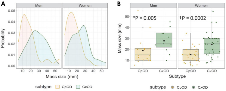
Radiographic measurements of the mass sizes of the tumors (Fig. 3) showed that CxODs were larger than CpODs in both male patients (median [IQR]: 25.0 [10.0-45.0] vs. 15.0 [6.0-55.0]; 95% CI: 5.0-16.0; P<0.05) and female patients (mean±SD: 24.5±10.9 vs. 15.4±7.4; 95% CI: 4.5-13.7; P<0.05). In fact, 73% (32/44) of CpODs exhibited a dimension of 10-30 mm, while 82% (38/46) of CxODs exhibited a dimension of 10-40 mm.
Fig. 3. Distribution of mass size (mm), stratified by sex, of complex and compound odontomas, where the masses were determined from panoramic radiographs. Data are illustrated by density line (A) and boxplot (B), in which the horizontal line and the black square (▮) correspond to the median and mean values, respectively. P-values are derived from the bootstrap method using median values (*) or the t-test (†) for 2-sample comparisons. CpOD: compound odontoma; CxOD: complex odontoma.
There was a significant positive correlation between the mass size of tumors and age in both male patients and female patients (P<0.05) (data not shown). Furthermore, the patients with CxOD were older and exhibited larger tumors than those with CpOD. Hence, multivariable linear regression analysis was conducted (Table 3). In male patients, there was an interaction between age and odontoma subtype (P<0.05), indicating that the association between age and mass size depended on the odontoma subtype. The mass size of CpOD tended to increase over time, while CxOD tended to show a gradual growth until the diagnosis (P<0.05). Collectively, these factors (i.e., age, odontoma subtype, and their interaction) explained 53% of the total variance in mass size.
Table 3. Association between age and mass size stratified by sex.
*: P<0.05
In female patients, there was no statistically significant association (P>0.05) between mass size and age, after adjusting for odontoma subtype and interaction effect. As expected, CxODs were significantly larger than CpODs (P<0.05). The 3 factors collectively accounted for 22% of the variance in mass size (Table 3).
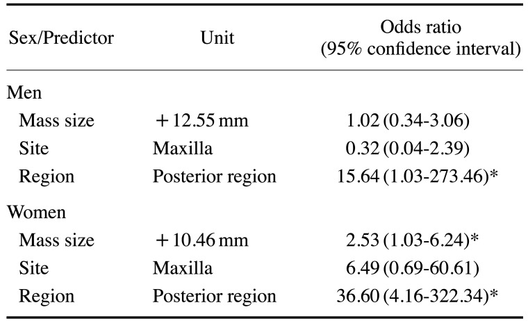
Multivariable logistic regression analysis with CxOD as the outcome (Table 4) showed a significant association between mass size and the odds of having complex odontoma in female patients (P<0.05), but not in male patients (P>0.05), controlling for site and region. An increase of 1 SD in mass size (i.e., 10.5 mm) increased the odds of having CxOD in female patients by about 3-fold. In both sexes, an odontoma located in the posterior region had odds of being CxOD of about 16-fold in male patients (P<0.05) and about 37-fold in female patients (P<0.05).
Table 4. Associations between diagnosed complex odontoma and mass size stratified by sex.
*: P<0.05
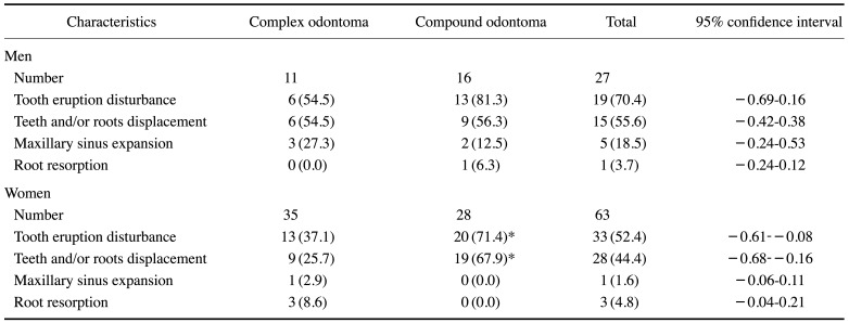
Concerning the impact of odontomas on adjacent teeth and surrounding anatomic structures determined in radiographic examination (Table 5), disturbances in tooth eruption (52/90, 58%) were the most common effect on the adjacent teeth, followed by displacement of teeth and roots (43/90, 48%) in all patients. Root resorption was found in only 4 cases (4%). Complications were caused more frequently by CpODs than by CxODs in both sexes, such as tooth eruption disturbance (0.8 vs. 0.4; 95% CI: 0.2-0.6; P<0.05), tooth and/or root displacement (0.6 vs. 0.3; 95% CI: 0.1-0.5; P<0.05). However, these differences showed statistical significance only in female patients (P<0.05), not in male patients (P>0.05).
Table 5. Distribution of complications in adjacent teeth and surrounding structures on radiography, stratified by sex (number [%]).
*: P<0.05 compared with complex odontoma by the chi-square test
Discussion
Many studies have reported that the mean age of patients at the time of odontoma diagnosis was between 10 and 30 years.4,12 In agreement with previous studies, the patients in this study were most frequently in the second and third decades of life. It was also observed that CxODs tended to occur at older ages (mean, 38.9 years) than CpOD (mean, 25.8 years). This outcome was consistent with some records,7,16 but in contrast with others.2,23 These studies were conducted on various populations with different sample sizes. Additionally, dental health care differs among countries and develops with time. These potential reasons could lead to inconsistencies between studies in the literature.
Sex predilection of odontomas and odontoma subtypes has appeared in various ways depending on the country and continent.2,4,7 In this study, the male-to-female ratio was 0.4 : 1, with a ratio of 0.3 : 1 for CxOD and 0.5 : 1 for CpOD. Although the literature has reported varying results regarding the sex predilection of odontomas, the findings of the present study showed a general agreement with some of the reported data.13,16
Odontomas display benign characteristics and a silent pattern of development in their location. Clinically, the majority of odontomas are frequently diagnosed through routine dental examinations.12,24 In line with previous studies, this study revealed that a routine radiographic examination was one of the most common reasons that led to odontoma diagnoses. The other main reasons for consultations that led to diagnoses were pain and/or swelling (about 72%). Routine dental examinations can be assumed to have been widely performed in Ho Chi Minh City due to the development of dental clinics and hospitals. However, the patients in this study came from various provinces, where public health care has not been fully investigated and regular attendance to dental clinics is relatively low among the population. Indeed, these patients asked for a consultation because they noticed symptoms.
Clinical characteristics are useful for the differential diagnosis of odontoma from other tumor lesions. In general, bone expansion at the tumor location was the most frequent clinical finding (68%), which is consistent with a previous study that reported this percentage at 65%.25 Because odontoma is a type of odontogenic tumor, the impacts related to teeth are usually evident in the oral cavity, such as the disturbance to tooth eruption in both the primary and permanent dentition.26 In this study, 47% of patients were missing permanent teeth, and this percentage was about 80% in the study by Iatrou et al.25 The present study also demonstrated that CxOD was characterized by bone expansion and pain more frequently than CpOD, the latter which was more frequently characterized by an anomaly in the dentition (Table 2). These findings are consistent with the patients’ reported reasons for consultations (Table 1): CxOD commonly manifested as pain and suppuration, while CxOD exhibited more signs or symptoms of inflammation. The prolonged existence of CxOD in the jaws is likely to be one of the causes that activate inflammation development.
Concerning location, odontomas present in both the maxilla and mandible, with variation depending on the studied population.1,15,16 In this patient cohort, 75% of CpODs occurred in the anterior region of both the maxilla and mandible, and 74% of CxODs occurred in the posterior mandible. These corresponding percentages in the study of Worawongvasu et al.13 were 78% and 42%, respectively. According to Kilinc et al.27 (2016), 52% of CpODs were found in the anterior regions of both jaw bones, and 58% of CxODs were found in the posterior region or ramus of the mandible. These location predilections of odontoma subtypes, combined with the younger age of diagnosis of CpOD compared to CxOD, suggest that tooth eruption disturbance in the anterior region and the subsequent aesthetic impairment lead to earlier diagnosis and treatment of CpOD than CxOD.
A previous study demonstrated that the majority of odontomas measured in the range of 10-30 mm,25 and occasionally, a CxOD exceeded 40 mm in diameter.10,11 These outcomes are in accordance with the results of the present study, in which 62% of odontomas had a size of 10-30 mm and only 8% of cases (7/90) showed a dimension of ≥40 mm. Furthermore, this study found that CxODs were larger than CpODs, which may be related to the older age at diagnosis of CxOD than CpOD (Fig. 2).
The positive association found between mass size and age is consistent with a recent study in the Colombian population,24 suggesting that the odontoma increases in size over time. However, the odontoma subtype appeared to be a confounder in this association. The present findings revealed that the mass size of CxOD was greater than that of CpOD in both male and female patients (Fig. 3). In a secondary analysis stratified by sex, accounting for subtype and interaction between age and subtype, the association between age and mass size remained statistically significant in male patients (Table 3). A possible explanation is that CpOD exhibits potential growth in young male patients, causes several complications related to tooth eruption, and is thus diagnosed and treated at an earlier time. In contrast, CxOD shows a moderate development over time, is found at a later stage, and thus reaches greater dimensions by the time of diagnosis, along with inflammatory signs. In female patients, although this association was no longer statistically significant, CxOD showed a significantly larger diameter than CpOD. The development of the specific odontoma subtype may be a result of their pathogenetic differences. CxOD, exhibiting a greater age at the time of diagnosis, may be considered the terminal stage of the ameloblastic fibro-odontoma lesion, whereas CpOD may be the result of malformation related to the process that produces hyperdontia because of local hyperactivity of the dental lamina.5,9 It should be considered that odontomas display different characteristics depending on sex. Sex-related physio-pathological mechanisms lead to these differences.
Because CxOD is larger on average than CpOD and the predilection of location is dependent on odontoma subtype, this study carefully adjusted for potential covariates (i.e., site and region) to examine whether mass size could be used as an indicator of odontoma subtypes (Table 4). Based on the findings of this analysis, a location in the posterior region may be a significant predictor for CxOD diagnosis in both sexes. This correlates with the radiographic observations that 85% of CxODs were found in the posterior region. An increase in mass size adequately reflects the CxOD subtype in female patients.
The delay of tooth eruption and other effects on teeth and surrounding anatomic structures have been meaningful for clinicians. This may originate from the current situation in which panoramic radiographs have been widely used for routine examinations. Many studies and case reports have revealed various complications in teeth and surrounding structures related to odontomas.12,17,28 Katz studied 396 cases of odontomas in all age groups and found that 41% were associated with unerupted teeth.28 A study on 73 Korean children reported that 62% of odontomas caused the impaction on permanent teeth.12 In the present study, about 58% (52/90) of odontomas resulted in tooth eruption disturbance (Table 5). Besides that, displacement of teeth and roots was identified in 43 of 90 cases (about 48%) in this study. The corresponding percentage in the study of An et al.12 was about 30%. Tooth displacement results from the continuous growth of the tumor, which expands or displaces the surrounding structures, such as the cortical jaw bone, mandibular canal, and even soft tissue. Furthermore, the incidence of such complications has been reported to vary between CxODs and CpODs. Bereket et al.18 demonstrated that 69% of CpODs were associated with unerupted teeth, while the corresponding percentage in CxODs was 50%. The same trend was found in the present study, given that CpODs resulted more frequently in these complications than CxODs. Although CpODs were characterized by smaller dimensions on panoramic radiography at the time of consultation, the contrasting greater frequency of complications could indicate that CpODs display more potential growth than CxOD over time.
Our findings should be considered within the context of its strengths and weaknesses. This study was based on a large number of cases (n=90) in order to be comparable to other studies and contribute to the literature on odontoma research. Despite the limitations of a retrospective study, an effort was made to perform a more complex analysis (multivariable linear regression, multivariable logistic regression) that enabled a deeper understanding of the association between age and mass size, and between odontoma subtype and mass size, accounting for different factors, stratified by sex. Indeed, these outcomes helped to confirm the cross-sectional observations and to explain the behaviors of odontomas in each sex. Additionally, the use of the same panoramic machines in both hospitals helped to avoid bias in data interpretation. These machines were adjusted at a 1:1 magnification to reflect the real dimensions of detected tumors and the impacts on the surrounding structures.
However, there were some limitations of this study, mainly related to the nature of the retrospective design, which does not account for longer-term follow-up, such as the postoperative treatment strategy, and its efficacy thereafter. The patients were closely followed for about 3-7 days postoperatively, until they checked out of the hospital. No severe complications were observed in any cases. The patients could choose to be re-examined at the hospitals of this study or at their provinces’ hospitals and continue with further treatment in large tumor cases, according to clinicians’ recommendations. Additionally, since the majority of the patients lived in the south of Vietnam, these outcomes should not be generalized to the whole Vietnamese population.
In summary, odontomas in the southern Vietnamese population were detected during young adulthood. Patients with CxOD were diagnosed at an older age and exhibited more severe clinical symptoms than patients with CpOD, which was generally detected through routine examinations. The differences in clinical and radiological features between CxOD and CpOD were related to their growth characteristics and location predilections, and were also influenced by sex. These results could support clinicians in obtaining a greater knowledge of odontomas, which would be useful for both the early detection and differential diagnosis of odontogenic masses. Further studies should be conducted with a prospective design to evaluate the efficacy of treatment not only postoperatively, but also in the long term, including the recurrence of odontomas.
Acknowledgments
The authors gratefully thank the staff of the records management office at the Hospital of Odonto-Stomatology and the National Hospital of Odonto-Stomatology in Ho Chi Minh City for help in the data collection and for providing informative support in this study.
Footnotes
Conflicts of Interest: None
References
- 1.Servato JP, Prieto-Oliveira P, de Faria PR, Loyola AM, Cardoso SV. Odontogenic tumours: 240 cases diagnosed over 31 years at a Brazilian university and a review of international literature. Int J Oral Maxillofac Surg. 2013;42:288–293. doi: 10.1016/j.ijom.2012.05.008. [DOI] [PubMed] [Google Scholar]
- 2.Siriwardena BS, Crane H, O’Neill N, Abdelkarim R, Brierley DJ, Franklin CD, et al. Odontogenic tumors and lesions treated in a single specialist oral and maxillofacial pathology unit in the United Kingdom in 1992-2016. Oral Surg Oral Med Oral Pathol Oral Radiol. 2019;127:151–166. doi: 10.1016/j.oooo.2018.09.011. [DOI] [PubMed] [Google Scholar]
- 3.Buchner A, Merrell PW, Carpenter WM. Relative frequency of central odontogenic tumors: a study of 1,088 cases from Northern California and comparison to studies from other parts of the world. J Oral Maxillofac Surg. 2006;64:1343–1352. doi: 10.1016/j.joms.2006.05.019. [DOI] [PubMed] [Google Scholar]
- 4.Soluk Tekkesin M, Pehlivan S, Olgac V, Aksakallı N, Alatli C. Clinical and histopathological investigation of odontomas: review of the literature and presentation of 160 cases. J Oral Maxillofac Surg. 2012;70:1358–1361. doi: 10.1016/j.joms.2011.05.024. [DOI] [PubMed] [Google Scholar]
- 5.El-Naggar AK, Chan JK, Rubin Grandis J, Takata T, Slootweg PJ. International agency for research on cancer. WHO classification of head and neck tumours. 4th ed. Lyon: International Agency for Research on Cancer; 2017. [Google Scholar]
- 6.Sekerci AE, Nazlim S, Etoz M, Deniz K, Yasa Y. Odontogenic tumors: a collaborative study of 218 cases diagnosed over 12 years and comprehensive review of the literature. Med Oral Patol Oral Cir Bucal. 2015;20:e34–e44. doi: 10.4317/medoral.19157. [DOI] [PMC free article] [PubMed] [Google Scholar]
- 7.Boffano P, Zavattero E, Roccia F, Gallesio C. Complex and compound odontomas. J Craniofac Surg. 2012;23:685–688. doi: 10.1097/SCS.0b013e31824dba1f. [DOI] [PubMed] [Google Scholar]
- 8.Kramer IR, Pindborg JJ, Shear M. The WHO histological typing of odontogenic tumours. A commentary on the second edition. Cancer. 1992;70:2988–2994. doi: 10.1002/1097-0142(19921215)70:12<2988::aid-cncr2820701242>3.0.co;2-v. [DOI] [PubMed] [Google Scholar]
- 9.Philipsen HP, Reichart PA, Praetorius F. Mixed odontogenic tumours and odontomas. Considerations on interrelationship. Review of the literature and presentation of 134 new cases of odontomas. Oral Oncol. 1997;33:86–99. doi: 10.1016/s0964-1955(96)00067-x. [DOI] [PubMed] [Google Scholar]
- 10.Bagewadi SB, Kukreja R, Suma GN, Yadav B, Sharma H. Unusually large erupted complex odontoma: a rare case report. Imaging Sci Dent. 2015;45:49–54. doi: 10.5624/isd.2015.45.1.49. [DOI] [PMC free article] [PubMed] [Google Scholar]
- 11.Perumal CJ, Mohamed A, Singh A, Noffke CE. Sequestrating giant complex odontoma: a case report and review of the literature. J Maxillofac Oral Surg. 2013;12:480–484. doi: 10.1007/s12663-010-0148-y. [DOI] [PMC free article] [PubMed] [Google Scholar]
- 12.An SY, An CH, Choi KS. Odontoma: a retrospective study of 73 cases. Imaging Sci Dent. 2012;42:77–81. doi: 10.5624/isd.2012.42.2.77. [DOI] [PMC free article] [PubMed] [Google Scholar]
- 13.Worawongvasu R, Tiensuwan M. Odontogenic tumors in Thailand: a study of 590 Thai patients. J Oral Maxillofac Surg Med Pathol. 2015;27:567–576. [Google Scholar]
- 14.Aregbesola B, Soyele O, Effiom O, Gbotolorun O, Taiwo O, Amole I. Odontogenic tumours in Nigeria: a multicentre study of 582 cases and review of the literature. Med Oral Patol Oral Cir Bucal. 2018;23:e761–e766. doi: 10.4317/medoral.22473. [DOI] [PMC free article] [PubMed] [Google Scholar]
- 15.Lima-Verde-Osterne R, Turatti E, Cordeiro-Teixeira R, Barroso-Cavalcante R. The relative frequency of odontogenic tumors: a study of 376 cases in a Brazilian population. Med Oral Patol Oral Cir Bucal. 2017;22:e193–e200. doi: 10.4317/medoral.21285. [DOI] [PMC free article] [PubMed] [Google Scholar]
- 16.Kämmerer PW, Schneider D, Schiegnitz E, Schneider S, Walter C, Frerich B, et al. Clinical parameter of odontoma with special emphasis on treatment of impacted teeth - a retrospective multicentre study and literature review. Clin Oral Investig. 2016;20:1827–1835. doi: 10.1007/s00784-015-1673-3. [DOI] [PubMed] [Google Scholar]
- 17.Srivastava A, Annaji AG, Nyamati SB, Singh G, Shivakumar GC, Sahana S. Complex odontoma in both the jaws: a rare case report. J Orofac Res. 2012;2:56–60. [Google Scholar]
- 18.Bereket C, Çakır-Özkan N, Şener İ, Bulut E, Tek M. Complex and compound odontomas: analysis of 69 cases and a rare case of erupted compound odontoma. Niger J Clin Pract. 2015;18:726–730. doi: 10.4103/1119-3077.154209. [DOI] [PubMed] [Google Scholar]
- 19.Worldometer [Internet]. Countries in the world by population. 2021. [cited 2023 Jan 10]. Available from: https://www.worldometers.info/world-population/population-by-country/
- 20.Efron B. Better bootstrap confidence intervals. J Am Stat Assoc. 1987;82:171–185. [Google Scholar]
- 21.Pattengale ND, Alipour M, Bininda-Emonds OR, Moret BM, Stamatakis A. How many bootstrap replicates are necessary? J Comput Biol. 2010;17:337–354. doi: 10.1089/cmb.2009.0179. [DOI] [PubMed] [Google Scholar]
- 22.R Development Core Team. R: a language and environment for statistical computing. Version 4.1.2 [software] 2021. Nov 01, [cited 2023 Jan 10]. Available from: https://www.r-project.org/
- 23.Gill S, Chawda J, Jani D. Odontogenic tumors in Western India (Gujarat): analysis of 209 cases. J Clin Exp Dent. 2011;3:e78–e83. [Google Scholar]
- 24.Levi-Duque F, Ardila CM. Association between odontoma size, age and gender: multivariate analysis of retrospective data. J Clin Exp Dent. 2019;11:e701–e706. doi: 10.4317/jced.55733. [DOI] [PMC free article] [PubMed] [Google Scholar]
- 25.Iatrou I, Vardas E, Theologie-Lygidakis N, Leventis M. A retrospective analysis of the characteristics, treatment and follow-up of 26 odontomas in Greek children. J Oral Sci. 2010;52:439–447. doi: 10.2334/josnusd.52.439. [DOI] [PubMed] [Google Scholar]
- 26.Tomizawa M, Otsuka Y, Noda T. Clinical observations of odontomas in Japanese children: 39 cases including one recurrent case. Int J Paediatr Dent. 2005;15:37–43. doi: 10.1111/j.1365-263X.2005.00607.x. [DOI] [PubMed] [Google Scholar]
- 27.Kilinc A, Saruhan N, Gundogdu B, Yalcin E, Ertas U, Urvasizoglu G. Benign tumors and tumor-like lesions of the oral cavity and jaws: an analysis of 709 cases. Niger J Clin Pract. 2017;20:1448–1454. doi: 10.4103/1119-3077.187309. [DOI] [PubMed] [Google Scholar]
- 28.Katz RW. An analysis of compound and complex odontomas. ASDC J Dent Child. 1989;56:445–449. [PubMed] [Google Scholar]


