Abstract
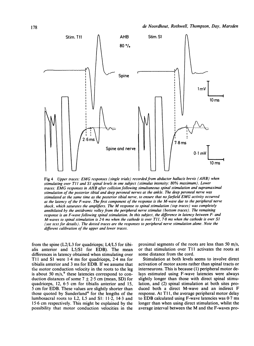
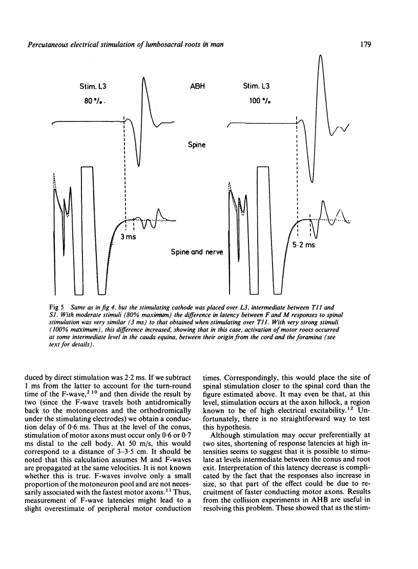
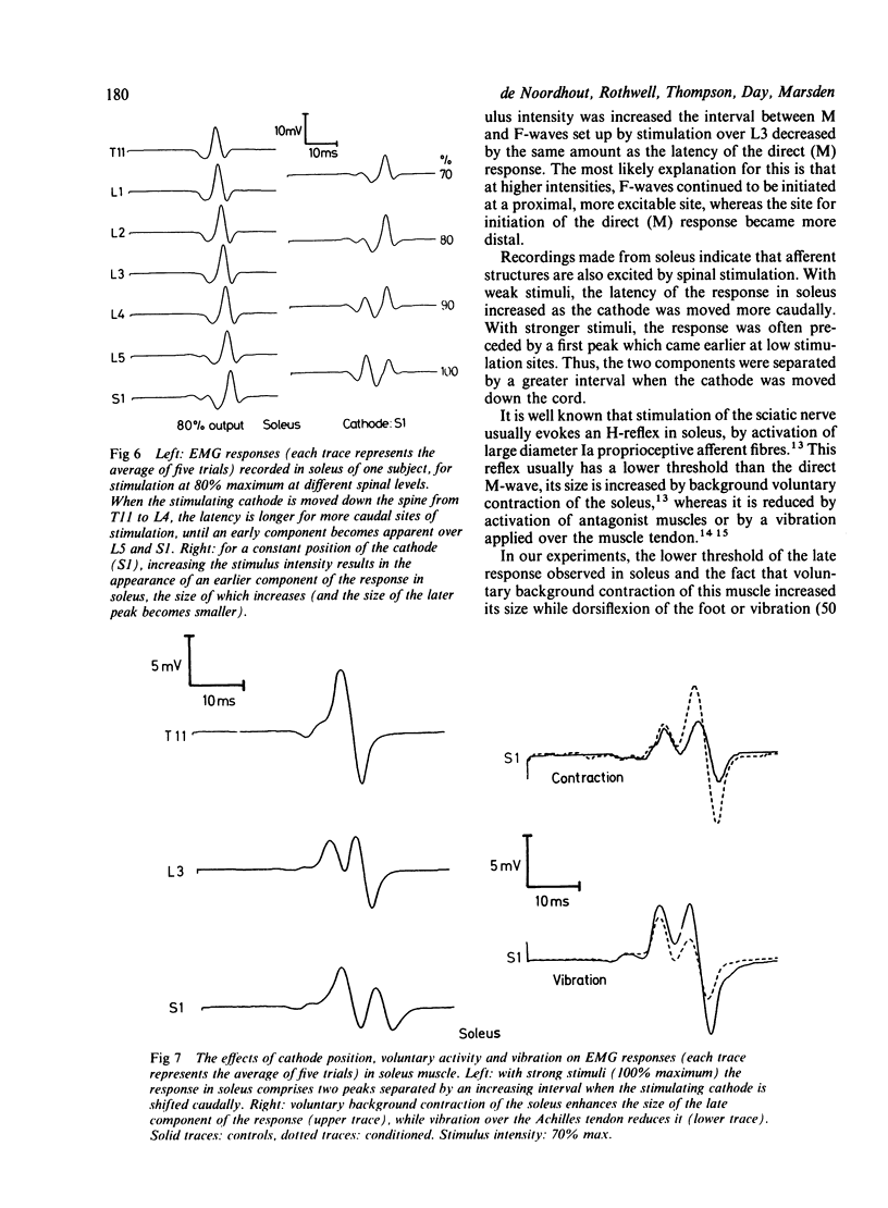
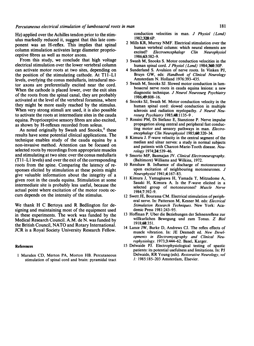
High voltage percutaneous electrical stimulation over the lumbosacral spinal column was used to assess conduction in the cauda equina of 13 normal subjects. Electromyographic activity elicited by such stimulation was recorded from various muscles of the lower limbs. The stimulating cathode was placed over the spinous process of each vertebral body and the anode kept on the iliac crest contralateral to the studied limb. Shifting the cathode in a rostro-caudal direction shortened the response latency in quadriceps, tibialis anterior and extensor digitorum brevis muscles. At moderate intensities (60% maximum), this occurred abruptly when the cathode was placed at levels corresponding to the exit sites from the spinal canal of the roots innervating these muscles. At these intensities, the size of the response in each muscle was largest when the cathode was placed over the conus medullaris or at or below the exit of the motor roots from the spine. Latencies were always equal to or shorter than those obtained with F-wave measurements, suggesting that peripheral motor axons, rather than intraspinal structures were activated by the stimulus. Collision experiments demonstrated that activation occurred at two sites: near the spinal cord and at the root exit site in the vertebral foramina. Recordings made from soleus indicated that larger diameter proprioceptive afferent fibres also could be activated. This technique might have useful clinical applications in the study of both proximal and distal lesions of the cauda equina and provide a non-invasive method of localising such lesions electrophysiologically.
Full text
PDF
Selected References
These references are in PubMed. This may not be the complete list of references from this article.
- Kimura J. F-wave velocity in the central segment of the median and ulnar nerves. A study in normal subjects and in patients with Charcot-Marie-Tooth disease. Neurology. 1974 Jun;24(6):539–546. doi: 10.1212/wnl.24.6.539. [DOI] [PubMed] [Google Scholar]
- Kimura J., Yanagisawa H., Yamada T., Mitsudome A., Sasaki H., Kimura A. Is the F wave elicited in a select group of motoneurons? Muscle Nerve. 1984 Jun;7(5):392–399. doi: 10.1002/mus.880070509. [DOI] [PubMed] [Google Scholar]
- Mills K. R., Murray N. M. Electrical stimulation over the human vertebral column: which neural elements are excited? Electroencephalogr Clin Neurophysiol. 1986 Jun;63(6):582–589. doi: 10.1016/0013-4694(86)90145-8. [DOI] [PubMed] [Google Scholar]
- Rossini P. M., Di Stefano E., Stanzione P. Nerve impulse propagation along central and peripheral fast conducting motor and sensory pathways in man. Electroencephalogr Clin Neurophysiol. 1985 Apr;60(4):320–334. doi: 10.1016/0013-4694(85)90006-9. [DOI] [PubMed] [Google Scholar]
- Snooks S. J., Swash M. Motor conduction velocity in the human spinal cord: slowed conduction in multiple sclerosis and radiation myelopathy. J Neurol Neurosurg Psychiatry. 1985 Nov;48(11):1135–1139. doi: 10.1136/jnnp.48.11.1135. [DOI] [PMC free article] [PubMed] [Google Scholar]
- Swash M., Snooks S. J. Slowed motor conduction in lumbosacral nerve roots in cauda equina lesions: a new diagnostic technique. J Neurol Neurosurg Psychiatry. 1986 Jul;49(7):808–816. doi: 10.1136/jnnp.49.7.808. [DOI] [PMC free article] [PubMed] [Google Scholar]


