Abstract
Background
The drug-eluting stent was a significant stride forward in the development of enhanced therapeutic therapy for coronary intervention, with three generations of increased advancement. VSTENT is a newly developed stent manufactured in Vietnam that aims to provide coronary artery patients with a safe, effective, and cost-efficient option. The purpose of this trial was to determine the efficacy and safety of a new bioresorbable polymer sirolimus-eluting stent called VSTENT.
Methods
This is a prospective, cohort, multicenter research in 5 centers of Vietnam. A prespecified subgroup received intravascular ultrasound (IVUS) or optical coherence tomography (OCT) imaging. We determined procedure success and complications during index hospitalization. We monitored all participants for a year. Six-month and 12-month rates of major cardiovascular events were reported. All patients had coronary angiography after 6 months to detect late lumen loss (LLL). Prespecified patients also had IVUS or OCT performed.
Results
The rate of device success was 100% (95% CI: 98.3–100%; P<0.001). Major cardiovascular events were 4.7% (95% CI: 1.9–9.4%; P<0.001). The LLL over quantitative coronary angiography (QCA) was 0.08±0.19 mm (95% CI: 0.05–0.10; P<0.001) in the in-stent segment and 0.07±0.31 mm (95% CI: 0.03–0.11; P=0.002) in 5 mm within the two ends of the stent segment. The LLL recorded by IVUS and OCT at 6 months was 0.12±0.35 mm (95% CI: 0.01–0.22; P=0.028) and 0.15±0.24 mm (95% CI: 0.02–0.28; P=0.024), respectively.
Conclusions
This study’s device success rates were perfect. IVUS and OCT findings on LLL were favorable at 6-month follow-up. One-year follow-up showed low in-stent restenosis (ISR) and target lesion revascularization (TLR) rates, reflecting few significant cardiovascular events. VSTENT’s safety and efficacy make it a promising percutaneous intervention option in developing nations.
Keywords: Revascularization, stent, bioresorbable, restenosis
Highlight box.
Key findings
• VSTENT has been shown to be a safe and effective tool for coronary intervention in daily practice. It is also an economical option in developing nations.
What is known and what is new?
• Drug-eluting stents remained a revolutionary stride in interventional practice, resulting in a considerable decrease in the re-stenosis rate as well as an improvement in life quality.
• VSTENT’s thin bioresorbable polymer and sirolimus dilution, which are improvements over the typical drug-eluting stent, fared well in terms of both safety and efficacy endpoints.
What is the implication, and what should change now?
• Interventionists should consider incorporating VSTENT into their regular practice, particularly in developing nations.
Introduction
In the development of advanced therapeutic therapy for coronary intervention, the drug eluting stent (DES) was a big step forward. It resulted in a considerable reduction in re-stenosis in clinical and imaging studies, improved life quality, and reduced the need for re-perfusion (1-4). Normally, a medication with an anti-proliferation attribute would be eluted. The most utilized ingredients for this purpose are sirolimus, paclitaxel, biolimus, zotarolimus and everolimus. The very first generation of drug-eluting stents consisted of a stainless steel base coated with a drug-eluting durable polymer (5,6). These devices permit the localized elution of neointimal inhibitory agents with antiproliferative properties, such as sirolimus and paclitaxel, over the duration of 1 month (7,8). In 2008, the next generation of DES was developed. These novel devices had enhanced strut thickness, deliverability, and pliability (5). All three aspects (platform, coating, and drug) of previous-generation stents (platform, coating, and drug) were modified (9). Despite this, stent thrombosis continues to be a substantial problem in the dilute stent population, even in the presence of prolonged anti-platelet medication treatment (10). As a result, various advancements in stent struts, which can be attributable to novel diluting elements, have been developed to increase the safety and effectiveness of the third-generation stents. In addition, the proportion of participation from developing nations, Vietnam in particular, remained low (4,11,12).
The VSTENT stent (made by USM Healthcare in Vietnam) demonstrates excellent characteristics such as thin strut, bioresorbable polymer and sirolimus dilution, which raises the possibility of spectacular clinical outcomes.
We conducted a prospective, multicenter study to assess the efficacy and safety of the VSTENT stent in patients with coronary artery disease. We hypothesized that the VSTENT stent performed well in terms of both safety and efficacy in Vietnamese patients. We present the following article in accordance with the STROBE reporting checklist (available at https://cdt.amegroups.com/article/view/10.21037/cdt-22-522/rc).
Methods
Patient population
This was a prospective cohort study, which the study population was recruited from five South Vietnam sites: University Medical Center Ho Chi Minh City, Thong Nhat Hospital, Khanh Hoa General Hospital, Kien Giang General Hospital and Ba Ria Hospital. Approvals for using patients data have been granted by all sites. The study started in June 2019 and the last patient follow up was in August 2021. All the patients must be at least 18 years old and have either chronic coronary syndrome or acute coronary syndrome as their presenting condition. The most important angiographic criteria were de novo lesions in one or two sites, anticipated diameters ranging from 2.5 to 3.5 mm, and a length of less than 33 mm. Those with severe left ventricular systolic dysfunction, as well as those who were allergic to aspirin, heparin, clopidogrel, ticlopidine, sirolimus or related drugs, cobalt, chromium, niken, molybden, or contrast agent were among the most common clinical exclusion criteria. Chronic total occlusion, severe calcified lesion, left main lesion, a significant thrombus burden, and tortuous target vessels were among the major angiographic exclusion criteria used in this study. The sample size was calculated using the prevalence sample size formula and was based on following criterias: standard normal variate was 0.41, which was based on previous study (2), and the precision of measurement with respect to the endpoint was 0.0656. Per calculation, 150 patients were required. Investigators were not blinded to the treatment allocation which could have introduced a bias.
The VSTENT coronary stent system
The VSTENT coronary stent system is comprised of a cobalt-chromium core with thin struts (65 mcm) and an open-cell architecture that allows for simple access to the lateral branches of the coronary artery. In this study, the system was diluted with 1.33 mcg/mm2 sirolimus, which was applied in layers of 3–5 mm thickness to form the thickest sirolimus coating of 300 mcg over the 4.5 mm × 48 mm stent.
Patient follow-up
Following the stenting procedure, all patients would be required to stay in the hospital for 1 to 2 days to assess the success rate before being discharged if they were stable. Anyone who experienced a failed procedure on day 1 would be reported and followed up as a failure, with the result that no case was enrolled as an alternate treatment.
All the participants had coronary angiogram (CAG) evaluated at day 1 (D1), and intravascular ultrasound (IVUS) and optical coherence tomography (OCT) were also done on predetermined subgroups that were selected depending on the availability of center resources. After 6 months, all the patients had CAG again, and predetermined subgroups also underwent IVUS or OCT. For IVUS, we used Boston Scientific with Image Viewer 1.6 and Boston Scientific with iLabTM Polaris version 2.8. For OCT, we used ILUMIEN System with AptiVue Software version D.3. For a period of 1-year, clinical follow-up would be undertaken on a monthly basis, and would include angina evaluation, medication monitoring, screening for the incidence of major cardiovascular events, and ordering additional examinations as needed.
Endpoints and definitions
Primary endpoints were procedural success rate; along with late lumen loss (LLL), consist of within the stent (in-stent LLL) or 5 mm from the stent ends (in-segment LLL), which was assessed 6 months after the day of percutaneous coronary intervention (PCI) using quantitative coronary angiography (QCA) in angiography, with IVUS or OCT. In-stent or in-segment re-stenosis rate at 6-month (by QCA, IVUS, OCT) and at 12 months (when clinically suspected) were also recorded. Major clinical outcomes include cardiovascular mortality within 12 months (death from acute myocardial infarction, heart failure, cardiac shock, or stroke; sudden cardiac death or other cardiovascular death).
At 6 months, angiographic proximal, distal, and in-segment minimum lumen diameter (MLD); IVUS-measured neointimal hyperplasia volume and% volume obstruction; and OCT-measured strut coverage and malapposition were analysed.
Statistical analysis
For continuous variables, the mean and standard deviation were reported, and for categorical variables, the number (percentage) was reported. The paired T test was used to compare quantitative variables with normal distributions, and the Wilcoxon sign-rank test was employed if the distribution was not normal. The association between LLL and re-stenosis was investigated using logistic regression, and the results were reported as predicted re-stenosis rate. A two-sided threshold of significance of 0.05 was considered significant. We used R version 4.0.5 for analysis purpose.
Ethical statement
The study was conducted in accordance with the Declaration of Helsinki (as revised in 2013). The study was approved by Ethics Boards of University of Medicine and Pharmacy at Ho Chi Minh City (No. 293/ĐHYD-HĐĐĐ), Thong Nhat Hospital (No. 02/BVTN-HDYD), Khanh Hoa General Hospital (No. 74/BVĐKT), Ba Ria Hospital (No. 2792/QĐ-BVBR), Kien Giang General Hospital (No. 03/HĐ-KHKT) and informed consent was taken from all individual participants.
Results
Clinical and angiographic characteristics
This study involved 150 patients from 5 centers (which coded “VN01”, “VN02”, VN03”, “VN04”, “VN05”) with 212 lesions. The mean age was 58.4±10.5 years (95% CI: 56.7–60.1; P<0.001), 67.3% (95% CI: 59.2–74.8; P<0.001) of participants were male, and the most prevalent co-morbidity was hypertension (74.0%) (95% CI: 66.2–80.8%; P<0.001). Acute coronary syndrome was diagnosed in 80% (95% CI: 72.7–86.1%; P<0.001) of patients, with the left anterior descending (LAD) artery being the most frequently targeted vessel (57.5%) (95% CI: 50.6–64.3%; P=0.033). The procedure and device success rate were both 100% (95% CI: 98.3–100%; P<0.001). All patients were discharged safely. Table 1 summarizes basic demographic features and major angiographic characteristics.
Table 1. Baseline clinical and PCI characteristics.
| Baseline clinical characteristics | Total (n=150) | 95% CI | P |
|---|---|---|---|
| Age (years), mean ± SD | 58.4±10.5 | 56.7–60.1 | 0.000 |
| Male, % | 67.3 | 59.2–74.8 | 0.000 |
| BMI >23 kg/m2, % | 48.7 | 40.4–57.0 | 0.806 |
| Risk factors | |||
| Ever smoked, % | 32.7 | 25.2–40.8 | 0.000 |
| Hypertension, % | 74.0 | 66.2–80.8 | 0.000 |
| Hyperlipidemia, % | 50.0 | 41.7–58.3 | 1.000 |
| Diabetes mellitus, % | 20.7 | 14.5–28.0 | 0.000 |
| History of coronary artery disease, % | 17.3 | 11.6–24.4 | 0.000 |
| Peripheral vascular disease, % | 0.7 | 0.0–3.7 | 0.000 |
| Clinical features | |||
| Acute coronary syndrome, % | 80.0 | 72.7–86.1 | 0.000 |
| Stable (chronic) coronary syndrome, % | 20.0 | 13.9–27.3 | 0.000 |
| CCS classification in chronic coronary syndrome (n=30) | |||
| CCS 2, % | 20.0 | 7.7–38.6 | 0.002 |
| CCS 3, % | 76.7 | 57.7–90.1 | 0.006 |
| CCS 4, % | 3.3 | 0.1–17.2 | 0.000 |
| Target lesions position | |||
| LAD, % | 57.5 | 50.6–64.3 | 0.033 |
| LCx, % | 12.3 | 8.2–17.5 | 0.000 |
| RCA, % | 30.2 | 24.1–36.9 | 0.000 |
| AHA classification | |||
| A, % | 4.7 | 2.3–8.5 | 0.000 |
| B, % | 55.7 | 48.7–62.5 | 0.114 |
| C, % | 39.6 | 33.0–46.5 | 0.003 |
| PCI characteristics | |||
| Pre-dilatation balloon, % | 76.4 | 70.1–82.0 | 0.000 |
| Post-dilatation balloon, % | 83.5 | 77.8–88.2 | 0.000 |
| Procedural success, % | 100.0 | 98.3–100 | 0.000 |
| Device success, % | 100.0 | 98.3–100 | 0.000 |
| No. of stents per patient, mean ± SD | 1.41±0.494 | 1.33–1.50 | 0.000 |
PCI, percutaneous coronary intervention; SD, standard deviation; BMI, body mass index; CCS, Canadian Cardiovascular Society; LAD, left anterior descending; LCx, the circumflex artery; RCA, right coronary artery; AHA, American Heart Association.
QCA findings
There was one in-stent and six in-segment re-stenosis among the 212 stents implanted during this research, accounting for 0.5% (95% CI: 0.0–2.8%; P<0.001) and 3% (95% CI: 1.1–6.4%; P<0.001), respectively. Overall, the rate of stent-related re-stenosis was 3.5% (95% CI: 1.4–7.1%; P<0.001) (n=7) in this study.
A total of 143 patients (199 lesions) were re-evaluated via QCA at 6 months after the initial evaluation. LLL was defined as the difference between the MLD measured on Day 1 (immediately following intervention) and the MLD measured after a 6-month follow-up. Table 2 has shown results regarding restenosis >50% rate at 6 months and also the LLL recorded at 6 months, which was also the primary enpoint of this study.
Table 2. Angiographic results post-PCI and CAG at 6 months.
| Total n=212 (lesions) | In-stent | In-segment | |||||
|---|---|---|---|---|---|---|---|
| Values | 95% CI | P | Values | 95% CI | P | ||
| Reference lumen diameter (mm) | 2.97±0.34 | 2.92–3.01 | 0.000 | 2.73±0.42 | 2.67–2.79 | 0.000 | |
| MLD (mm) | |||||||
| Pre-PCI | 0.71±0.35 | 0.66–0.76 | 0.000 | – | – | ||
| Post-PCI | 2.68±0.32 | 2.64–2.73 | 0.000 | 2.38±0.42 | 2.32–2.44 | 0.000 | |
| At 6 months | 2.61±0.38 | 2.56–2.67 | 0.000 | 2.31±0.45 | 2.25–2.37 | 0.000 | |
| LLL at 6 months | |||||||
| LLL, mm | 0.08±0.19 | 0.05–0.10 | 0.000 | 0.07±0.31 | 0.03–0.11 | 0.002 | |
| Index LLL, % | 2.93±7.26 | 1.91–3.94 | 0.000 | 2.36±12.99 | 0.55–4.18 | 0.011 | |
| Restenosis > 50% at 6 months, % | 0.5 | 0.0–2.8 | 0.000 | 3 | 1.1–6.4 | 0.000 | |
Data are shown as mean ± SD or %. PCI, percutaneous coronary intervention; CAG, coronary angiogram; MLD, minimum lumen diameter; LLL, late lumen loss; SD, standard deviation.
IVUS and OCT evaluation
VSTENT stents were put in a variety of vascular sizes, ranging from modest 2.5 mm (11.3%) (95% CI: 7.4–16.4; P<0.001) to larger 2.75 mm (24.1%) (95% CI: 18.5–30.4; P<0.001) to larger ones with widths greater than 3 mm (64.6%) (95% CI: 57.8–71.0; P<0.001). The length of the stents ranged from 15 to 38 mm, with 78.3% (95% CI: 72.1–83.7%; P<0.001) of the patients having a stent length over 20 mm. By IVUS and OCT (Table 3), it has been determined that 92.3% (95% CI: 74.9–99.1%; P<0.001) of all stents had good apposition (Figures 1-4).
Table 3. IVUS and OCT results at D1 (the day of PCI) and at D180 (6 months).
| Results | D1 | D180 | |||||
|---|---|---|---|---|---|---|---|
| Values | 95% CI | P | Values | 95% CI | P | ||
| IVUS results* | |||||||
| MLD (mm), mean ± SD | 2.9±0.4 | 2.8–3.1 | 0.000 | 2.8±0.5 | 2.7–2.9 | 0.000 | |
| Minimum lumen area (mm2), mean ± SD | 7.8±2.1 | 7.2–8.4 | 0.000 | 7.0±2.3 | 6.4–7.6 | 0.000 | |
| Plague burden (%) | 40.7±10.3 | 37.9–43.5 | 0.000 | 47.5±14.1 | 43.5–51.5 | 0.000 | |
| Stent apposition, n (%) | 56 (100.0) | 93.6–100 | 0.000 | 50 (100.0) | 92.9–100 | 0.000 | |
| LLL (mm), mean ± SD (min, max) | – | – | 0.12±0.35 (−0.92, 0.73) | 0.01–0.2 | 0.028 | ||
| LLL (%), mean ± SD (min, max) | – | – | 3.19±12.72 (−44.88, 23.25) | −0.5 to 7.0 | 0.096 | ||
| OCT results# | |||||||
| MLD (mm), mean ± SD | 2.6±0.3 | 2.5–2.8 | 0.000 | 2.5±0.3 | 2.3–2.7 | 0.000 | |
| Minimum lumen area (mm2), mean ± SD | 6.7±1.7 | 5.9–7.5 | 0.000 | 5.6±1.4 | 4.9–6.2 | 0.000 | |
| Stent apposition, n (%) | 24 (92.3) | 74.9–99.1 | 0.000 | 20 (100.0) | 83.2–100 | 0.000 | |
| LLL (mm), mean ± SD (min, max) | – | – | 0.15±0.24 (−0.03, 1.02) | 0.02–0.28 | 0.024 | ||
| LLL (%), mean ± SD (min, max) | – | – | 5.56±8.45 (−1.51, 35.17) | 1.05–10.06 | 0.019 | ||
*, D1 N=56 lesions, D180 N=50 lesions; #, D1 N=26 lesions, D180 N=20 lesions. IVUS, intravascular ultrasound; OCT, optical coherence tomography; PCI, percutaneous coronary intervention; MLD, minimum lumen diameter; SD, standard deviation; LLL, late lumen loss.
Figure 1.
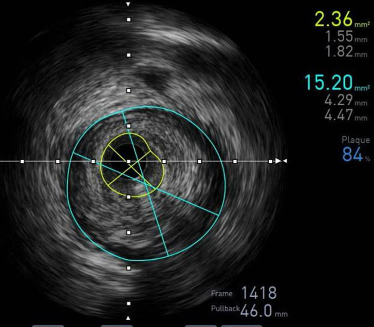
IVUS of the lesion before intervention. IVUS, intravascular ultrasound.
Figure 2.
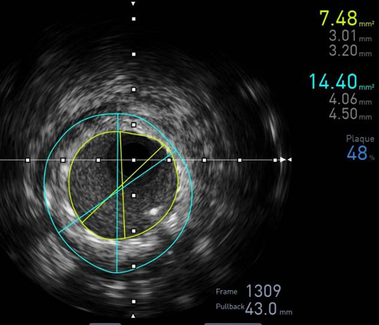
IVUS after intervention with VSTENT (D1). IVUS, intravascular ultrasound.
Figure 3.
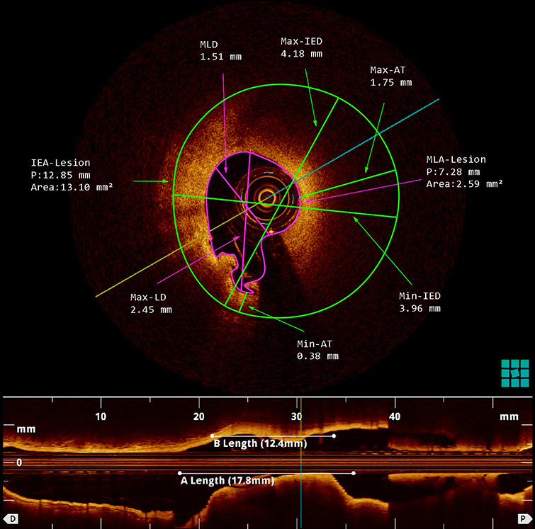
OCT of the lesion before intervention. OCT, optical coherence tomography.
Figure 4.
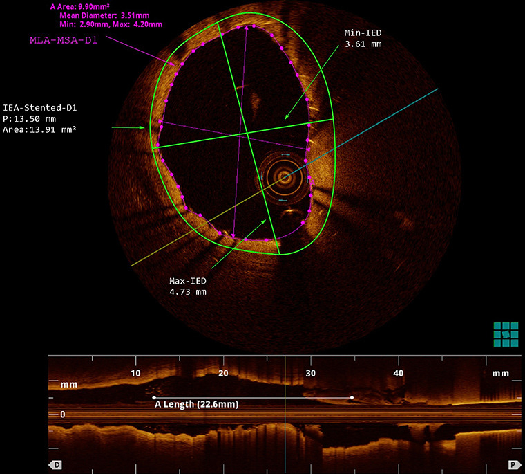
OCT after intervention with VSTENT (D1). OCT, optical coherence tomography.
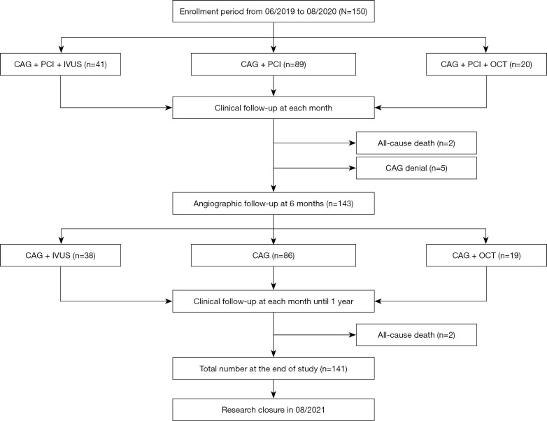
IVUS was intended to be performed in VN01 center with 25 prespecified patients, which ended up being 41 due to flexible regulation (Figure 5). To assure recruitment rate, 5 patients planned for the OCT group at the VN02 center were shifted to the VN01 center for IVUS. In addition, the VN04 center performed 11 additional IVUS procedures due to the availability of resources. All the regulations have been approved by the Committees. Thirty-eight of total 41 IVUS patients were assessed again at 6 months. The results at D180 showed no ruptured atheroschlerosis plaque, no dissection, thrombosis or aneurysm incidents recorded with all the stents were in accurate position (Figure 6). Neointimal hyperplasia area at minimum arteria lumen was 1.3±0.7 mm2 (95% CI: 1.1–1.5; P<0.001). Several IVUS parameters at D1 and D180 are presented in Table 3.
Figure 5.
Research patient flow. CAG, coronary angiogram; PCI, percutaneous coronary intervention; IVUS, intravascular ultrasound; OCT, optical coherence tomography.
Figure 6.
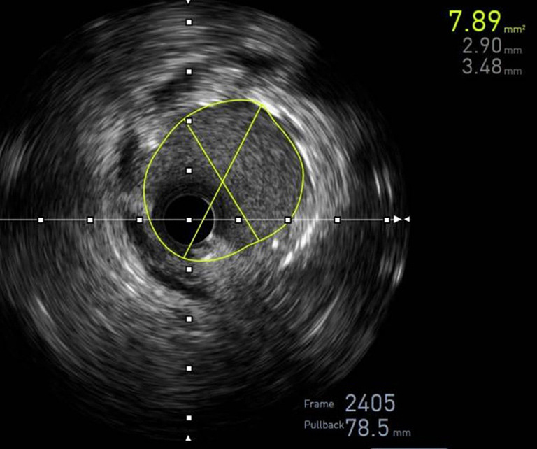
IVUS at 3-month follow-up (D180). IVUS, intravascular ultrasound.
On the other hand, OCT prespified group originally contained 25 patients, then reduced to 20 patients at the beginning of the study and 19 of them were assessed again at 6 months (Figure 5). At 180-day period, OCT showed all the stents were in proper position and no incidents were recorded (Figure 7). There were 2 lesions with plaques due to increased neointimal hyperplasia rate, but not translated in reduced blood flow nor patient’s clinical function. Several OCT parameters are presented in Table 3.
Figure 7.
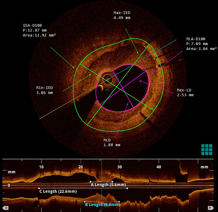
OCT at 3-month follow-up (D180). OCT, optical coherence tomography.
Clinical outcomes
All the recruited participants were followed up and proceeded with examination as planned, with laboratory orders when needed. 150 patients recalled 6 times reduction of angina episodes (from 100% when recruited to 17.3%), meanwhile dyspnea rate was 2 times reduction from 44.7% to 20% right after intervention. Table 4 showed clinical outcomes at 6-month and 1 year follow-up, in which major adverse clinical events were composite endpoint of target lesion revascularization (TLR), myocardial infarction (MI) related target vessel and cardiovascular death.
Table 4. Clinical outcomes at follow-up.
| Clinical outcomes | 6 months | 1 year | |||||
|---|---|---|---|---|---|---|---|
| N=150, n (%) | 95% CI | P | N=150, n (%) | 95% CI | P | ||
| All death | 2 (1.33) | 0.20–4.7 | 0.000 | 4 (2.67) | 0.7–6.7 | 0.000 | |
| Cardiovascular death (stroke) | 1 (0.67) | 0.00–3.7 | 0.000 | 1 (0.67) | 0.00–3.7 | 0.000 | |
| Target vessel myocardial infarction | 0 (0.00) | 0.00–2.4 | 0.000 | 0 (0.00) | 0.00–2.4 | 0.000 | |
| TLR | 6 (4.00) | 1.5–8.5 | 0.000 | 6 (4.00) | 1.5–8.5 | 0.000 | |
| MACE | 7 (4.67) | 1.9–9.4 | 0.000 | 7 (4.67) | 1.9–9.4 | 0.000 | |
| Stent thrombosis | 0 (0.00) | 0.00–2.4 | 0.000 | 0 (0.00) | 0.00–2.4 | 0.000 | |
TLR, target lesion revascularization; MACE, major adverse clinical events.
In-hospital outcomes
After a non-event in-hospital stay, all of the subjects fully recovered and were discharged.
Six-month follow-up
All patients underwent clinical examinations at 6-month and 1-year intervals. After 6 months, we documented two all-cause deaths, one of which was cardiovascular in nature (due to stroke). Additionally, six patients in QCA required revascularization of the target vessel. There was no evidence of stent thrombosis.
One-year follow-up
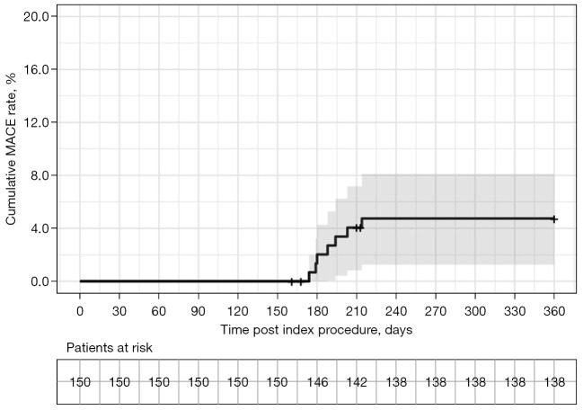
All patients were monitored for a period of 1 year. Between a 6-month and a 1-year period, we recorded two all-cause deaths, none of which were cardiovascular. During this time frame, we did not notice any revascularization of the target vessel or stent thrombosis. Thus, cardiovascular events occurred in seven instances after discharge for a period of 1 year (Table 4, Figure 8).
Figure 8.
Kaplan-Meier cumulative event curves of MACE for the sirolimus-eluting VSTENT stent. MACE, major adverse cardiac events.
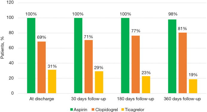
After 1 year, no patients recalled symptoms of angina or heart failure. Antiplatelet medications prescribed upon discharge, 30, 180, and 360 days are described in Figure 9.
Figure 9.
Antiplatelet medications prescribed upon discharge, 30, 180 and 360 days.
Discussion
This is a prospective, multicenter study designed to evaluate the safety and efficacy of a newly developed drug-diluting stent made in a developing country. The following are the major findings: the VSTENT stent demonstrated favorable efficacy, with minimal LLL accessed at 6-month follow-up. We recorded no myocardial infarction associated with the target vessel, no in-stent thrombosis, and a low rate of target vessel revascularization. At 6-month period, the VSTENT stent achieved complete stent apposition (100%) (95% CI: 93.6–100%; P<0.001) in IVUS and OCT cases. In-stent and in-segment re-stenosis rate were equivalent to those seen in prior investigations, which were low.
Our study cohort is a good representation of the percutaneous intervention population, with a male predominance and numerous major risk factors such as hypertension and dyslipidemia. The rejuvenation of ischemic heart disease was also evident in our investigation, as the mean age was significantly younger than that of most previous studies conducted decades ago (4,11,12), and comparable to that of the MERIT-2 experiment (13). Notable features of the VSTENT stent included the fact that 80% (95% CI: 72.7–86.1%; P<0.001) of patients had an acute coronary syndrome, which resulted in a higher rate of lumen stenosis prior to intervention than in earlier studies (4,12). Additionally, the lesions treated in this study were relatively lengthy, 23 mm on average, reflecting the reality of coronary intervention in Vietnam. As a result, the average stent length employed in this study was 27.6±7.3 mm (95% CI: 26.6–28.6; P<0.001), with 58.5% (95% CI: 51.5–65.2%; P=0.16) of all stents exceeding 28mm.
In terms of LLL, our study found that implanting the VSTENT stent resulted in a minor in-stent and in-segment LLL of 0.08±0.19 mm (95% CI: 0.05–0.10; P<0.001) and 0.07±0.31 mm (95% CI: 0.03–0.11; P=0.002). These findings were consistent with those of the meriT-V study, which indicated an in-stent LLL of 0.15±0.27 mm for the BioMime sirolimus-eluting stent (14). The stuty aimed to evaluate the safety and efficacy of a new sirolimus-eluting stent in comparison with a everolimus-eluting stent, finding no significant difference in in-stent LLL between the two stents. Another sirolimus-eluting stent with bioresorbable polymer (BP-SES) was compared against an everolimus-eluting, permanent polymer stent (PP-EES) in the CENTURY II study (4). According to this study, in-stent LLL for BP-SES and PP-EES were 0.26 and 0.18 mm, respectively (P=0.003), while in-segment LLL were 0.09 and 0.10 mm (P=0.59). The observed variations in lumen loss between the two stents did not translate into greater restenosis rates [in-stent restenosis (ISR): 1.21 vs. 1.27, P=0.96] nor in any measurable difference in revascularization rates (4.97% vs. 8.22%, P=0.08). Additionally, our study discovered a low rate of binary in-stent and in-segment restenosis of 0.5% (95% CI: 0.0–2.8%; P<0.001) and 3% (95% CI: 1.1–6.4%; P<0.001) and 3%, respectively. This finding reaffirmed that lumen loss within the stent is highly correlated with restenosis rate and the requirement for re-intervention (15).
Even though the initial generation of DES stents was found to be superior to bare metal stents in terms of minimizing in-stent re-stenosis, various occurrences emerged after a longer period of observation, including delayed re-stenosis and stent thrombosis (16,17). There have been several hypotheses put forward, including the possibility that bigger struts and a less biocompatible polymer could trigger a more severe inflammatory response (18). To overcome these drawbacks, the bioresorbable polymer DES was developed (19). The major advances for the next generation of stents would be early effects that are equivalent to those of durable polymer DES while being safer following complete polymer resorption of the stent. This is primarily accomplished using the VSTENT stent, which has cobalt-chromium struts and diluted sirolimus with bioresorbable polymer. Furthermore, with the assistance of a dilation balloon before and after stenting, we were able to thoroughly optimize the process and deliver the most effective treatment possible in the case of coronary artery disease. According to the VSTENT study, the rate of pre-dilation balloon and post-dilation balloon dilation was 76.4% (95% CI: 70.1–82.0%; P<0.001) and 83.5% (95% CI: 77.8–88.2%; P<0.001), respectively. Similar results had been seen in other studies (4,12,20). During their hospital stay, all the subjects were completely recovered. There have been no incidences of cardiac arrest, coronary artery perforation, side branch loss, or entrance hematoma recorded. The mean remaining lumen stenosis was 9.6% (95% CI: 9.2–10.1%; P<0.001), and there was no residual stenosis more than 30%, resulting in a 100% (95% CI: 98.3–100%; P<0.001) device success rate. These findings significantly support the use of the VSTENT stent with sirolimus dilution agents in percutaneous coronary intervention.
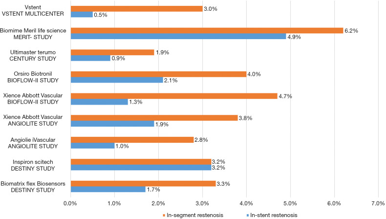
Other similar trials have shown a re-stenosis rate between 2.8% and 6.2% (11,12,14,20) (Figure 10), but our study found only a 3.5% (95% CI: 1.4–7.1%; P<0.001) incidence, which was considered to be a low re-stenosis rate given the small sample size. As a result, one of the most essential objectives of our study, which was to reduce the rate of re-stenosis, was achieved. Patient characteristics, stent quality, and intervention technique could all be factors in the development of binary lumen stenosis over a 6-month period. Using target-vessel re-vascularization, we were able to treat these individuals. All of them were in good health at the 12-month follow-up, demonstrating the effectiveness of VSTENT in lowering the re-stenosis rate to less than 5%.
Figure 10.
Re-stenosis rate in major diluted stent trials (4,11,20-22).
In terms of major cardiovascular events, the VSTENT study rate was comparatively low when compared to the rates of previous trials. During the study, there were no cases of myocardial infarction and just one case of cardiovascular mortality due to stroke. Furthermore, no stent thrombosis was observed after 12 months, which was consistent with the findings of other trials. In this investigation, acute coronary syndrome (ACS) was present in 80% (95% CI: 72.7–86.1%; P<0.001) of the participants, resulting in increased ischemia burden. Additionally, AHA type C complex lesions accounted for up to 39.6% of all lesions, and the adaptation of a longer stent made it more difficult to optimize the treatment during the study. Despite this, when compared to other similar diluted sirolimus stents, such as the ORSIRO (11), Ultimaster (4), Angiolite (12), and Biomime (21), the VSTENT stent nevertheless produced comparable MACE results, which were low.
Study limitations
There are some drawbacks to this study. First and foremost, those with chronic total occlusion, severe calcified lesion, lesion at the left main artery, significant thrombus burden, or tortuous target vessels were eliminated from the study. As a result, the results can only be applied to the previously indicated specific demographic. Apart from that, this study was not designed as randomization; instead, we compared the outcomes to data from previous trials. Even though these limitations are significant, none of them is severe enough to call into question the validity of the primary study findings.
Conclusions
This study validated a new generation of DES and contributed to the body of knowledge, promoting the goal of globalizing interventional practice with this type of stent. This also aims to give patients the best equipment. Procedure and device success rates were excellent. IVUS and OCT showed excellent LLL at 6 months. At 1 year follow-up, the new VSTENT had low angiographic ISR and TLR, resulting in minimal major cardiovascular events. The VSTENT trial shows the efficacy and safety of sirolimus-eluting polymer bioresorbable VSTENT stent in percutaneous coronary intervention with intravascular imaging findings over a 6-month follow-up. This could be an affordable interventional cardiology treatment in underdeveloped nations.
Impact on daily practice
This study made a substantial contribution to the body of information, furthering the objective of globalizing the use of a new stent in interventional field. In developing nations, the VSTENT stent is not only an effective and safe option, but also an economical one.
Supplementary
The article’s supplementary files as
Acknowledgments
We are grateful to our colleagues at University Medical Center Ho Chi Minh City, Khanh Hoa General Hospital, Ba Ria Hospital, Kien Giang General Hospital and Thong Nhat Hospital for their great assistance.
Funding: This study was supported by USM Healthcare, Vietnam and Ho Chi Minh City Department of Science and Technology, Vietnam.
Ethical Statement: The authors are accountable for all aspects of the work in ensuring that questions related to the accuracy or integrity of any part of the work are appropriately investigated and resolved. The study was conducted in accordance with the Declaration of Helsinki (as revised in 2013). The study was approved by Ethics Boards of University of Medicine and Pharmacy at Ho Chi Minh City (No. 293/ĐHYD-HĐĐĐ), Thong Nhat Hospital (No. 02/BVTN-HDYD), Khanh Hoa General Hospital (No. 74/BVĐKT), Ba Ria Hospital (No. 2792/QĐ-BVBR), Kien Giang General Hospital (No. 03/HĐ-KHKT) and informed consent was taken from all individual participants.
Footnotes
Reporting Checklist: The authors have completed the STROBE reporting checklist. Available at https://cdt.amegroups.com/article/view/10.21037/cdt-22-522/rc
Data Sharing Statement: Available at https://cdt.amegroups.com/article/view/10.21037/cdt-22-522/dss
Conflicts of Interest: All authors have completed the ICMJE uniform disclosure form (available at https://cdt.amegroups.com/article/view/10.21037/cdt-22-522/coif). The authors have no conflicts of interest to declare.
References
- 1.Moses JW, Leon MB, Popma JJ, et al. Sirolimus-eluting stents versus standard stents in patients with stenosis in a native coronary artery. N Engl J Med 2003;349:1315-23. 10.1056/NEJMoa035071 [DOI] [PubMed] [Google Scholar]
- 2.Stone GW, Midei M, Newman W, et al. Comparison of an everolimus-eluting stent and a paclitaxel-eluting stent in patients with coronary artery disease: a randomized trial. JAMA 2008;299:1903-13. 10.1001/jama.299.16.1903 [DOI] [PubMed] [Google Scholar]
- 3.Jeremias A, Kirtane A. Balancing efficacy and safety of drug-eluting stents in patients undergoing percutaneous coronary intervention. Ann Intern Med 2008;148:234-8. 10.7326/0003-4819-148-3-200802050-00199 [DOI] [PubMed] [Google Scholar]
- 4.Barbato E, Salinger-Martinovic S, Sagic D, et al. A first-in-man clinical evaluation of Ultimaster, a new drug-eluting coronary stent system: CENTURY study. EuroIntervention 2015;11:541-8. 10.4244/EIJY14M08_06 [DOI] [PubMed] [Google Scholar]
- 5.Ho MY, Chen CC, Wang CY, et al. The Development of Coronary Artery Stents: From Bare-Metal to Bio-Resorbable Types. Metals 2016;6:168. 10.3390/met6070168 [DOI] [Google Scholar]
- 6.Ako J, Bonneau HN, Honda Y, et al. Design criteria for the ideal drug-eluting stent. Am J Cardiol 2007;100:3M-9M. 10.1016/j.amjcard.2007.08.016 [DOI] [PubMed] [Google Scholar]
- 7.Pendyala LK, Yin X, Li J, et al. The first-generation drug-eluting stents and coronary endothelial dysfunction. JACC Cardiovasc Interv 2009;2:1169-77. 10.1016/j.jcin.2009.10.004 [DOI] [PubMed] [Google Scholar]
- 8.Ali RM, Abdul Kader MASK, Wan Ahmad WA, et al. Treatment of Coronary Drug-Eluting Stent Restenosis by a Sirolimus- or Paclitaxel-Coated Balloon. JACC Cardiovasc Interv 2019;12:558-66. 10.1016/j.jcin.2018.11.040 [DOI] [PubMed] [Google Scholar]
- 9.Kobo O, Saada M, Meisel SR, et al. Modern Stents: Where Are We Going? Rambam Maimonides Med J 2020;11:e0017. 10.5041/RMMJ.10403 [DOI] [PMC free article] [PubMed] [Google Scholar]
- 10.Palmerini T, Biondi-Zoccai G, Della Riva D, et al. Clinical outcomes with bioabsorbable polymer- versus durable polymer-based drug-eluting and bare-metal stents: evidence from a comprehensive network meta-analysis. J Am Coll Cardiol 2014;63:299-307. 10.1016/j.jacc.2013.09.061 [DOI] [PubMed] [Google Scholar]
- 11.Kandzari DE, Mauri L, Koolen JJ, et al. Ultrathin, bioresorbable polymer sirolimus-eluting stents versus thin, durable polymer everolimus-eluting stents in patients undergoing coronary revascularisation (BIOFLOW V): a randomised trial. Lancet 2017;390:1843-52. 10.1016/S0140-6736(17)32249-3 [DOI] [PubMed] [Google Scholar]
- 12.Puri R, Otaegui I, Sabaté M, et al. Three- and 6-month optical coherence tomographic surveillance following percutaneous coronary intervention with the Angiolite® drug-eluting stent: The ANCHOR study. Catheter Cardiovasc Interv 2018;91:435-43. 10.1002/ccd.27189 [DOI] [PubMed] [Google Scholar]
- 13.Patted SV, Jain RK, Jiwani PA, et al. Clinical Outcomes of Novel Long-Tapered Sirolimus-Eluting Coronary Stent System in Real-World Patients With Long Diffused De Novo Coronary Lesions. Cardiol Res 2018;9:350-7. 10.14740/cr795 [DOI] [PMC free article] [PubMed] [Google Scholar]
- 14.Abizaid A, Kedev S, Kedhi E, et al. Randomised comparison of a biodegradable polymer ultra-thin sirolimus-eluting stent versus a durable polymer everolimus-eluting stent in patients with de novo native coronary artery lesions: the meriT-V trial. EuroIntervention 2018;14:e1207-14. 10.4244/EIJ-D-18-00762 [DOI] [PubMed] [Google Scholar]
- 15.Mauri L, Orav EJ, Candia SC, et al. Robustness of late lumen loss in discriminating drug-eluting stents across variable observational and randomized trials. Circulation 2005;112:2833-9. 10.1161/CIRCULATIONAHA105.570093 [DOI] [PubMed] [Google Scholar]
- 16.Liu MW, Roubin GS, King SB, 3rd. Restenosis after coronary angioplasty. Potential biologic determinants and role of intimal hyperplasia. Circulation 1989;79:1374-87. 10.1161/01.CIR.79.6.1374 [DOI] [PubMed] [Google Scholar]
- 17.Byrne RA, Joner M, Kastrati A. Stent thrombosis and restenosis: what have we learned and where are we going? The Andreas Grüntzig Lecture ESC 2014. Eur Heart J 2015;36:3320-31. 10.1093/eurheartj/ehv511 [DOI] [PMC free article] [PubMed] [Google Scholar]
- 18.Sollott SJ, Cheng L, Pauly RR, et al. Taxol inhibits neointimal smooth muscle cell accumulation after angioplasty in the rat. J Clin Invest 1995;95:1869-76. 10.1172/JCI117867 [DOI] [PMC free article] [PubMed] [Google Scholar]
- 19.Souza CF, El Mouallem AM, de Brito Júnior FS, et al. Safety and efficacy of biolimus-eluting stent with biodegradable polymer: insights from EINSTEIN (Evaluation of Next-generation drug-eluting STEnt IN patients with coronary artery disease) Registry. Einstein (Sao Paulo) 2013;11:350-6. 10.1590/S1679-45082013000300015 [DOI] [PMC free article] [PubMed] [Google Scholar]
- 20.Prado GFA, Jr, Abizaid AAC, Meireles GC, et al. Comparative clinical performance of two types of drug-eluting stents with abluminal biodegradable polymer coating: Five-year results of the DESTINY randomized trial. Rev Port Cardiol (Engl Ed) 2021;40:71-6. 10.1016/j.repc.2020.05.017 [DOI] [PubMed] [Google Scholar]
- 21.Dani S, Costa RA, Joshi H, et al. First-in-human evaluation of the novel BioMime sirolimus-eluting coronary stent with bioabsorbable polymer for the treatment of single de novo lesions located in native coronary vessels - results from the meriT-1 trial. EuroIntervention 2013;9:493-500. 10.4244/EIJV9I4A79 [DOI] [PubMed] [Google Scholar]
- 22.Moreu J, Moreno-Gómez R, Pérez de Prado A, et al. First-in-man randomised comparison of the Angiolite durable fluoroacrylate polymer-based sirolimus-eluting stent versus a durable fluoropolymer-based everolimus-eluting stent in patients with coronary artery disease: the ANGIOLITE trial. EuroIntervention 2019;15:e1081-9. 10.4244/EIJ-D-19-00206 [DOI] [PubMed] [Google Scholar]
Associated Data
This section collects any data citations, data availability statements, or supplementary materials included in this article.
Supplementary Materials
The article’s supplementary files as


