

Abstract
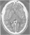
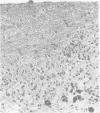
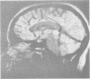
Seven cases of clinically symptomatic benign glial cyst of the pineal gland are reported. The cysts' size ranged from 1.0-4.5 cm in diameter. They were characterised by a golden or, less frequently, brown-reddish proteinaceous or haemorrhagic fluid content. The cyst wall, up to 2 mm thick, consisted of clusters of normal pineal parenchymal cells, often compressed and distorted, surrounded by reactive gliotic tissue which sometimes contained Rosenthal fibres. The presenting clinical features included headache (6/7), signs of raised intracranial pressure, partial or complete Parinaud's syndrome (5/7), cerebellar deficits (2/7), corticospinal and corticopontine fibre (2/7) or sensory (1/7) deficits, and emotional disturbances (2/7). CT and MRI (in 2/7 cases) scans showed a hypodense or nonhomogeneous lesion in the region of the pineal gland, with or without contrast enhancement. Surgical excision resulted in complete clearance of the symptoms in 5/7 patients. The previous literature is reviewed and the clinicopathological correlations and the possible pathogenetic mechanisms are discussed. The need to distinguish this benign lesion from other mass lesions of the pineal region, in particular from pinealocytoma, is stressed.
Full text
PDF


Images in this article

Selected References
These references are in PubMed. This may not be the complete list of references from this article.
- ARIETI S. The pineal gland in old age. J Neuropathol Exp Neurol. 1954 Jul;13(3):482–491. doi: 10.1097/00005072-195407000-00009. [DOI] [PubMed] [Google Scholar]
- Apuzzo M. L., Davey L. M., Manuelidis E. E. Pineal apoplexy associated with anticoagulant therapy. Case report. J Neurosurg. 1976 Aug;45(2):223–226. doi: 10.3171/jns.1976.45.2.0223. [DOI] [PubMed] [Google Scholar]
- Chik C. L., Talalla A., Brown G. M. Effect of pinealectomy on serum melatonin, luteinizing hormone and prolactin: a case report. Clin Endocrinol (Oxf) 1985 Oct;23(4):367–372. doi: 10.1111/j.1365-2265.1985.tb01093.x. [DOI] [PubMed] [Google Scholar]
- Cooper E. R. The Human Pineal Gland and Pineal Cysts. J Anat. 1932 Oct;67(Pt 1):28–46. [PMC free article] [PubMed] [Google Scholar]
- Hasegawa A., Ohtsubo K., Mori W. Pineal gland in old age; quantitative and qualitative morphological study of 168 human autopsy cases. Brain Res. 1987 Apr 21;409(2):343–349. doi: 10.1016/0006-8993(87)90720-7. [DOI] [PubMed] [Google Scholar]
- Higashi K., Katayama S., Orita T. Pineal apoplexy. J Neurol Neurosurg Psychiatry. 1979 Nov;42(11):1050–1053. doi: 10.1136/jnnp.42.11.1050. [DOI] [PMC free article] [PubMed] [Google Scholar]
- Kabuto M., Hayashi M., Kawano H., Kobayashi H., Ishii H., Shirasaki N., Noguchi Y., Hirose S. [A case of non-neoplastic pineal cyst presenting Parinaud's syndrome]. No Shinkei Geka. 1987 Mar;15(3):335–338. [PubMed] [Google Scholar]
- Lee D. H., Norman D., Newton T. H. MR imaging of pineal cysts. J Comput Assist Tomogr. 1987 Jul-Aug;11(4):586–590. doi: 10.1097/00004728-198707000-00005. [DOI] [PubMed] [Google Scholar]
- Lum G. B., Williams J. P., Machen B. C., Akkaraju V. Benign cystic pineal lesions by magnetic resonance imaging. J Comput Tomogr. 1987 Jul;11(3):228–235. doi: 10.1016/0149-936x(87)90087-7. [DOI] [PubMed] [Google Scholar]
- RINGERTZ N., NORDENSTAM H., FLYGER G. Tumors of the pineal region. J Neuropathol Exp Neurol. 1954 Oct;13(4):540–561. doi: 10.1097/00005072-195410000-00004. [DOI] [PubMed] [Google Scholar]
- Richardson J. K., Hirsch C. S. Sudden, unexpected death due to "pineal apoplexy". Am J Forensic Med Pathol. 1986 Mar;7(1):64–68. doi: 10.1097/00000433-198603000-00014. [DOI] [PubMed] [Google Scholar]
- SEVITT S., SCHORSTEIN J. A case of pineal cyst. Br Med J. 1947 Sep 27;2(4525):490–490. doi: 10.1136/bmj.2.4525.490. [DOI] [PMC free article] [PubMed] [Google Scholar]