Abstract
Background
Tinea pedis is one of the most common superficial fungal infections of the skin, with various clinical manifestations. This review aims to familiarize physicians with the clinical features, diagnosis and management of tinea pedis.
Methods
A search was conducted in April 2023 in PubMed Clinical Queries using the key terms ‘tinea pedis’ OR ‘athlete’s foot’. The search strategy included all clinical trials, observational studies and reviews published in English within the past 10 years.
Results
Tinea pedis is most often caused by Trichophyton rubrum and Trichophyton interdigitale. It is estimated that approximately 3% of the world population have tinea pedis. The prevalence is higher in adolescents and adults than in children. The peak age incidence is between 16 and 45 years of age. Tinea pedis is more common amongst males than females. Transmission amongst family members is the most common route, and transmission can also occur through indirect contact with contaminated belongings of the affected patient. Three main clinical forms of tinea pedis are recognized: interdigital, hyperkeratotic (moccasin-type) and vesiculobullous (inflammatory). The accuracy of clinical diagnosis of tinea pedis is low. A KOH wet-mount examination of skin scrapings of the active border of the lesion is recommended as a point-of-care testing. The diagnosis can be confirmed, if necessary, by fungal culture or culture-independent molecular tools of skin scrapings. Superficial or localized tinea pedis usually responds to topical antifungal therapy. Oral antifungal therapy should be reserved for severe disease, failed topical antifungal therapy, concomitant presence of onychomycosis or in immunocompromised patients.
Conclusion
Topical antifungal therapy (once to twice daily for 1–6 weeks) is the mainstay of treatment for superficial or localized tinea pedis. Examples of topical antifungal agents include allylamines (e.g. terbinafine), azoles (e.g. ketoconazole), benzylamine, ciclopirox, tolnaftate and amorolfine. Oral antifungal agents used for the treatment of tinea pedis include terbinafine, itraconazole and fluconazole. Combined therapy with topical and oral antifungals may increase the cure rate. The prognosis is good with appropriate antifungal treatment. Untreated, the lesions may persist and progress.
Keywords: athlete’s foot, dermatophytosis, interdigital, moccasin, Trichophyton interdigitale, Trichophyton rubrum, vesiculobullous
Introduction
Tinea pedis (commonly known as ‘athlete’s foot’) refers to a superficial fungal infection of the skin on the foot caused predominantly by dermatophytes. Worldwide, tinea pedis is one of the most common, if not the most common, superficial fungal infections of the skin after puberty and its prevalence is increasing.1–9 The condition was first described by Pellizzari in 1888.10 Tinea pedis is a public health concern because it is contagious and recurrent and serves as an important reservoir for dermatophytosis elsewhere in the body.
Methods
A search was conducted in April 2023 in PubMed Clinical Queries using the key terms “tinea pedis” OR “athlete’s foot”. The search strategy included all clinical trials (including open trials, non-randomized controlled trials and randomized controlled trials), observational studies (including case reports and case series) and reviews (including narrative reviews, clinical guidelines and meta-analyses) published within the past 10 years. Google, UpToDate and Wikipedia were also searched to enrich the review. Only papers published in English were included in this review. The information retrieved from the search was used in the compilation of the present article.
Review
Aetiology
The most common etiological agents are Trichophyton rubrum and Trichophyton interdigitale.11–18 The predominating dermatophytes may vary with geographic locations (related to climate characteristics and social factors) and change over time.19 Other less common causative dermatophytes include Epidermophyton floccosum, Trichophyton tonsurans, Trichophyton soudanense, Trichophyton violaceum and Microsporum audouinii.20,21 Non-dermatophyte moulds, such as Neoscytalidium hyalinum, Neoscytalidium dimidiatum, Scopulariopsis brevicaulis and Fusarium species, are uncommon causes of tinea pedis.14,20,22 Rarely, Cylindrocarpon lichenicola and yeasts primarily Candida species may also cause tinea pedis.14,20,22
Epidemiology
It is estimated that the global prevalence of tinea pedis is approximately 3%.2,23 The lifetime risk is up to 70%.2,23 The prevalence is higher in adolescents and adults than in prepubertal children.24,25 In the past decades, there has been an increase in prevalence due, at least in part, to an increase in recreational activities and growing urbanization.26 The peak age of incidence is between 16 and 45 years, when working and leisure activities are at a maximum.27 The condition is more common in industrial countries.28,29 The male to female ratio is approximately 3:1.30,31 Tinea pedis affects individuals of all races. In the United States, the condition is more common amongst Blacks and Hispanics.32 Humans may become infected through close contact with infected persons, animals (in particular, house pets), contaminated fomites or soil.25 Transmission amongst family members is the most common route; children often become infected by direct contact with spores of the causative organism or infected skin fragments shed by household contact.25 On the other hand, transmission can also occur through indirect contact with contaminated belongings (e.g. shoes, socks, bedding) of an affected individual.33,34 Autoinfection by dermatophytes elsewhere in the body may also occur.25 The transmission of tinea pedis is facilitated by prolonged wearing of occlusive footwear (promoting a moist, warm local environment and a high dew point of footwear).11,13,30,31 The condition is more prevalent amongst athletes (especially common amongst elite athletes who walk barefoot in swimming pool facilities or locker rooms), military personnel, manual laborers, individuals in long-term care facilities and homeless individuals.35–45 In one study of the 169 employees at 21 swimming pools in the Netanya area, Israel, 78 (46%) of the employees had concurrent onychomycosis and tinea pedis and 50 (30%) had tinea pedis only; the diagnosis was based on KOH microscopy and fungal culture.46 Past history of tinea pedis, concurrent tinea pedis amongst family members, hot humid climates, hyperhidrosis (especially plantar hyperhidrosis), prolonged exposure of the feet to water, communal bathing/sharing washing facilities, use of public swimming pools, insufficient foot care, poor personal hygiene, maceration or breaks in the pedal skin, diabetes mellitus, peripheral vascular disease, atopic dermatitis, psoriasis, obesity, immunodeficiency, depression, schizophrenia, and genetic predisposition or susceptibility are other predisposing factors.47–58
Pathophysiology
The causative fungus produces and releases enzymes, such as proteases, that digest keratin and keratinase that penetrates keratinized tissue.59 The hyphae then invade the stratum corneum and keratin and spread centrifugally outward. Infection is usually cutaneous and limited to the non-living cornified layers because the fungus is not able to penetrate the deeper tissue of a healthy immunocompetent host.59 Scaling results from increased epidermal replacement following inflammation. The wall of the fungus also contains mannans, which suppress the body’s immune system, decrease lymphoproliferative response and reduce the proliferation of keratinocytes. The latter results in a decreased rate of sloughing of the affected skin and prolongs the infection. Sweating and warmth promote growth of the fungus.
Histopathology
Histological findings include hyperkeratosis, acanthosis and presence of neutrophils in the dermis, hyphae between the lower layer of the parakeratotic stratum corneum and the upper normal basket-weave stratum corneum (‘sandwich sign’).2 Hyphae may be more readily seen on haematoxylin and eosin staining.60
Clinical manifestations
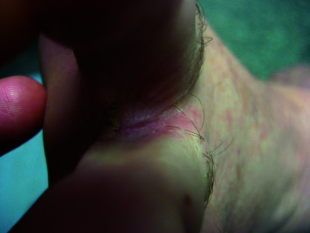
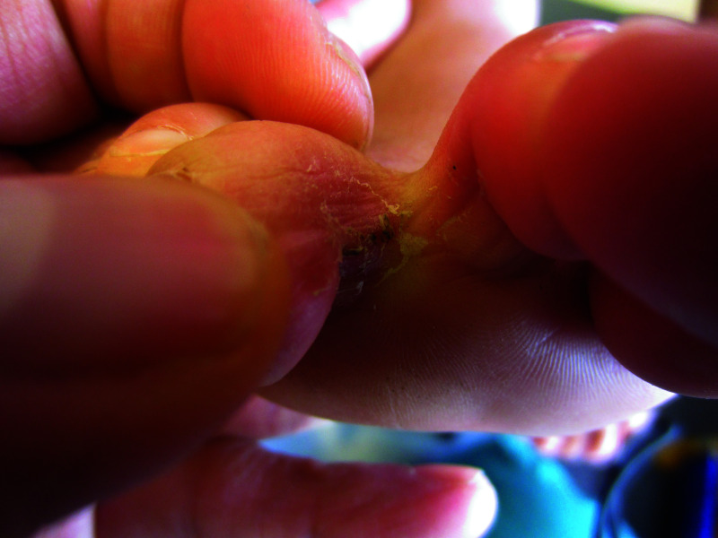
Three main clinical forms of tinea pedis are recognized: interdigital tinea pedis, hyperkeratotic (moccasin-type) tinea pedis and vesiculobullous (inflammatory) tinea pedis.6,35,43 Interdigital tinea pedis, the most common form (especially in children), presents with erythema, silvery white scaling, peeling and maceration in the web spaces, typically in the web space between the fourth and fifth toes (most common) (Figures 1 and 2).61–63 The web space may become white and soggy.64 Presumably, the narrow interdigital spaces in children’s feet may predispose them to this form of tinea pedis.65 Pruritus is the most common symptom.64,66 Often, there is peripheral fissuring, which may cause pain and burning sensation.2,19 Adjacent areas such as the sole may also be affected (Figure 2). Secondary bacterial infections with gram-positive bacteria (Staphylococcus aureus, streptococci) and gram-negative bacteria (Escherichia coli, Klebsiella species, Proteus species, Pseudomonas aeruginosa) in the interdigital areas can result in foul odour, maceration, erosions and crusting – a condition referred to as dermatophytosis complex.67
Figure 1.
Interdigital tinea pedis presenting with maceration, desquamation and fissuring between the toes.
Figure 2.
Interdigital tinea pedis presenting with erythema and scaling between the toes and the adjacent skin.
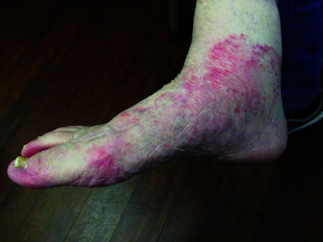
Hyperkeratotic (moccasin-type) tinea pedis, the second most common form of tinea pedis, is characterized by chronic, hyperkeratotic, scaling plaques with varying degrees of underlying erythema on the heels, soles, lateral and medial sides and back of the foot, and distal dorsum of the foot (corresponding to the skin covered by this type of footwear) (Figure 3).6,19,68 The dorsum of the foot, except the distal portion, is typically spared.4,66 Erythema is more prominent on the distal dorsum of one or both feet.19 A collarette of scale can be seen along the border of the feet in a moccasin-type distribution.26 The leading edge of the eruption may be arcuate, annular and slightly elevated.69 Occasionally, fissures may develop. Hyperkeratotic tinea pedis can be slightly pruritic but most often is asymptomatic.35,43 The condition is usually chronic and quite resistant to treatment.35,43
Figure 3.
Diffuse scale and erythema on the sole and medial aspect of the foot in a patient with hyperkeratotic (moccasin-type) tinea pedis.
Vesiculobullous (inflammatory) tinea pedis (often acquired from animals) typically presents with intensely pruritic (sometimes painful) vesicles and/or bullae on a background of erythema (Figures 4 and 5). Vesicles typically range from 1 to 3 mm in diameter. Coalescence of vesicles leads to the formation of bulla.2 Sites of predilection include the instep or medial plantar surface of the foot.68,70 The lesions develop far more rapidly than the other forms of tinea pedis.26 Rupture of the vesicles and/or bullae releases serous or purulent fluid with exposure of a red, raw and oozing surface.2,71
Figure 4.
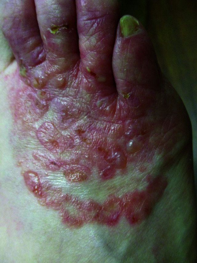
Vesiculobullous (inflammatory) tinea pedis presenting with pruritic vesicles and bullae over a background of erythema on the dorsal surface of the right foot.
Figure 5.
Vesiculobullous (inflammatory) tinea pedis presenting with pruritic vesicles and bullae over a background of erythema on the medial plantar surface of the left foot.
Other clinical variants include occult tinea pedis, ulcerative tinea pedis and tinea incognito. Occult tinea pedis is common, particularly amongst elite athletes.72,73 In one study of 150 regular swimmers, 22 (15%) swimmers had positive cultures for dermatophytes from toe-web samples;72 8 (36%) of these patients had no lesions.72 In another study of 405 marathon runners, scrapings were taken from the fourth toe-web space of both feet and sent for fungal culture.73 Cultures for dermatophytes were positive in 89 (22%) marathon runners, 43 (10.6%) of whom had no lesions. Individuals with occult tinea pedis are asymptomatic and they are often unaware of tinea pedis.2 The diagnosis of occult tinea pedis is easily missed unless it is specifically sought. The majority of occult tinea pedis cases are caused by Trichophyton interdigitale.2,72
Ulcerative tinea pedis is rare and typically presents with rapidly spreading interdigital erosions and ulcers that are more severe than those seen in vesiculobullous tinea pedis.26,66 The condition is more commonly seen in patients with peripheral vascular disease, diabetes mellitus or immunodeficiency.74 Ulcerative tinea pedis is often complicated by secondary bacterial infection, which can be severe and debilitating.26
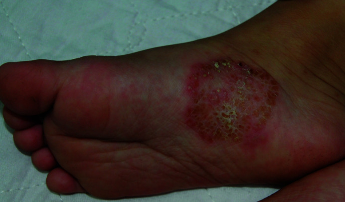
Tinea incognito (also referred to as tinea atypica) (Figure 6) may result if tinea pedis is mistakenly treated with immunosuppressive agents, notably calcineurin inhibitors or corticosteroids, leading to loss of typical appearance of the lesion. Pruritus may be minimal or absent.75,76 The active margin of the lesion may be lost, erythema and scale may be attenuated, and papules and pustules may be present.4
Figure 6.
Tinea incognito in a patient with tinea pedis treated with topical corticosteroid resulting in loss of typical appearance of the lesion.
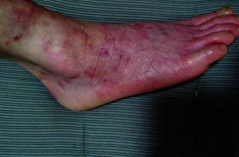
Very rarely, tinea pedis presents as scattered, asymptomatic, purpuric papules on the plantar surface of the foot in the absence of pruritus, erythema, maceration, desquamation or vesicles/bullae.77 Physicians should be aware of the rare variant so that appropriate treatment can be initiated. Untreated tinea pedis poses a public health hazard as the condition is contagious.
A concomitant secondary eruption, usually in the form of symmetric pruritic vesicles or papules, may occur at distant sites, such as the palmar aspect of the hands and sides of the fingers (referred to as a dermatophytid reaction), presumably due to an immunological reaction to the fungus.78,79 It is estimated 7–17% of patients with tinea pedis develop a dermatophytid reaction;2,79 the eruption does not contain fungal elements and eradication of tinea pedis results in a cure of the eruption.66
Diagnosis
Many physicians make the diagnosis of tinea pedis clinically. However, the accuracy of clinical diagnosis of tinea pedis is low. In one study, the specificities and sensitivities of clinical diagnosis were 0.94 and 0.47 for tinea pedis in the plantar area, respectively, and 0.95 and 0.37, respectively, for tinea pedis in the interdigital area.80
Reflectance confocal microscopy, a non-invasive, real-time imaging of the superficial layers of the skin, can be used as a tool for the diagnosis of tinea pedis, though this is seldom performed at present.81 With tinea pedis, reflectance confocal microscopy typically shows branching hyphae and inflammatory infiltrate in the stratum corneum.81
To avoid misdiagnosis, especially in the setting of tinea incognito, a KOH wet-mount examination of skin scrapings of the active border of the lesion or the roof of a vesicle taken with a sterile instrument can be performed.82,83 A drop of 10–20% solution of KOH, with or without dimethyl sulfoxide, is added to the scrapings on a microscopic slide. The specimen is then gently heated to accelerate the destruction of the squamous cells. KOH dissolves the epithelial tissue, leaving behind easily visualized dichotomously branched, septate hyphae and spores. The addition of 20–40% of dimethyl sulfoxide to the KOH preparation allows rapid examination of the scrapings even without heating. Direct microscopic examination of a KOH wet-mount preparation of skin scrapings of the active border of the lesion or the roof of a vesicle, however, cannot distinguish pathogenic species of dermatophytes and whether the dermatophyte is alive. On the other hand, the procedure is easily performed, the cost is low and the result is readily available (usually less than 2 hours). A pooled analysis of data from 460 patients showed that the specificity and sensitivity of the test was 42.5% (95% CI 36.6–48.6) and 73.3% (95% CI 66.3–79.5), respectively.84 Some authors suggest repetition of the test if the initial result is negative, and the clinical picture is suggestive of tinea pedis.85
Although fungal culture is the gold standard to diagnosing dermatophytosis and the pathogenic species, culture is rarely needed, unless the diagnosis is still in doubt after KOH wet-mount examination of skin scrapings of the active border of the lesion or roof of a vesicle, or the infection is severe, widespread or resistant to antifungal treatment. Culture is expensive and it takes 7–14 days for results to be available. The most common culture medium is Sabouraud dextrose agar, which contains 10 g/L peptone, 40 g/L dextrose and 15–20 g/L agar, at pH 5.6. As contamination or coinfection with Gram-negative bacteria may result in decreased sensitivity of cultures, gentamycin or chloramphenicol (0.005%) is added to reduce bacterial contamination.35,60 Cycloheximide should not be added to the culture medium because it inhibits growth of non-dermatophyte moulds, which may be a cause of tinea pedis. A skin biopsy for histopathology is rarely necessary but, when performed, may show fungal elements and neutrophils within the stratum corneum.
More recently, a DNA-chip assay of the clinical specimen has been developed that allows rapid diagnosis of tinea pedis.86 In the DNA-chip assay, fungal DNA is amplified by using multiplex polymerase chain reaction with subsequent determination by hybridization on a microarray chip.86 The DNA-chip assay has a high sensitivity (90.7%).86 The sensitivity can be further increased to 96.3% by factoring results from microscopic examination of KOH wet-mount preparation or direct immunofluorescence microscopy.86 However, the DNA-chip assay is expensive and unavailable in most physicians’ offices or laboratories.
Differential diagnosis
Tinea pedis is very common and mimics many other plantar dermatoses. The differential diagnosis includes intertrigo from Candida or bacterial infection, atopic dermatitis, xerosis, irritant contact dermatitis, allergic contact dermatitis, interdigital erythrasma, impetigo, friction blisters, pemphigus, cellulitis, herpes simplex, acute palmoplantar (dyshidrotic) eczema, scabies, pitted keratolysis, juvenile plantar dermatosis, palmoplantar psoriasis, palmoplantar pustulosis, palmoplantar keratoderma, keratolysis exfoliative (lamellar dyshidrosis sicca), mycosis fungoides and pityriasis rubra pilaris.87–95
Complications
Bacterial superinfection such as cellulitis (most common), pyoderma and lymphangitis may complicate tinea pedis as this may provide a portal of entry for bacteria, especially in patients with vesiculobullous tinea pedis and ulcerative tinea pedis.96–99 The fungus can spread to other parts of the body such as the nails (onychomycosis), groin (tinea cruris), hands (tinea manuum), bearded areas (tinea barbae) and face (tinea faciei).100 The fungus can invade the hair follicle, leading to the formation of perifollicular granulomatous inflammation (Majocchi granuloma).12 Invasion of subcutaneous tissue results in subcutaneous dermatophytosis.101
It is estimated that approximately 84% of patients with tinea manuum have concomitant tinea pedis.102 Tinea manuum is often unilateral, whereas coexisting tinea pedis is bilateral (referred to as two feet-one hand syndrome).102–105 Two feet-one hand syndrome likely results from contact (usually scratching) of the feet with the hand.102–105 In a study of 80 patients (72 men and 8 women) with mycologically confirmed two feet-one hand syndrome, tinea was found to begin in the feet.106 Occasionally, tinea manuum and concurrent tinea pedis can be bilateral (referred to as two feet-two hands syndrome).107
Concurrent tinea pedis with onychomycosis is a frequent phenomenon.108–113 Untreated or inadequately treated tinea pedis is a predisposing factor for the development of onychomycosis. In a 2022 systematic review of 44 articles including 2382 children with onychomycosis, 527 (22%) children had concurrent tinea pedis.114 Whilst onychomycosis can serve as a reservoir for the dermatophyte causing tinea pedis, the dermatophyte causing tinea pedis can also spread to the nail, resulting in onychomycosis.115
Treatment
General measures
Because fungi thrive best in moist warm environments, patients should be advised to wear non-occlusive, clean, natural fibre or cotton socks and shoes (ideally, sandals) and dry their feet thoroughly after bathing.14 Prolonged use of occlusive footwear should be avoided. The importance of proper washing of the feet, good personal hygiene, avoidance of footwear sharing and proper disinfection of footwear of affected patients cannot be over-emphasized.66 In one study, Trichophyton species was detected from the footwear of 47% of patients with tinea pedis.116 If necessary, footwear of affected individuals can be sterilized by ultraviolet-C (UVC) sanitizing devices and ozone gas.117,118 Some authors suggest disinfection of socks by soaking in quaternary ammonium compounds.119 Hot washing of socks or running shoes can also kill fungus. Application of desiccating, antiseptic powders to the feet (particularly between toes) as well as the footwear may help.14,64 Hyperhidrosis of the feet should be treated appropriately.
Topical antifungal therapy
Topical antifungal therapy is the mainstay of treatment for superficial or localized tinea pedis. Topical antifungal agents are generally applied once to twice daily for 1–6 weeks (usually 2–4 weeks) depending on the severity of the lesion, the type of medication used and the response of the lesions to treatment.2,21 A variety of topical antifungal agents are available for the treatment of tinea pedis, including azoles (e.g. econazole, ketoconazole, sertaconazole, efinaconazole, miconazole, clotrimazole, luliconazole, isoconazole, oxiconazole, sulconazole, tioconazole), allylamines (e.g. naftifine, terbinafine), benzylamine (e.g. butenafine), ciclopirox, tolnaftate and amorolfine.120–140 It has been shown that patients with interdigital tinea pedis usually respond to 1 week of treatment with topical terbinafine whereas those with hyperkeratotic tinea pedis may require treatment for 4 weeks.2 In a mixed-treatment comparison (head-to-head trials and trials with a common comparator) meta-analysis involving 14 topical antifungal treatments, there was no significant difference in the efficacy amongst the antifungal agents.141 A 2022 systematic review of seven randomized controlled trials (n=680) showed that the likelihood of successful treatment of tinea pedis with topical terbinafine and topical butenafine was 3.9 (95% CI 2.0–7.8) and 5.3 (95% CI 1.4–19.6) times that of patients treated with a placebo, respectively.142 Topical terbinafine and topical butenafine had similar efficacy.142 Topical nystatin is not effective for the treatment of tinea pedis and is therefore not recommended.68 As topical ciclopirox has antidermatophytic, anticandidal and antibacterial activities, the medication is particularly effective for the treatment of dermatophytosis complex.2,143 Other topical antifungal agents that have antibacterial activity include miconazole, sulconazole and naftifine.144 Topical antifungal agents are well tolerated.140 Side-effects are uncommon and consist mainly of pruritus, stinging and burning sensation at the site of applications.140,145 Most of the relapses are a result of poor compliance, which is more commonly seen in patients/parents who are working long hours or individuals taking multiple medications; these individuals tend to discontinue the medication prematurely if there is no response (not enough time for the medication to work) or with partial cure.146 In this regard, topical antifungals, such as sertaconazole, terbinafine and econazole, which can be used once daily, may help to improve compliance.35,43 Terbinafine 1% cream might be the best strategy for maintaining remission, especially for the treatment of interdigital tinea pedis.56,141
Resistance to terbinafine therapy may be due to infection with non-dermatophyte moulds or infection with a dermatophyte that has acquired resistance to terbinafine.19 Acquired resistance to terbinafine may be due to a missense mutation in the squalene epoxidase-encoding gene.19,147 Squalene epoxidase is an enzyme involved in ergosterol synthesis, which is a target for terbinafine.19,148
Use of a special carrier system where topical antifungal agents are attached to carriers (e.g. noisomes, liposomes, ethosomes, nanostructured lipid) to enhance their ability to penetrate the stratum corneum and to increase their bioavailability opens the door for future research in this area.149,150 New formulations of topical antifungal agents that may enhance the efficacy and, potentially, compliance include BB2603 (a nano-formulation of terbinafine with the polymer polyhexamethylene biguanide to enhance solubility and drug delivery to the skin)123 and a miconazole–urea combination (claimed to potentiate the spectrum of antifungal activity whilst reducing the chance of drug resistance).151
Systemic antifungal therapy
Systemic treatment should be considered if the condition is extensive, recurrent, chronic or resistant to topical antifungal treatment, if the patient is immunocompromised, or if there is evidence of concomitant onychomycosis.61,83 Oral antifungal agents used for the treatment of tinea pedis include terbinafine (62.5 mg/day for body weight 10–20 kg, 125 mg/day for body weight >20 kg and <40 kg, and 250 mg/day for body weight ≥40 kg for 2 weeks), itraconazole (3–5 mg/kg/day divided into two doses for 1 week; maximum 200 mg twice daily), and fluconazole (6 mg/kg once weekly; maximum 150 mg once weekly for 2–6 weeks).25,68 These oral antifungal agents and their adverse events are listed in Table 1.25,68,103,144,152–155 Oral ketoconazole has fallen out of favour and is rarely used now because of adverse events, notably hepatotoxicity.144,156 Other adverse events include neurotoxicity, renal toxicity, pancytopenia, suppression of testosterone synthesis, gynecomastia, drug–drug interactions, gastrointestinal upset, urticaria, rhabdomyolysis, headache, asthenia, fatigue and suicidal ideation.156 The use of oral ketoconazole for the treatment of tinea pedis has been replaced by other oral antifungal agents that have greater efficacy and less adverse events.
Table 1.
Oral antifungal agents for the treatment of tinea pedis and side-effects.
| Antifungal agent | Dosage | Duration of treatment | Adverse events |
|---|---|---|---|
| Terbinafine | 62.5 mg/day for body weight 10–20 kg; 125 mg/day for body weight >20 kg but <40 kg; 250 mg/day for body weight ≥40 kg | 2 weeks | Headache, difficulty to concentrate, dysgeusia, ageusia, anorexia, nausea, vomiting, abdominal cramps, diarrhoea, pruritus, drug eruption, visual disturbance, drug–drug interactions (uncommon), mild transaminitis, fulminant hepatic failure (rare), neutropenia (rare), depression (rare), Steven–Johnson syndrome (rare) |
| Itraconazole | 3–5 mg/kg/day divided into two doses; maximum: 200 mg bid | 1 week | Drug–drug interactions (common), pruritus, pyrexia, peripheral oedema, hypertension, hypokalaemia, dermatitis, hepatic dysfunction, headache, dizziness, nausea, vomiting, abdominal pain, diarrhoea, dyspnoea, cardiac failure (rare), thrombocytopenia (rare) |
| Fluconazole | 6 mg/kg once weekly; maximum 150 mg once weekly | 2–6 weeks | Drug–drug interactions (common), pyrexia, nausea, vomiting, diarrhoea, headache, pruritus, transaminitis, drug eruption, serum sickness-like reaction (uncommon), Steven–Johnson syndrome (rare), prolongation of QT-interval (rare), Torsades de pointes (rare) |
In a meta-analysis of 15 randomized controlled trials (n=1438) comparing the efficacy of various oral antifungals, no significant difference in efficacy was detected between terbinafine and itraconazole, fluconazole and itraconazole, and fluconazole and ketoconazole in the treatment of tinea pedis.25 A 2022 systematic review showed that oral terbinafine was more efficacious than oral itraconazole in the treatment of tinea pedis with a risk ratio of 1.3 (95% CI 1.1–1.5).142 Oral terbinafine was found to be more effective than oral griseofulvin with a risk ratio of 2.26 (95% CI 1.49–3.44).25 Additionally, oral griseofulvin has more adverse events (photosensitivity, fixed drug eruption, gastrointestinal upset, hepatic dysfunction) than other oral antifungal agents as well as its poor and highly variable bioavailability and contraindication for use in pregnancy; therefore, the medication is rarely used nowadays.157–160 Combined therapy with topical and oral antifungals likely increases the cure rate.
Alternative and complementary therapies
In some cultures, complementary and alternative therapies are popular for the treatment of tinea pedis. A wide variety of products from medicinal plants have been shown to have antifungal activity and some therapeutic effects on tinea pedis. These include red onion (Allium cepa L.) extract, Griseococcin extracted from Bovistella radicata (a species of puffball mushroom), extract from the root of Impatiens tinctoria A. (an herbal plant), extract from the leaves of Cestrum schlechtendalii (a plant in the Solanaceae family), extract from the leaves and twigs of Isodan flavidus (a Miao medicinal plant), extract from the leaves of Eucalyptus globulus labill (a species of flowering plant in the family Myrtaceae), extract from Coptis (a flowering plant in the family of Ranunculaceae), soleshine (a polyherbal preparation containing extracts of resin of Sal tree, leaves of neem, henna, sesame oil and castor oil), and essential oils extracted from Lavandula luisieri (a Portuguese lavender) and Cymbopogon citratus (lemon grass).161–169 These products have not been subjected to rigorous studies or randomized clinical trials. Well-designed, large-scale, multicentre, randomized, placebo-controlled trials are needed before the use of these products can be routinely recommended.
Antibiotic use should be considered for secondary bacterial infection. Interdigital tinea pedis may benefit from placing a separator between the affected toes to ensure better aeration and dryness, in turn increasing the efficacy of antifungal therapy.170 Hyperkeratotic tinea pedis may benefit from combining antifungal treatment with a topical keratolytic such as salicylic acid, lactic acid and urea.83,151,171–173 Topical keratolytics reduce the thickness of the horny layer, thereby facilitating absorption of topical antifungal agents.173
Prognosis
The prognosis is good with appropriate antifungal treatment. Untreated, the lesions of tinea pedis may persist and progress.68 Underlying conditions such as immunodeficiency, hyperhidrosis and diabetes mellitus may have an adverse effect on the prognosis.
Conclusion
Tinea pedis is one of the most common superficial fungal infections of the skin worldwide. The most common aetiological agents are T. rubrum and T. interdigitale. The accuracy of clinical diagnosis of tinea pedis is low. KOH wet-mount examination of skin scrapings of the active border of the lesion is recommended as a point-of-care testing to identify fungal hyphae and spores before antifungal treatment is initiated. The diagnosis can be confirmed, if necessary, by fungal culture or culture-independent molecular tools of skin scrapings. Accurate diagnosis and proper antifungal treatment are important to alleviate pruritus, prevent the fungus from spreading to other parts of the body and to other individuals, reduce the risk of complications that may arise, and improve quality of life. Topical antifungal therapy is the mainstay of treatment for superficial or localized tinea pedis. Oral antifungal therapy should be reserved for severe disease, failed topical antifungal therapy, with concomitant onychomycosis, or for immunocompromised patients. Patient compliance is often an issue, as patients tend to cease treatment too early when clinical symptoms have improved, which may lead to recurrence of the disease.
Acknowledgements
None.
Footnotes
Contributions: AKCL is the principal author. BB, JML, KFL and KLH are co-authors who contributed and helped with the drafting of this manuscript. All named authors meet the International Committee of Medical Journal Editors (ICMJE) criteria for authorship for this article, take responsibility for the integrity of the work and have given their approval for this version to be published.
Disclosure and potential conflicts of interest: AKCL and KLH are associate editors of Drugs in Context and confirm that this article has no other conflicts of interest otherwise. This manuscript was sent out for independent peer review. The International Committee of Medical Journal Editors (ICMJE) Potential Conflicts of Interests form for the authors is available for download at: https://www.drugsincontext.com/wp-content/uploads/2023/06/dic.2023-5-1-COI.pdf
Funding declaration: There was no funding associated with the preparation of this article.
Correct attribution: Copyright © 2023 Leung AKC, Barankin B, Lam JM, Leong KF, Hon KL. https://doi.org/10.7573/dic.2023-5-1. Published by Drugs in Context under Creative Commons License Deed CC BY NC ND 4.0.
Provenance: Invited; externally peer reviewed.
Drugs in Context is published by BioExcel Publishing Ltd. Registered office: 6 Green Lane Business Park, 238 Green Lane, New Eltham, London, SE9 3TL, UK.
BioExcel Publishing Limited is registered in England Number 10038393. VAT GB 252 7720 07.
For all manuscript and submissions enquiries, contact the Editorial office editorial@drugsincontext.com
For all permissions, rights, and reprints, contact David Hughes david.hughes@bioexcelpublishing.com
References
- 1.Farokhipor S, Ghiasian SA, Nazeri H, Kord M, Didehdar M. Characterizing the clinical isolates of dermatophytes in Hamadan city, Central west of Iran, using PCR-RLFP method. J Mycol Med. 2018;28(1):101–105. doi: 10.1016/j.mycmed.2017.11.009. [DOI] [PubMed] [Google Scholar]
- 2.Ilkit M, Durdu M. Tinea pedis: the etiology and global epidemiology of a common fungal infection. Crit Rev Microbiol. 2015;41(3):374–388. doi: 10.3109/1040841X.2013.856853. [DOI] [PubMed] [Google Scholar]
- 3.Kaštelan M, Utješinović-Gudelj V, Prpić-Massari L, Brajac I. Dermatophyte infections in Primorsko-Goranska County, Croatia: a 21-year survey. Acta Dermatovenerol Croat. 2014;22(3):175–179. [PubMed] [Google Scholar]
- 4.Kovitwanichkanont T, Chong AH. Superficial fungal infections. Aust J Gen Pract. 2019;48(10):706–711. doi: 10.31128/AJGP-05-19-4930. [DOI] [PubMed] [Google Scholar]
- 5.Kromer C, Celis D, Hipler UC, Zampeli VA, Mößner R, Lippert U. Dermatophyte infections in children compared to adults in Germany: a retrospective multicenter study in Germany. J Dtsch Dermatol Ges. 2021;19(7):993–1001. doi: 10.1111/ddg.14432. [DOI] [PubMed] [Google Scholar]
- 6.Leung AKC, Barankin B. Tinea pedis. Aperito J Dermatol. 2015;2:109. doi: 10.14437/AJD-2-109. [DOI] [Google Scholar]
- 7.Nenoff P, Krüger C, Schaller J, Ginter-Hanselmayer G, Schulte-Beerbühl R, Tietz HJ. Mycology - an update part 2: dermatomycoses: clinical picture and diagnostics. J Dtsch Dermatol Ges. 2014;12(9):749–777. doi: 10.1111/ddg.12420. [DOI] [PubMed] [Google Scholar]
- 8.Parish LC, Parish JL, Routh HB, et al. A randomized, double-blind, vehicle-controlled efficacy and safety study of naftifine 2% cream in the treatment of tinea pedis. J Drugs Dermatol. 2011;10(11):1282–1288. [PubMed] [Google Scholar]
- 9.Zamani S, Sadeghi G, Yazdinia F, et al. Epidemiological trends of dermatophytosis in Tehran, Iran: a five-year retrospective study. J Mycol Med. 2016;26(4):351–358. doi: 10.1016/j.mycmed.2016.06.007. [DOI] [PubMed] [Google Scholar]
- 10.Pellizzari C. Recherche sur Trichophyton tonsurans. G Ital Mal Veneree. 1888;29:8. [Google Scholar]
- 11.Andrews MD, Burns M. Common tinea infections in children. Am Fam Physician. 2008;77(10):1415–1420. [PubMed] [Google Scholar]
- 12.Cai W, Lu C, Li X, et al. Epidemiology of superficial fungal infections in Guangdong, Southern China: a retrospective study from 2004 to 2014. Mycopathologia. 2016;181(5–6):387–395. doi: 10.1007/s11046-016-9986-6. [DOI] [PubMed] [Google Scholar]
- 13.de Oliveira Pereira F, Gomes SM, Lima da Silva S, Paula de Castro Teixeira A, Lima IO. The prevalence of dermatophytoses in Brazil: a systematic review. J Med Microbiol. 2021;70(3) doi: 10.1099/jmm.0.001321. [DOI] [PubMed] [Google Scholar]
- 14.Gupta AK, Chow M, Daniel CR, Aly R. Treatments of tinea pedis. Dermatol Clin. 2003;21(3):431–462. doi: 10.1016/s0733-8635(03)00032-9. [DOI] [PubMed] [Google Scholar]
- 15.López-Martínez R, Manzano-Gayosso P, Hernández-Hernández F, Bazán-Mora E, Méndez-Tovar LJ. Dynamics of dermatophytosis frequency in Mexico: an analysis of 2084 cases. Med Mycol. 2010;48(3):476–479. doi: 10.3109/13693780903219006. [DOI] [PubMed] [Google Scholar]
- 16.Song G, Zhang M, Liu W, Liang G. Changing face of epidemiology of dermatophytoses in Chinese Mainland: a 30 years nationwide retrospective study from 1991 to 2020. Mycoses. 2022;65(4):440–448. doi: 10.1111/myc.13425. [DOI] [PubMed] [Google Scholar]
- 17.Taghipour S, Pchelin IM, Zarei Mahmoudabadi A, et al. Trichophyton mentagrophytes and T interdigitale genotypes are associated with particular geographic areas and clinical manifestations. Mycoses. 2019;62(11):1084–1091. doi: 10.1111/myc.12993. [DOI] [PubMed] [Google Scholar]
- 18.Takenaka M, Murota H, Nishimoto K. Epidemiological survey of 42 403 dermatophytosis cases examined at Nagasaki University Hospital from 1966 to 2015. J Dermatol. 2020;47(6):615–621. doi: 10.1111/1346-8138.15340. [DOI] [PubMed] [Google Scholar]
- 19.Nowicka D, Nawrot U. Tinea pedis – an embarrassing problem for health and beauty – a narrative review. Mycoses. 2021;64(10):1140–1150. doi: 10.1111/myc.13340. [DOI] [PubMed] [Google Scholar]
- 20.Mistik S, Ferahbas A, Koc AN, Ayangil D, Ozturk A. What defines the quality of patient care in tinea pedis? J Eur Acad Dermatol Venereol. 2006;20(2):158–165. doi: 10.1111/j.1468-3083.2006.01396.x. [DOI] [PubMed] [Google Scholar]
- 21.Nigam PK, Saleh D. StatPearls [Internet] Treasure Island, FL: StatPearls Publishing; 2022. [Accessed June 7, 2023]. Tinea pedis. https://www.ncbi.nlm.nih.gov/books/NBK470421/ [Google Scholar]
- 22.Diongue K, Diallo MA, Seck MC, et al. Tinea pedis due to Cylindrocarpon lichenicola beginning onycholysis. Med Mycol Case Rep. 2016;11:13–15. doi: 10.1016/j.mmcr.2016.02.002. [DOI] [PMC free article] [PubMed] [Google Scholar]
- 23.Perea S, Ramos MJ, Garau M, Gonzalez A, Noriega AR, del Palacio A. Prevalence and risk factors of tinea unguium and tinea pedis in the general population in Spain. J Clin Microbiol. 2000;38(9):3226–3230. doi: 10.1128/JCM.38.9.3226-3230.2000. [DOI] [PMC free article] [PubMed] [Google Scholar]
- 24.Balci E, Gulgun M, Babacan O, et al. Prevalence and risk factors of tinea capitis and tinea pedis in school children in Turkey. J Pak Med Assoc. 2014;64(5):514–518. [PubMed] [Google Scholar]
- 25.Bell-Syer SE, Khan SM, Torgerson DJ. Oral treatments for fungal infections of the skin of the foot. Cochrane Database Syst Rev . 2012;10(10):CD003584. doi: 10.1002/14651858.CD003584.pub2. [DOI] [PMC free article] [PubMed] [Google Scholar]
- 26.Canavan TN, Elewski BE. Identifying signs of tinea pedis: a key to understanding clinical variables. J Drugs Dermatol. 2015;14(Suppl 10):s42–s47. [PubMed] [Google Scholar]
- 27.Pau M, Atzori L, Aste N, Tamponi R, Aste N. Epidemiology of Tinea pedis in Cagliari, Italy. G Ital Dermatol Venereol. 2010;145(1):1–5. [PubMed] [Google Scholar]
- 28.Diongue K, Ndiaye M, Diallo MA, et al. Fungal interdigital tinea pedis in Dakar (Senegal) J Mycol Med. 2016;26(4):312–316. doi: 10.1016/j.mycmed.2016.04.002. [DOI] [PubMed] [Google Scholar]
- 29.Sakka N, Shemer A, Barzilai A, Farhi R, Daniel R. Occult tinea pedis in an Israeli population and predisposing factors for the acquisition of the disease. Int J Dermatol. 2015;54(2):146–149. doi: 10.1111/ijd.12506. [DOI] [PubMed] [Google Scholar]
- 30.Al-Mahmood A, Al-Sharifi E. Epidemiological characteristics and risk factors of tinea pedis disease among adults attending Tikrit Teaching Hospital/Iraq. Infect Disord Drug Targets. 2021;21(3):384–388. doi: 10.2174/1871526520666200707114509. [DOI] [PubMed] [Google Scholar]
- 31.Sasagawa Y. Internal environment of footwear is a risk factor for tinea pedis. J Dermatol. 2019;46(11):940–946. doi: 10.1111/1346-8138.15060. [DOI] [PMC free article] [PubMed] [Google Scholar]
- 32.Moseley I, Ragi SD, Ouellette S, Rao B. Tinea pedis in underrepresented groups: All of Us database analysis. Mycoses. 2023;66(1):29–34. doi: 10.1111/myc.13522. [DOI] [PubMed] [Google Scholar]
- 33.Akhoundi M, Nasrallah J, Marteau A, Chebbah D, Izri A, Brun S. Effect of household laundering, heat drying, and freezing on the survival of dermatophyte conidia. J Fungi. 2022;8(5):546. doi: 10.3390/jof8050546. [DOI] [PMC free article] [PubMed] [Google Scholar]
- 34.Jazdarehee A, Malekafzali L, Lee J, Lewis R, Mukovozov I. Transmission of onychomycosis and dermatophytosis between household members: a scoping review. J Fungi. 2022;8(1):60. doi: 10.3390/jof8010060. [DOI] [PMC free article] [PubMed] [Google Scholar]
- 35.Field LA, Adams BB. Tinea pedis in athletes. Int J Dermatol. 2008;47(5):485–492. doi: 10.1111/j.1365-4632.2008.03443.x. [DOI] [PubMed] [Google Scholar]
- 36.Kintsurashvili N, Kvlividze O, Galdava G. Prevalence and risk factors of tinea pedis in Georgian Defense Forces. BMJ Mil Health. 2021;167(6):433–436. doi: 10.1136/bmjmilitary-2019-001397. [DOI] [PubMed] [Google Scholar]
- 37.Liebich C, Wegin VV, Marquart C, et al. Skin diseases in elite athletes. Int J Sports Med. 2021;42(14):1297–1304. doi: 10.1055/a-1446-9828. [DOI] [PubMed] [Google Scholar]
- 38.Ongsri P, Bunyaratavej S, Leeyaphan C, et al. Prevalence and clinical correlation of superficial fungal foot infection in Thai naval rating cadets. Mil Med. 2018;183(9–10):e633–e637. doi: 10.1093/milmed/usx187. [DOI] [PubMed] [Google Scholar]
- 39.Pichardo-Geisinger R, Mora DC, Newman JC, Arcury TA, Feldman SR, Quandt SA. Comorbidity of tinea pedis and onychomycosis and evaluation of risk factors in Latino immigrant poultry processing and other manual laborers. South Med J. 2014;107(6):374–379. doi: 10.14423/01.SMJ.0000450705.67259.26. [DOI] [PubMed] [Google Scholar]
- 40.Şenel E, Doğruer Şenel S, Salmanoğlu M. Prevalence of skin diseases in civilian and military population in a Turkish military hospital in the central Black Sea region. J R Army Med Corps. 2015;161(2):112–115. doi: 10.1136/jramc-2014-000267. [DOI] [PubMed] [Google Scholar]
- 41.Suzuki S, Mano Y, Furuya N, Fujitani K. Molecular epidemiological analysis of the spreading conditions of Trichophyton in long-term care facilities in Japan. Jpn J Infect Dis. 2018;71(6):462–466. doi: 10.7883/yoken.JJID.2018.090. [DOI] [PubMed] [Google Scholar]
- 42.To MJ, Brothers TD, Van Zoost C. Foot conditions among homeless persons: a systematic review. PLoS One. 2016;11(12):e0167463. doi: 10.1371/journal.pone.0167463. [DOI] [PMC free article] [PubMed] [Google Scholar]
- 43.Weinberg JM, Koestenblatt EK. Treatment of interdigital tinea pedis: once-daily therapy with sertaconazole nitrate. J Drugs Dermatol. 2011;10(10):1135–1140. [PubMed] [Google Scholar]
- 44.Williams VF, Stahlman S, McNellis MG. Brief report: Tinea pedis, active component, U.S. Armed Forces, 2000–2016. MSMR. 2017;24(5):19–21. [PubMed] [Google Scholar]
- 45.Zhou Z, Liu T, Zhang Z. Skin disease in United Nations peacekeepers in Lebanon. J R Army Med Corps. 2017;163(1):27–30. doi: 10.1136/jramc-2015-000601. [DOI] [PubMed] [Google Scholar]
- 46.Shemer A, Grunwald MH, Davidovici B, Nathansohn N, Amichai B. A novel two-step kit for topical treatment of tinea pedis — an open study. J Eur Acad Dermatol Venereol. 2010;24(9):1099–1101. doi: 10.1111/j.1468-3083.2010.03576.x. [DOI] [PubMed] [Google Scholar]
- 47.Abdel-Rahman SM. Genetic predictors of susceptibility to dermatophytoses. Mycopathologia. 2017;182(1–2):67–76. doi: 10.1007/s11046-016-0046-z. [DOI] [PubMed] [Google Scholar]
- 48.Akkus G, Evran M, Gungor D, Karakas M, Sert M, Tetiker T. Tinea pedis and onychomycosis frequency in diabetes mellitus patients and diabetic foot ulcers. A cross sectional - observational study. Pak J Med Sci. 2016;32(4):891–895. doi: 10.12669/pjms.324.10027. [DOI] [PMC free article] [PubMed] [Google Scholar]
- 49.Aragón-Sánchez J, López-Valverde ME, Víquez-Molina G, Milagro-Beamonte A, Torres-Sopena L. Onychomycosis and tinea pedis in the feet of patients with diabetes. Int J Low Extrem Wounds. 2023;22(2):321–327. doi: 10.1177/15347346211009409. [DOI] [PubMed] [Google Scholar]
- 50.Costa JE, Neves RP, Delgado MM, Lima-Neto RG, Morais VM, Coêlho MR. Dermatophytosis in patients with human immunodeficiency virus infection: clinical aspects and etiologic agents. Acta Trop. 2015;150:111–115. doi: 10.1016/j.actatropica.2015.07.012. [DOI] [PubMed] [Google Scholar]
- 51.Dubljanin E, Džamić A, Vujčić I, et al. Epidemiology of onychomycosis in Serbia: a laboratory-based survey and risk factor identification. Mycoses. 2017;60(1):25–32. doi: 10.1111/myc.12537. [DOI] [PubMed] [Google Scholar]
- 52.Gursu M, Uzun S, Topcuoğlu D, et al. Skin disorders in peritoneal dialysis patients: an underdiagnosed subject. World J Nephrol. 2016;5(4):372–377. doi: 10.5527/wjn.v5.i4.372. [DOI] [PMC free article] [PubMed] [Google Scholar]
- 53.Kawai M, Suzuki T, Hiruma M, Ikeda S. A retrospective cohort study of tinea pedis and tinea unguium in inpatients in a psychiatric hospital. Med Mycol J. 2014;55(2):E35–E41. doi: 10.3314/mmj.55.e35. [DOI] [PubMed] [Google Scholar]
- 54.Leibovici V, Ramot Y, Siam R, et al. Prevalence of tinea pedis in psoriasis, compared to atopic dermatitis and normal controls — a prospective study. Mycoses. 2014;57(12):754–758. doi: 10.1111/myc.12227. [DOI] [PubMed] [Google Scholar]
- 55.Lenefsky M, Rice ZP. Hyperhidrosis and its impact on those living with it. Am J Manag Care. 2018;24(Suppl 23):S491–S495. [PubMed] [Google Scholar]
- 56.Oz Y, Qoraan I, Oz A, Balta I. Prevalence and epidemiology of tinea pedis and toenail onychomycosis and antifungal susceptibility of the causative agents in patients with type 2 diabetes in Turkey. Int J Dermatol. 2017;56(1):68–74. doi: 10.1111/ijd.13402. [DOI] [PubMed] [Google Scholar]
- 57.Toukabri N, Dhieb C, El Euch D, Rouissi M, Mokni M, Sadfi-Zouaoui N. Prevalence, etiology, and risk factors of Tinea Pedis and Tinea Unguium in Tunisia. Can J Infect Dis Med Microbiol. 2017;2017:6835725. doi: 10.1155/2017/6835725. [DOI] [PMC free article] [PubMed] [Google Scholar]
- 58.Vlahovic TC. Plantar hyperhidrosis: an overview. Clin Podiatr Med Surg. 2016;33(3):441–451. doi: 10.1016/j.cpm.2016.02.010. [DOI] [PubMed] [Google Scholar]
- 59.Surendran K, Bhat RM, Boloor R, Nandakishore B, Sukumar D. A clinical and mycological study of dermatophytic infections. Indian J Dermatol. 2014;59(3):262–267. doi: 10.4103/0019-5154.131391. [DOI] [PMC free article] [PubMed] [Google Scholar]
- 60.Sahoo AK, Mahajan R. Management of tinea corporis, tinea cruris, and tinea pedis: a comprehensive review. Indian Dermatol Online J. 2016;7(2):77–86. doi: 10.4103/2229-5178.178099. [DOI] [PMC free article] [PubMed] [Google Scholar]
- 61.Ely JW, Rosenfeld S, Seabury Stone M. Diagnosis and management of tinea infections. Am Fam Physician. 2014;90(10):702–710. [PubMed] [Google Scholar]
- 62.Flint WW, Cain JD. Nail and skin disorders of the foot. Med Clin North Am. 2014;98(2):213–225. doi: 10.1016/j.mcna.2013.11.002. [DOI] [PubMed] [Google Scholar]
- 63.Rosvailer MS, Carvalho VO, Robl R, Uber M, Abagge KT, Marinoni LP. Not only athlete’s foot survives in feet. Arch Dis Child Educ Pract Ed. 2015;100(2):112. doi: 10.1136/archdischild-2014-307341a. [DOI] [PubMed] [Google Scholar]
- 64.Kaushik N, Pujalte GG, Reese ST. Superficial fungal infections. Prim Care. 2015;42(4):501–516. doi: 10.1016/j.pop.2015.08.004. [DOI] [PubMed] [Google Scholar]
- 65.Ishizaki S, Watanabe S, Sawada M, et al. A case of tinea pedis in a child caused by Trichophyton interdigitale with two different colony phenotypes on primary culture. Med Mycol J. 2019;60(4):91–94. doi: 10.3314/mmj.19-00011. [DOI] [PubMed] [Google Scholar]
- 66.Alter SJ, McDonald MB, Schloemer J, Simon R, Trevino J. Common child and adolescent cutaneous infestations and fungal infections. Curr Probl Pediatr Adolesc Health Care. 2018;48(1):3–25. doi: 10.1016/j.cppeds.2017.11.001. [DOI] [PubMed] [Google Scholar]
- 67.Leyden JL. Tinea pedis pathophysiology and treatment. J Am Acad Dermatol. 1994;31(3 Pt 2):S31–S33. doi: 10.1016/s0190-9622(08)81264-9. [DOI] [PubMed] [Google Scholar]
- 68.Goldstein AO, Goldstein BG. Dermatophyte (tinea) infections. In: Dellavalle RP, Levy ML, Rosen T, editors. UpToDate. Waltham, MA: [Accessed April 1, 2023]. https://www.uptodate.com/contents/dermatophyte-tinea-infections . [Google Scholar]
- 69.Rasner CJ, Kullberg SA, Pearson DR, Boull CL. Diagnosis and management of plantar dermatoses. J Am Board Fam Med. 2022;35(2):435–442. doi: 10.3122/jabfm.2022.02.200410. [DOI] [PubMed] [Google Scholar]
- 70.Hawkins DM, Smidt AC. Superficial fungal infections in children. Pediatr Clin North Am. 2014;61(2):443–455. doi: 10.1016/j.pcl.2013.12.003. [DOI] [PubMed] [Google Scholar]
- 71.Xie F, Lehman JS. Bullous tinea pedis. Mayo Clin Proc. 2022;97(7):1396–1397. doi: 10.1016/j.mayocp.2022.05.007. [DOI] [PubMed] [Google Scholar]
- 72.Attye A, Auger P, Joly J. Incidence of occult athlete’s foot in swimmers. Eur J Epidemiol. 1990;6(3):244–247. doi: 10.1007/BF00150426. [DOI] [PubMed] [Google Scholar]
- 73.Auger P, Marquis G, Joly J, Attye A. Epidemiology of tinea pedis in marathon runners: prevalence of occult athlete’s foot. Mycoses. 1993;36(1–2):35–41. doi: 10.1111/j.1439-0507.1993.tb00685.x. [DOI] [PubMed] [Google Scholar]
- 74.Legge BS, Grady JF, Lacey AM. The incidence of tinea pedis in diabetic versus nondiabetic patients with interdigital macerations: a prospective study. J Am Podiatr Med Assoc. 2008;98(5):353–356. doi: 10.7547/0980353. [DOI] [PubMed] [Google Scholar]
- 75.Spinicci M, Rinaldi F, Zammarchi L, Bartoloni A. Steroid modified tinea. Intern Emerg Med. 2020;15(6):1107–1108. doi: 10.1007/s11739-020-02311-5. [DOI] [PubMed] [Google Scholar]
- 76.Starace M, Alessandrini A, Piraccini BM. Tinea incognita following the use of an antipsoriatic Gel. Skin Appendage Disord. 2016;1(3):123–125. doi: 10.1159/000441193. [DOI] [PMC free article] [PubMed] [Google Scholar]
- 77.Chen JY, Stroz MJ, Adam DN. Tinea pedis presenting as asymmetric purpuric papules on the sole of the foot: a case report. Case Rep Dermatol. 2015;7(1):36–38. doi: 10.1159/000380848. [DOI] [PMC free article] [PubMed] [Google Scholar]
- 78.Bertoli MJ, Schwartz RA, Janniger CK. Autoeczematization: a strange id reaction of the skin. Cutis. 2021;108(3):163–166. doi: 10.12788/cutis.0342. [DOI] [PubMed] [Google Scholar]
- 79.Veien NK, Hattel T, Laurberg G. Plantar Trichophyton rubrum infections may cause dermatophytids on the hands. Acta Derm Venereol. 1994;74(5):403–404. doi: 10.2340/0001555574403404. [DOI] [PubMed] [Google Scholar]
- 80.Goto T, Nakagami G, Takehara K, et al. Examining the accuracy of visual diagnosis of tinea pedis and tinea unguium in aged care facilities. J Wound Care. 2017;26(4):179–183. doi: 10.12968/jowc.2017.26.4.179. [DOI] [PubMed] [Google Scholar]
- 81.Cantelli M, Capasso G, Costanzo L, Fabbrocini G, Gallo L. Tinea pedis in a child: how reflectance confocal microscopy can help in diagnosis of dermatophytosis. Pediatr Dermatol. 2021;38(2):522–523. doi: 10.1111/pde.14487. [DOI] [PubMed] [Google Scholar]
- 82.Kutlubay Z, Yardımcı G, Kantarcıoğlu AS, Serdaroğlu S. Acral manifestations of fungal infections. Clin Dermatol. 2017;35(1):28–39. doi: 10.1016/j.clindermatol.2016.09.005. [DOI] [PubMed] [Google Scholar]
- 83.Rajagopalan M, Inamadar A, Mittal A, et al. Expert consensus on the management of dermatophytosis in India (ECTODERM India) BMC Dermatol. 2018;18(1):6. doi: 10.1186/s12895-018-0073-1. [DOI] [PMC free article] [PubMed] [Google Scholar]
- 84.Levitt JO, Levitt BH, Akhavan A, Yanofsky H. The sensitivity and specificity of potassium hydroxide smear and fungal culture relative to clinical assessment in the evaluation of tinea pedis: a pooled analysis. Dermatol Res Pract. 2010;2010:764843. doi: 10.1155/2010/764843. [DOI] [PMC free article] [PubMed] [Google Scholar]
- 85.Karaman BF, Topal SG, Aksungur VL, Ünal İ, İlkit M. Successive potassium hydroxide testing for improved diagnosis of tinea pedis. Cutis. 2017;100(2):110–114. [PubMed] [Google Scholar]
- 86.Bieber K, Harder M, Ständer S, et al. DNA chip-based diagnosis of onychomycosis and tinea pedis. J Dtsch Dermatol Ges. 2022;20(8):1112–1121. doi: 10.1111/ddg.14819. [DOI] [PubMed] [Google Scholar]
- 87.Chang YY, van der Velden J, van der Wier G, et al. Keratolysis exfoliativa (dyshidrosis lamellosa sicca): a distinct peeling entity. Br J Dermatol. 2012;167(5):1076–1084. doi: 10.1111/j.1365-2133.2012.11175.x. [DOI] [PubMed] [Google Scholar]
- 88.Hanna SA, Kirchhof MG. Mycosis fungoides mimicking tinea pedis. CMAJ. 2016;188(17–18):E539. doi: 10.1503/cmaj.160038. [DOI] [PMC free article] [PubMed] [Google Scholar]
- 89.Hon KL, Leung AKC, Leung TNH, Lee VWY. Investigational drugs for atopic dermatitis. Expert Opin Investig Drugs. 2018;27(8):637–647. doi: 10.1080/13543784.2018.1494723. [DOI] [PubMed] [Google Scholar]
- 90.Leung AK, Hon KL, Robson WL. Atopic dermatitis. Adv Pediatr. 2007;54:241–273. doi: 10.1016/j.yapd.2007.03.013. [DOI] [PubMed] [Google Scholar]
- 91.Leung AK, Barankin B. Pitted Keratolysis. J Pediatr. 2015;167(5):1165. doi: 10.1016/j.jpeds.2015.07.056. [DOI] [PubMed] [Google Scholar]
- 92.Leung AKC, Lam JM, Leong KF. Scabies: a neglected global disease. Curr Pediatr Rev. 2020;16(1):33–42. doi: 10.2174/1573396315666190717114131. [DOI] [PubMed] [Google Scholar]
- 93.Lin JY, Shih YL, Ho HC. Foot bacterial intertrigo mimicking interdigital tinea pedis. Chang Gung Med J. 2011;34(1):44–49. [PubMed] [Google Scholar]
- 94.Polat M, İlhan MN. The prevalence of interdigital erythrasma: a prospective study from an outpatient clinic in Turkey. J Am Podiatr Med Assoc. 2015;105(2):121–124. doi: 10.7547/0003-0538-105.2.121. [DOI] [PubMed] [Google Scholar]
- 95.Sweeney SM, Wiss K, Mallory SB. Inflammatory tinea pedis/manuum masquerading as bacterial cellulitis. Arch Pediatr Adolesc Med. 2002;156(11):1149–1152. doi: 10.1001/archpedi.156.11.1149. [DOI] [PubMed] [Google Scholar]
- 96.Brodell LA, Brodell JD, Brodell RT. Recurrent lymphangitic cellulitis syndrome: a quintessential example of an immunocompromised district. Clin Dermatol. 2014;32(5):621–627. doi: 10.1016/j.clindermatol.2014.04.009. [DOI] [PubMed] [Google Scholar]
- 97.Bystritsky RJ. Cellulitis. Infect Dis Clin North Am. 2021;35(1):49–60. doi: 10.1016/j.idc.2020.10.002. [DOI] [PubMed] [Google Scholar]
- 98.Korecka K, Mikiel D, Banaszak A, Neneman A. Fungal infections of the feet in patients with erysipelas of the lower limb: is it a significant clinical problem? Infection. 2021;49(4):671–676. doi: 10.1007/s15010-021-01582-0. [DOI] [PubMed] [Google Scholar]
- 99.Volkow-Fernández P, Islas-Muñoz B, Alatorre-Fernández P, Cornejo-Juárez P. Cellulitis in patients with chronic lower-limb lymphedema due to HIV-related Kaposi sarcoma. Int J STD AIDS. 2022;33(3):296–303. doi: 10.1177/09564624211059359. [DOI] [PubMed] [Google Scholar]
- 100.Vazheva G, Zisova L. Tinea barbae profunda caused by Trichophyton rubrum - an autoinoculation from a primary tinea pedis. Folia Med. 2021;63(2):292–296. doi: 10.3897/folmed.63.e54559. [DOI] [PubMed] [Google Scholar]
- 101.Zhu P, Shao J, Yu J. Subcutaneous dermatophytosis caused by Trichophyton rubrum of tinea pedis in an immunocompetent patient. Mycopathologia. 2021;186(4):565–567. doi: 10.1007/s11046-021-00572-y. [DOI] [PubMed] [Google Scholar]
- 102.Zhan P, Geng C, Li Z, et al. The epidemiology of tinea manuum in Nanchang area, South China. Mycopathologia. 2013;176(1–2):83–88. doi: 10.1007/s11046-013-9673-9. [DOI] [PubMed] [Google Scholar]
- 103.Leung AKC, Lam JM, Leong KF, et al. Onychomycosis: an updated review. Recent Pat Inflamm Allergy Drug Disco. 2020;14(1):32–45. doi: 10.2174/1872213X13666191026090713. [DOI] [PMC free article] [PubMed] [Google Scholar]
- 104.Mizumoto J. Two feet-one hand syndrome. Cureus. 2021;13(12):e20758. doi: 10.7759/cureus.20758. [DOI] [PMC free article] [PubMed] [Google Scholar]
- 105.Zhan P, Ge YP, Lu XL, She XD, Li ZH, Liu WD. A case-control analysis and laboratory study of the two feet-one hand syndrome in two dermatology hospitals in China. Clin Exp Dermatol. 2010;35(5):468–472. doi: 10.1111/j.1365-2230.2009.03458.x. [DOI] [PubMed] [Google Scholar]
- 106.Daniel CR, 3rd, Gupta AK, Daniel MP, Daniel CM. Two feet-one hand syndrome: a retrospective multicenter survey. Int J Dermatol. 1997;36(9):658–660. doi: 10.1046/j.1365-4362.1997.00237.x. [DOI] [PubMed] [Google Scholar]
- 107.Wilson M, Bender S, Lynfield Y, Finelli LJ. Two feet-one hand syndrome. J Am Podiatr Med Assoc. 1988;78(5):250–253. doi: 10.7547/87507315-78-5-250. [DOI] [PubMed] [Google Scholar]
- 108.Ali-Shtayeh MS, Yaish S, Jamous RM, Arda H, Husein EI. Updating the epidemiology of dermatophyte infections in Palestine with special reference to concomitant dermatophytosis. J Mycol Med. 2015;25(2):116–122. doi: 10.1016/j.mycmed.2015.02.046. [DOI] [PubMed] [Google Scholar]
- 109.Eichenfield LF, Friedlander SF. Pediatric onychomycosis: the emerging role of topical therapy. J Drugs Dermatol. 2017;16(2):105–109. [PubMed] [Google Scholar]
- 110.Lipner SR, Scher RK. Management of onychomycosis and co-existing tinea pedis. J Drugs Dermatol. 2015;14(5):492–494. [PubMed] [Google Scholar]
- 111.Navarro-Bielsa A, Gracia-Cazaña T, Robres P, et al. Combination of photodynamic therapy and oral antifungals for the treatment of onychomycosis. Pharmaceuticals. 2022;15(6):722. doi: 10.3390/ph15060722. [DOI] [PMC free article] [PubMed] [Google Scholar]
- 112.Piraccini BM, Starace M, Rubin AI, et al. and A working group of the European Nail Society. Onychomycosis: recommendations for diagnosis, assessment of treatment efficacy, and specialist referral. The CONSONANCE Consensus Project. Dermatol Ther. 2022;12(4):885–898. doi: 10.1007/s13555-022-00698-x. [DOI] [PMC free article] [PubMed] [Google Scholar]
- 113.Vestergaard-Jensen S, Mansouri A, Jensen LH, Jemec GBE, Saunte DML. Systematic review of the prevalence of onychomycosis in children. Pediatr Dermatol. 2022;39(6):855–865. doi: 10.1111/pde.15100. [DOI] [PMC free article] [PubMed] [Google Scholar]
- 114.Song G, Zhang M, Liu W, Liang G. Children onychomycosis, a neglected dermatophytosis: a retrospective study of epidemiology and treatment. Mycoses. 2023;66(5):448–454. doi: 10.1111/myc.13571. [DOI] [PubMed] [Google Scholar]
- 115.Wriedt TR, Skaastrup KN, Andersen PL, Simmelsgaard L, Jemec GBE, Saunte DML. Patients with tinea pedis and onychomycosis are more likely to use disinfectants when washing textiles than controls. APMIS. 2023 doi: 10.1111/apm.13293. [DOI] [PubMed] [Google Scholar]
- 116.Ishijima SA, Hiruma M, Sekimizu K, Abe S. Detection of Trichophyton spp. from footwear of patients with tinea pedis. Drug Discov Ther. 2019;13(4):207–211. doi: 10.5582/ddt.2019.01060. [DOI] [PubMed] [Google Scholar]
- 117.Ghannoum MA, Isham N, Long L. Optimization of an infected shoe model for the evaluation of an ultraviolet shoe sanitizer device. J Am Podiatr Med Assoc. 2012;102(4):309–313. doi: 10.7547/1020309. [DOI] [PubMed] [Google Scholar]
- 118.Gupta AK, Brintnell WC. Sanitization of contaminated footwear from onychomycosis patients using ozone gas: a novel adjunct therapy for treating onychomycosis and tinea pedis? J Cutan Med Surg. 2013;17(4):243–249. doi: 10.2310/7750.2012.12068. [DOI] [PubMed] [Google Scholar]
- 119.Skaastrup KN, Astvad KMT, Arendrup MC, Jemec GBE, Saunte DML. Disinfection trials with terbinafine-susceptible and terbinafine-resistant dermatophytes. Mycoses. 2022;65(7):741–746. doi: 10.1111/myc.13468. [DOI] [PMC free article] [PubMed] [Google Scholar]
- 120.Draelos ZD, Vlahovic TC, Gold MH, Parish LC, Korotzer A. A randomized, double-blind, vehicle-controlled trial of luliconazole cream 1% in the treatment of interdigital tinea pedis. J Clin Aesthet Dermatol. 2014;7(10):20–27. [PMC free article] [PubMed] [Google Scholar]
- 121.Elewski BE, Vlahovic TC. Econazole nitrate foam 1% for the treatment of tinea pedis: results from two double-blind, vehicle-controlled, phase 3 clinical trials. J Drugs Dermatol. 2014;13(7):803–808. [PubMed] [Google Scholar]
- 122.Fleischer AB, Jr, Raymond I. Econazole nitrate foam 1% improves the itch of tinea pedis. J Drugs Dermatol. 2016;15(9):1111–1114. [PubMed] [Google Scholar]
- 123.Fuhr R, Cook D, Ridden J, et al. Results from a Phase 1/2 trial of BB2603, a terbinafine-based topical nano-formulation, in onychomycosis and tinea pedis. Mycoses. 2022;65(6):661–669. doi: 10.1111/myc.13448. [DOI] [PubMed] [Google Scholar]
- 124.Gobbato AAM, Gobbato CARS, Moreno RA, Antunes NJ, De Nucci G. Dapaconazole versus ketoconazole in the treatment of interdigital tinea pedis. Int J Clin Pharmacol Ther. 2018;56(1):31–33. doi: 10.5414/CP203124. [DOI] [PubMed] [Google Scholar]
- 125.Gold MH, Olin JT. Once-daily luliconazole cream 1% for the treatment of interdigital tinea pedis. Expert Rev Anti Infect Ther. 2015;13(12):1433–1440. doi: 10.1586/14787210.2015.1116939. [DOI] [PubMed] [Google Scholar]
- 126.Gupta AK, Cvetković D, Abramovits W, Vincent KD. LUZU (luliconazole) 1% cream. Skinmed. 2014;12(2):90–93. [PubMed] [Google Scholar]
- 127.Gupta AK, Daigle D. A critical appraisal of once-daily topical luliconazole for the treatment of superficial fungal infections. Infect Drug Resist. 2016;9:1–6. doi: 10.2147/IDR.S61998. [DOI] [PMC free article] [PubMed] [Google Scholar]
- 128.Hoffman LK, Raymond I, Kircik L. Treatment of signs and symptoms (pruritus) of interdigital tinea pedis with econazole nitrate foam, 1. J Drugs Dermatol. 2018;17(2):229–232. [PubMed] [Google Scholar]
- 129.Iwanaga T, Ushigami T, Anzawa K, Mochizuki T. Viability of pathogenic dermatophytes during a 4-week treatment with 1% topical luliconazole for tinea pedis. Med Mycol. 2020;58(3):401–403. doi: 10.1093/mmy/myz056. [DOI] [PMC free article] [PubMed] [Google Scholar]
- 130.Jarratt M, Jones T, Adelglass J, et al. Efficacy and safety of once-daily luliconazole 1% cream in patients ≥12 years of age with interdigital tinea pedis: a phase 3, randomized, double-blind, vehicle-controlled study. J Drugs Dermatol. 2014;13(7):838–846. [PubMed] [Google Scholar]
- 131.Khanna D, Bharti S. Luliconazole for the treatment of fungal infections: an evidence-based review. Core Evid. 2014;9:113–124. doi: 10.2147/CE.S49629. [DOI] [PMC free article] [PubMed] [Google Scholar]
- 132.Li RY, Wang AP, Xu JH, et al. Efficacy and safety of 1% terbinafine film-forming solution in Chinese patients with tinea pedis: a randomized, double-blind, placebo-controlled, multicenter, parallel-group study. Clin Drug Investig. 2014;34(3):223–230. doi: 10.1007/s40261-014-0171-8. [DOI] [PMC free article] [PubMed] [Google Scholar]
- 133.Markinson BC, Caldwell BD. Efinaconazole topical solution, 10%: efficacy in patients with onychomycosis and coexisting tinea pedis. J Am Podiatr Med Assoc. 2015;105(5):407–411. doi: 10.7547/14-088. [DOI] [PubMed] [Google Scholar]
- 134.Pires CA, Cruz NF, Lobato AM, Sousa PO, Carneiro FR, Mendes AM. Clinical, epidemiological, and therapeutic profile of dermatophytosis. An Bras Dermatol. 2014;89(2):259–264. doi: 10.1590/abd1806-4841.20142569. [DOI] [PMC free article] [PubMed] [Google Scholar]
- 135.Rosen T. Tinea and onychomycosis. Semin Cutan Med Surg. 2016;35(Suppl 6):S110–S113. doi: 10.12788/j.sder.2016.035. [DOI] [PubMed] [Google Scholar]
- 136.Routt ET, Jim SC, Zeichner JA, Kircik LH. What is new in fungal pharmacotherapeutics? J Drugs Dermatol. 2014;13(4):391–395. quiz 396. [PubMed] [Google Scholar]
- 137.Salehi Z, Fatahi N, Taran M, et al. Comparison of in vitro antifungal activity of novel triazoles with available antifungal agents against dermatophyte species caused tinea pedis. J Mycol Med. 2020;30(2):100935. doi: 10.1016/j.mycmed.2020.100935. [DOI] [PubMed] [Google Scholar]
- 138.Saunders J, Maki K, Koski R, Nybo SE. Tavaborole, efinaconazole, and luliconazole: three new antimycotic agents for the treatment of dermatophytic fungi. J Pharm Pract. 2017;30(6):621–630. doi: 10.1177/0897190016660487. [DOI] [PubMed] [Google Scholar]
- 139.Stein Gold LF, Vlahovic T, Verma A, Olayinka B, Fleischer AB., Jr Naftifine hydrochloride gel 2%: an effective topical treatment for moccasin-type tinea pedis. J Drugs Dermatol. 2015;14(10):1138–1144. [PubMed] [Google Scholar]
- 140.Verma A, Olayinka B, Fleischer AB., Jr An open-label, multi-center, multiple-application pharmacokinetic study of naftifine HCl Gel 2% in pediatric subjects with tinea pedis. J Drugs Dermatol. 2015;14(7):686–691. [PubMed] [Google Scholar]
- 141.Rotta I, Ziegelmann PK, Otuki MF, Riveros BS, Bernardo NL, Correr CJ. Efficacy of topical antifungals in the treatment of dermatophytosis: a mixed-treatment comparison meta-analysis involving 14 treatments. JAMA Dermatol. 2013;149(3):341–349. doi: 10.1001/jamadermatol.2013.1721. [DOI] [PubMed] [Google Scholar]
- 142.Ward H, Parkes N, Smith C, Kluzek S, Pearson R. Consensus for the treatment of tinea pedis: a systematic review of randomised controlled trials. J Fungi. 2022;8(4):351. doi: 10.3390/jof8040351. [DOI] [PMC free article] [PubMed] [Google Scholar]
- 143.Gupta AK, Skinner AR, Cooper EA. Evaluation of the efficacy of ciclopirox 0.77% gel in the treatment of tinea pedis interdigitalis (dermatophytosis complex) in a randomized, double-blind, placebo-controlled trial. Int J Dermatol. 2005;44(7):590–593. doi: 10.1111/j.1365-4632.2004.02559.x. [DOI] [PubMed] [Google Scholar]
- 144.Gupta AK, Foley KA, Versteeg SG. New antifungal agents and new formulations against dermatophytes. Mycopathologia. 2017;182(1–2):127–141. doi: 10.1007/s11046-016-0045-0. [DOI] [PubMed] [Google Scholar]
- 145.Thomas B, Falk J, Allan GM. Topical management of tinea pedis. Can Fam Physician. 2021;67(1):30. doi: 10.46747/cfp.670130. [DOI] [PMC free article] [PubMed] [Google Scholar]
- 146.Tsunemi Y, Abe S, Kobayashi M, et al. Adherence to oral and topical medication in 445 patients with tinea pedis as assessed by the Morisky Medication Adherence Scale-8. Eur J Dermatol. 2015;25(6):570–577. doi: 10.1684/ejd.2015.2650. [DOI] [PubMed] [Google Scholar]
- 147.Hiruma J, Noguchi H, Hase M, et al. Epidemiological study of terbinafine-resistant dermatophytes isolated from Japanese patients. J Dermatol. 2021;48(4):564–567. doi: 10.1111/1346-8138.15745. [DOI] [PubMed] [Google Scholar]
- 148.Yamada T, Maeda M, Alshahni MM, et al. Terbinafine resistance of Trichophyton clinical isolates caused by specific point mutations in the squalene epoxidase gene. Antimicrob Agents Chemother. 2017;61(7):e00115–e00117. doi: 10.1128/AAC.00115-17. [DOI] [PMC free article] [PubMed] [Google Scholar]
- 149.Kumar N, Goindi S. Statistically designed nonionic surfactant vesicles for dermal delivery of itraconazole: characterization and in vivo evaluation using a standardized Tinea pedis infection model. Int J Pharm. 2014;472(1–2):224–240. doi: 10.1016/j.ijpharm.2014.06.030. [DOI] [PubMed] [Google Scholar]
- 150.Singh S, Singh M, Tripathi CB, Arya M, Saraf SA. Development and evaluation of ultra-small nanostructured lipid carriers: novel topical delivery system for athlete’s foot. Drug Deliv Transl Res. 2016;6(1):38–47. doi: 10.1007/s13346-015-0263-x. [DOI] [PubMed] [Google Scholar]
- 151.Mady OY, Al-Madboly LA, Donia AA. Preparation, and assessment of antidermatophyte activity of miconazole-urea water-soluble film. Front Microbiol. 2020;11:385. doi: 10.3389/fmicb.2020.00385. [DOI] [PMC free article] [PubMed] [Google Scholar]
- 152.Cohen PR, Erickson CP, Calame A. Terbinafine-induced lichenoid drug eruption: case report and review of terbinafine-associated cutaneous adverse events. Dermatol Online J. 2020;26(7) 13030/qt9jh9p0xp. [PubMed] [Google Scholar]
- 153.Fávero MLD, Bonetti AF, Domingos EL, Tonin FS, Pontarolo R. Oral antifungal therapies for toenail onychomycosis: a systematic review with network meta-analysis toenail mycosis: network meta-analysis. J Dermatolog Treat. 2022;33(1):121–130. doi: 10.1080/09546634.2020.1729336. [DOI] [PubMed] [Google Scholar]
- 154.Maxfield L, Preuss CV, Bermudez R. StatPearls [Internet] Treasure Island, FL: StatPearls Publishing; 2023. [Accessed June 7, 2023]. Terbinafine. https://www.ncbi.nlm.nih.gov/books/NBK545218/ [Google Scholar]
- 155.Wang Y, Lipner SR. Retrospective analysis of adverse events with systemic onychomycosis medications reported to the United States Food and Drug Administration. J Dermatolog Treat. 2021;32(7):783–787. doi: 10.1080/09546634.2019.1708242. [DOI] [PubMed] [Google Scholar]
- 156.Gupta AK, Daigle D, Foley KA. Drug safety assessment of oral formulations of ketoconazole. Expert Opin Drug Saf. 2015;14(2):325–334. doi: 10.1517/14740338.2015.983071. [DOI] [PubMed] [Google Scholar]
- 157.Echuri H, Sivesind TE, Dellavalle RP. From the Cochrane library: oral treatments for fungal infections of the skin of the foot. J Am Acad Dermatol. 2022;87(5):e183–e184. doi: 10.1016/j.jaad.2022.07.006. [DOI] [PubMed] [Google Scholar]
- 158.Ma L, Guo S, Piao J, Piao M. Preparation and evaluation of a microsponge dermal stratum corneum retention drug delivery system for griseofulvin. AAPS PharmSciTech. 2022;23(6):199. doi: 10.1208/s12249-022-02362-1. [DOI] [PubMed] [Google Scholar]
- 159.Griseofulvin. LiverTox: Clinical and Research Information on Drug-Induced Liver Injury [Internet] Bethesda, MD: National Institute of Diabetes and Digestive and Kidney Diseases; 2012. [Accessed June 7, 2023]. https://pubmed.ncbi.nlm.nih.gov/31643505/ [Google Scholar]
- 160.Olson JM, Troxell T. StatPearls [Internet] Treasure Island, FL: StatPearls Publishing; 2023. [Accessed June 7, 2023]. Griseofulvin. https://www.ncbi.nlm.nih.gov/books/NBK537323/ [Google Scholar]
- 161.Degu S, Abebe A, Gemeda N, Bitew A. Evaluation of antibacterial and acute oral toxicity of Impatiens tinctoria A. Rich root extracts. PLoS One. 2021;16(8):e0255932. doi: 10.1371/journal.pone.0255932. [DOI] [PMC free article] [PubMed] [Google Scholar]
- 162.Dias N, Dias MC, Cavaleiro C, Sousa MC, Lima N, Machado M. Oxygenated monoterpenes-rich volatile oils as potential antifungal agents for dermatophytes. Nat Prod Res. 2017;31(4):460–464. doi: 10.1080/14786419.2016.1195379. [DOI] [PubMed] [Google Scholar]
- 163.Genatrika E, Sundhani E, Oktaviana MI. Gel potential of red onion (Allium cepa L.) ethanol extract as antifungal cause tinea pedis. J Pharm Bioallied Sci. 2020;12(Suppl 2):S733–S736. doi: 10.4103/jpbs.JPBS_256_19. [DOI] [PMC free article] [PubMed] [Google Scholar]
- 164.Li JX, Li QJ, Guan YF, et al. Discovery of antifungal constituents from the Miao medicinal plant Isodon flavidus. J Ethnopharmacol. 2016;191:372–378. doi: 10.1016/j.jep.2016.06.046. [DOI] [PubMed] [Google Scholar]
- 165.Luo N, Jin L, Yang C, et al. Antifungal activity and potential mechanism of magnoflorine against Trichophyton rubrum. J Antibiot. 2021;74(3):206–214. doi: 10.1038/s41429-020-00380-4. [DOI] [PubMed] [Google Scholar]
- 166.Simhadri Vsdna N, Muniappan M, Kannan I, Viswanathan S. Phytochemical analysis and docking study of compounds present in a polyherbal preparation used in the treatment of dermatophytosis. Curr Med Mycol. 2017;3(4):6–14. doi: 10.29252/cmm.3.4.6. [DOI] [PMC free article] [PubMed] [Google Scholar]
- 167.Ta CA, Guerrero-Analco JA, Roberts E, et al. Antifungal saponins from the Maya medicinal plant Cestrum schlechtendahlii G. Don (Solanaceae) Phytother Res. 2016;30(3):439–446. doi: 10.1002/ptr.5545. [DOI] [PubMed] [Google Scholar]
- 168.Wong JH, Lau KM, Wu YO, et al. Antifungal mode of action of macrocarpal C extracted from Eucalyptus globulus Labill (Lan An) towards the dermatophyte Trichophyton mentagrophytes. Chin Med. 2015;10:34. doi: 10.1186/s13020-015-0068-3. [DOI] [PMC free article] [PubMed] [Google Scholar]
- 169.Ye Y, Zeng Q, Zeng Q. Griseococcin (1) from Bovistella radicata (Mont.) Pat and antifungal activity. BMC Microbiol. 2020;20(1):276. doi: 10.1186/s12866-020-01961-x. [DOI] [PMC free article] [PubMed] [Google Scholar]
- 170.Kaliyadan F, Ashique KT. A simple interdigital separator to aid the management of tinea pedis. Indian Dermatol Online J. 2019;10(3):347–348. doi: 10.4103/idoj.IDOJ_69_19. [DOI] [PMC free article] [PubMed] [Google Scholar]
- 171.Kircik LH, Onumah N. Use of naftifine hydrochloride 2% cream and 39% urea cream in the treatment of tinea pedis complicated by hyperkeratosis. J Drugs Dermatol. 2014;13(2):162–165. [PubMed] [Google Scholar]
- 172.Shemer A, Gupta AK, Amichai B, et al. Increased risk of tinea pedis and onychomycosis among swimming pool employees in Netanya Area, Israel. Mycopathologia. 2016;181(11–12):851–856. doi: 10.1007/s11046-016-0040-5. [DOI] [PubMed] [Google Scholar]
- 173.Shi TW, Zhang JA, Zhang XW, Yu HX, Tang YB, Yu JB. Combination treatment of oral terbinafine with topical terbinafine and 10% urea ointment in hyperkeratotic type tinea pedis. Mycoses. 2014;57(9):560–564. doi: 10.1111/myc.12198. [DOI] [PubMed] [Google Scholar]


