

Abstract
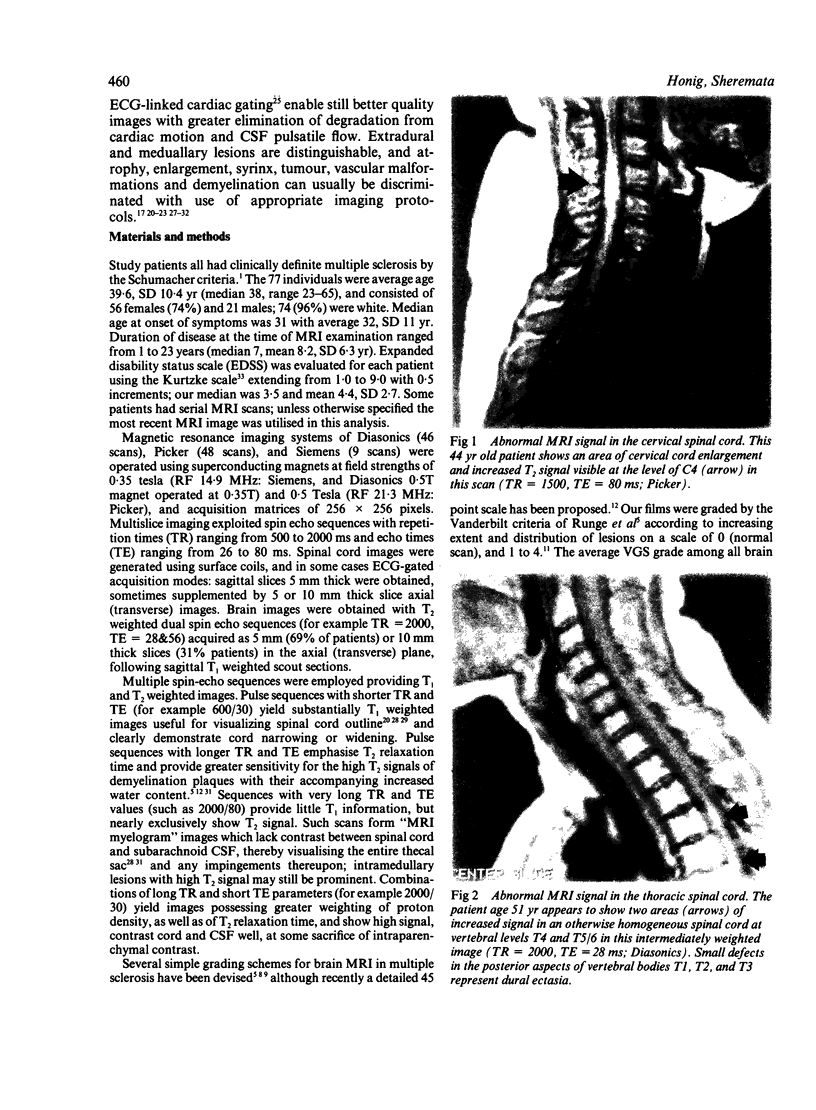
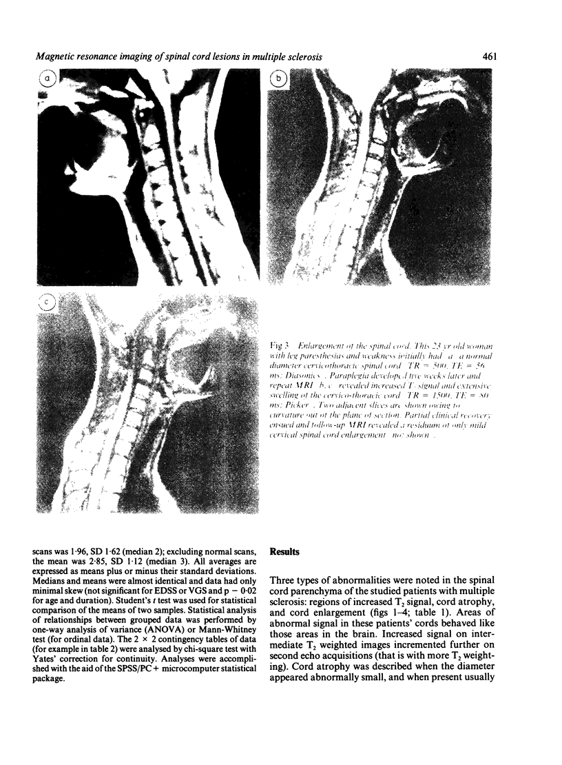
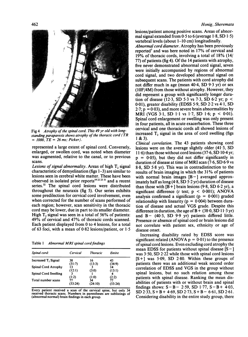
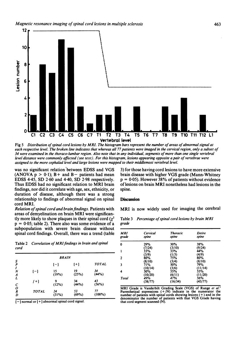
The clinical and pathological manifestations of multiple sclerosis are due to areas of demyelination which occur throughout the white matter of the central nervous system. MRI of the brain frequently shows abnormalities in the hemispheric subcortical white matter; these are demonstrable in the majority of patients and support the clinical diagnosis of multiple sclerosis. Our studies have shown that while MRI identifies such cerebral lesions in nearly all clinically definite multiple sclerosis patients with illness of duration greater than 10 years, these areas of abnormal T2 signal are present less often in the brains of patients studied within 3 years of disease onset. However, symptoms referable to the long tracts of the spinal cord are prominent in many of these patients. Imaging of the spinal cord has presented technical problems because of the small size of the cord, patient body, heart and respiratory movements, and limitations of surface coil technology. The spinal cord of 77 patients with multiple sclerosis have been imaged, revealing three types of abnormalities: (1) approximately half the cords show regions of abnormal T2 weighted signal; (2) during acute exacerbation, spinal cord enlargement (swelling) may be observed; (3) spinal cord atrophy (narrowing) is found particularly in patients with disease of longer duration and greater disability. Unlike the presence of brain lesions, the existence of spinal cord lesions of high T2 signal is not associated with increasing duration of disease but is correlated with disability status. Of patients with such lesions about one fifth did not exhibit brain lesions discernible by MRI.
Full text
PDF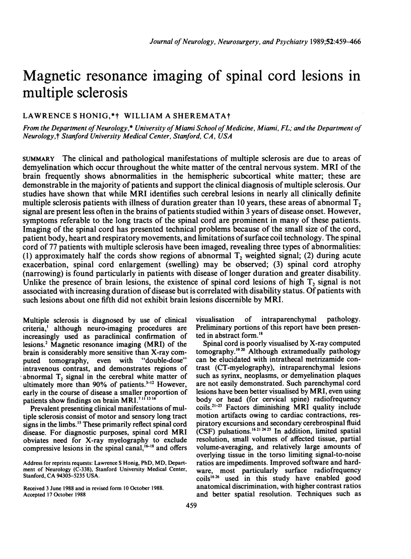
Images in this article
Selected References
These references are in PubMed. This may not be the complete list of references from this article.
- ADAMS R. D., KUBIK C. S. The morbid anatomy of the demyelinative disease. Am J Med. 1952 May;12(5):510–546. doi: 10.1016/0002-9343(52)90234-9. [DOI] [PubMed] [Google Scholar]
- Bydder G. M., Brown J., Niendorf H. P., Young I. R. Enhancement of cervical intraspinal tumors in MR imaging with intravenous gadolinium-DTPA. J Comput Assist Tomogr. 1985 Sep-Oct;9(5):847–851. doi: 10.1097/00004728-198509000-00001. [DOI] [PubMed] [Google Scholar]
- Dee G. J., Bello J. A., Hilal S. K. High field, thin section nuclear magnetic resonance imaging of the cervical spine. Cardiovasc Intervent Radiol. 1986;8(5-6):283–291. doi: 10.1007/BF02552364. [DOI] [PubMed] [Google Scholar]
- Edelman R. R., Shoukimas G. M., Stark D. D., Davis K. R., New P. F., Saini S., Rosenthal D. I., Wismer G. L., Brady T. J. High-resolution surface-coil imaging of lumbar disk disease. AJR Am J Roentgenol. 1985 Jun;144(6):1123–1129. doi: 10.2214/ajr.144.6.1123. [DOI] [PubMed] [Google Scholar]
- Enzmann D. R., Rubin J. B., DeLaPaz R., Wright A. Cerebrospinal fluid pulsation: benefits and pitfalls in MR imaging. Radiology. 1986 Dec;161(3):773–778. doi: 10.1148/radiology.161.3.3786731. [DOI] [PubMed] [Google Scholar]
- Enzmann D. R., Rubin J. B., Wright A. Use of cerebrospinal fluid gating to improve T2-weighted images. Part I. The spinal cord. Radiology. 1987 Mar;162(3):763–767. doi: 10.1148/radiology.162.3.3809491. [DOI] [PubMed] [Google Scholar]
- Gebarski S. S., Gabrielsen T. O., Gilman S., Knake J. E., Latack J. T., Aisen A. M. The initial diagnosis of multiple sclerosis: clinical impact of magnetic resonance imaging. Ann Neurol. 1985 May;17(5):469–474. doi: 10.1002/ana.410170509. [DOI] [PubMed] [Google Scholar]
- Giesser B. S., Kurtzberg D., Vaughan H. G., Jr, Arezzo J. C., Aisen M. L., Smith C. R., LaRocca N. G., Scheinberg L. C. Trimodal evoked potentials compared with magnetic resonance imaging in the diagnosis of multiple sclerosis. Arch Neurol. 1987 Mar;44(3):281–284. doi: 10.1001/archneur.1987.00520150035017. [DOI] [PubMed] [Google Scholar]
- Gonzalez-Scarano F., Grossman R. I., Galetta S., Atlas S. W., Silberberg D. H. Multiple sclerosis disease activity correlates with gadolinium-enhanced magnetic resonance imaging. Ann Neurol. 1987 Mar;21(3):300–306. doi: 10.1002/ana.410210312. [DOI] [PubMed] [Google Scholar]
- Han J. S., Kaufman B., El Yousef S. J., Benson J. E., Bonstelle C. T., Alfidi R. J., Haaga J. R., Yeung H., Huss R. G. NMR imaging of the spine. AJR Am J Roentgenol. 1983 Dec;141(6):1137–1145. doi: 10.2214/ajr.141.6.1137. [DOI] [PubMed] [Google Scholar]
- Honig L. S., Siddharthan R., Sheremata W. A., Sheldon J. J., Sazant A. Multiple sclerosis: correlation of magnetic resonance imaging with cerebrospinal fluid findings. J Neurol Neurosurg Psychiatry. 1988 Feb;51(2):277–280. doi: 10.1136/jnnp.51.2.277. [DOI] [PMC free article] [PubMed] [Google Scholar]
- Hyman R. A., Edwards J. H., Vacirca S. J., Stein H. L. 0.6 T MR imaging of the cervical spine: multislice and multiecho techniques. AJNR Am J Neuroradiol. 1985 Mar-Apr;6(2):229–236. [PMC free article] [PubMed] [Google Scholar]
- Ikuta F., Zimmerman H. M. Distribution of plaques in seventy autopsy cases of multiple sclerosis in the United States. Neurology. 1976 Jun;26(6 Pt 2):26–28. doi: 10.1212/wnl.26.6_part_2.26. [DOI] [PubMed] [Google Scholar]
- Jackson J. A., Leake D. R., Schneiders N. J., Rolak L. A., Kelley G. R., Ford J. J., Appel S. H., Bryan R. N. Magnetic resonance imaging in multiple sclerosis: results in 32 cases. AJNR Am J Neuroradiol. 1985 Mar-Apr;6(2):171–176. [PMC free article] [PubMed] [Google Scholar]
- Jacobs L., Kinkel W. R., Polachini I., Kinkel R. P. Correlations of nuclear magnetic resonance imaging, computerized tomography, and clinical profiles in multiple sclerosis. Neurology. 1986 Jan;36(1):27–34. doi: 10.1212/wnl.36.1.27. [DOI] [PubMed] [Google Scholar]
- Kulkarni M. V., Burks D. D., Price A. C., Cobb C., Allen J. H. Diagnosis of spinal arteriovenous malformation in a pregnant patient by MR imaging. J Comput Assist Tomogr. 1985 Jan-Feb;9(1):171–173. doi: 10.1097/00004728-198501000-00029. [DOI] [PubMed] [Google Scholar]
- Kurtzke J. F. Rating neurologic impairment in multiple sclerosis: an expanded disability status scale (EDSS). Neurology. 1983 Nov;33(11):1444–1452. doi: 10.1212/wnl.33.11.1444. [DOI] [PubMed] [Google Scholar]
- Maravilla K. R., Weinreb J. C., Suss R., Nunnally R. L. Magnetic resonance demonstration of multiple sclerosis plaques in the cervical cord. AJR Am J Roentgenol. 1985 Feb;144(2):381–385. doi: 10.2214/ajr.144.2.381. [DOI] [PubMed] [Google Scholar]
- Masaryk T. J., Modic M. T., Geisinger M. A., Standefer J., Hardy R. W., Boumphrey F., Duchesneau P. M. Cervical myelopathy: a comparison of magnetic resonance and myelography. J Comput Assist Tomogr. 1986 Mar-Apr;10(2):184–194. [PubMed] [Google Scholar]
- Miller D. H., McDonald W. I., Blumhardt L. D., du Boulay G. H., Halliday A. M., Johnson G., Kendall B. E., Kingsley D. P., MacManus D. G., Moseley I. F. Magnetic resonance imaging in isolated noncompressive spinal cord syndromes. Ann Neurol. 1987 Dec;22(6):714–723. doi: 10.1002/ana.410220607. [DOI] [PubMed] [Google Scholar]
- Modic M. T., Hardy R. W., Jr, Weinstein M. A., Duchesneau P. M., Paushter D. M., Boumphrey F. Nuclear magnetic resonance of the spine: clinical potential and limitation. Neurosurgery. 1984 Oct;15(4):583–592. doi: 10.1227/00006123-198410000-00022. [DOI] [PubMed] [Google Scholar]
- Norman D., Mills C. M., Brant-Zawadzki M., Yeates A., Crooks L. E., Kaufman L. Magnetic resonance imaging of the spinal cord and canal: potentials and limitations. AJR Am J Roentgenol. 1983 Dec;141(6):1147–1152. doi: 10.2214/ajr.141.6.1147. [DOI] [PubMed] [Google Scholar]
- Ormerod I. E., Miller D. H., McDonald W. I., du Boulay E. P., Rudge P., Kendall B. E., Moseley I. F., Johnson G., Tofts P. S., Halliday A. M. The role of NMR imaging in the assessment of multiple sclerosis and isolated neurological lesions. A quantitative study. Brain. 1987 Dec;110(Pt 6):1579–1616. doi: 10.1093/brain/110.6.1579. [DOI] [PubMed] [Google Scholar]
- Poser C. M., Paty D. W., Scheinberg L., McDonald W. I., Davis F. A., Ebers G. C., Johnson K. P., Sibley W. A., Silberberg D. H., Tourtellotte W. W. New diagnostic criteria for multiple sclerosis: guidelines for research protocols. Ann Neurol. 1983 Mar;13(3):227–231. doi: 10.1002/ana.410130302. [DOI] [PubMed] [Google Scholar]
- Rossi D. R., Charney A. S. Magnetic resonance imaging of the spine. Semin Neurol. 1986 Mar;6(1):84–93. doi: 10.1055/s-2008-1041451. [DOI] [PubMed] [Google Scholar]
- Runge V. M., Clanton J. A., Lukehart C. M., Partain C. L., James A. E., Jr Paramagnetic agents for contrast-enhanced NMR imaging: a review. AJR Am J Roentgenol. 1983 Dec;141(6):1209–1215. doi: 10.2214/ajr.141.6.1209. [DOI] [PubMed] [Google Scholar]
- Runge V. M., Price A. C., Kirshner H. S., Allen J. H., Partain C. L., James A. E., Jr Magnetic resonance imaging of multiple sclerosis: a study of pulse-technique efficacy. AJR Am J Roentgenol. 1984 Nov;143(5):1015–1026. doi: 10.2214/ajr.143.5.1015. [DOI] [PubMed] [Google Scholar]
- SCHUMACHER G. A., BEEBE G., KIBLER R. F., KURLAND L. T., KURTZKE J. F., MCDOWELL F., NAGLER B., SIBLEY W. A., TOURTELLOTTE W. W., WILLMON T. L. PROBLEMS OF EXPERIMENTAL TRIALS OF THERAPY IN MULTIPLE SCLEROSIS: REPORT BY THE PANEL ON THE EVALUATION OF EXPERIMENTAL TRIALS OF THERAPY IN MULTIPLE SCLEROSIS. Ann N Y Acad Sci. 1965 Mar 31;122:552–568. doi: 10.1111/j.1749-6632.1965.tb20235.x. [DOI] [PubMed] [Google Scholar]
- Schenck J. F., Foster T. H., Henkes J. L., Adams W. J., Hayes C., Hart H. R., Jr, Edelstein W. A., Bottomley P. A., Wehrli F. W. High-field surface-coil MR imaging of localized anatomy. AJNR Am J Neuroradiol. 1985 Mar-Apr;6(2):181–186. [PMC free article] [PubMed] [Google Scholar]
- Stevens J. C., Farlow M. R., Edwards M. K., Yu P. L. Magnetic resonance imaging. Clinical correlation in 64 patients with multiple sclerosis. Arch Neurol. 1986 Nov;43(11):1145–1148. doi: 10.1001/archneur.1986.00520110039011. [DOI] [PubMed] [Google Scholar]
- Young I. R., Hall A. S., Pallis C. A., Legg N. J., Bydder G. M., Steiner R. E. Nuclear magnetic resonance imaging of the brain in multiple sclerosis. Lancet. 1981 Nov 14;2(8255):1063–1066. doi: 10.1016/s0140-6736(81)91273-3. [DOI] [PubMed] [Google Scholar]