Abstract
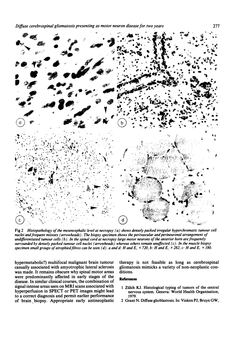
A patient with symptoms and signs of motor neuron disease for 2 years finally developed sensory disturbances and increased intracranial pressure. MRI and CT showed enlargement of the right side of the cerebellum, the brainstem and parts of the cerebral hemisphere with focal hyperperfusion demonstrated by SPECT. Necropsy revealed a diffuse cerebrospinal gliomatosis with loss of spinal motor neurons in tumour infiltration of the anterior horns. This type of spinal cord involvement is considered responsible for the unusual clinical presentation of the neoplasm.
Full text
PDF


Images in this article
Selected References
These references are in PubMed. This may not be the complete list of references from this article.
- Artigas J., Cervos-Navarro J., Iglesias J. R., Ebhardt G. Gliomatosis cerebri: clinical and histological findings. Clin Neuropathol. 1985 Jul-Aug;4(4):135–148. [PubMed] [Google Scholar]
- Kawano N., Miyasaka Y., Yada K., Atari H., Sasaki K. Diffuse cerebrospinal gliomatosis. Case report. J Neurosurg. 1978 Aug;49(2):303–307. doi: 10.3171/jns.1978.49.2.0303. [DOI] [PubMed] [Google Scholar]
- MOORE M. T. Diffuse cerebrospinal gliomatosis, masked by syphilis. J Neuropathol Exp Neurol. 1954 Jan;13(1):129–143. doi: 10.1097/00005072-195401000-00010. [DOI] [PubMed] [Google Scholar]
- Wechsler L. R., Gross R. A., Miller D. C. Meningeal gliomatosis with "negative" CSF cytology: the value of GFAP staining. Neurology. 1984 Dec;34(12):1611–1615. doi: 10.1212/wnl.34.12.1611. [DOI] [PubMed] [Google Scholar]


