Abstract
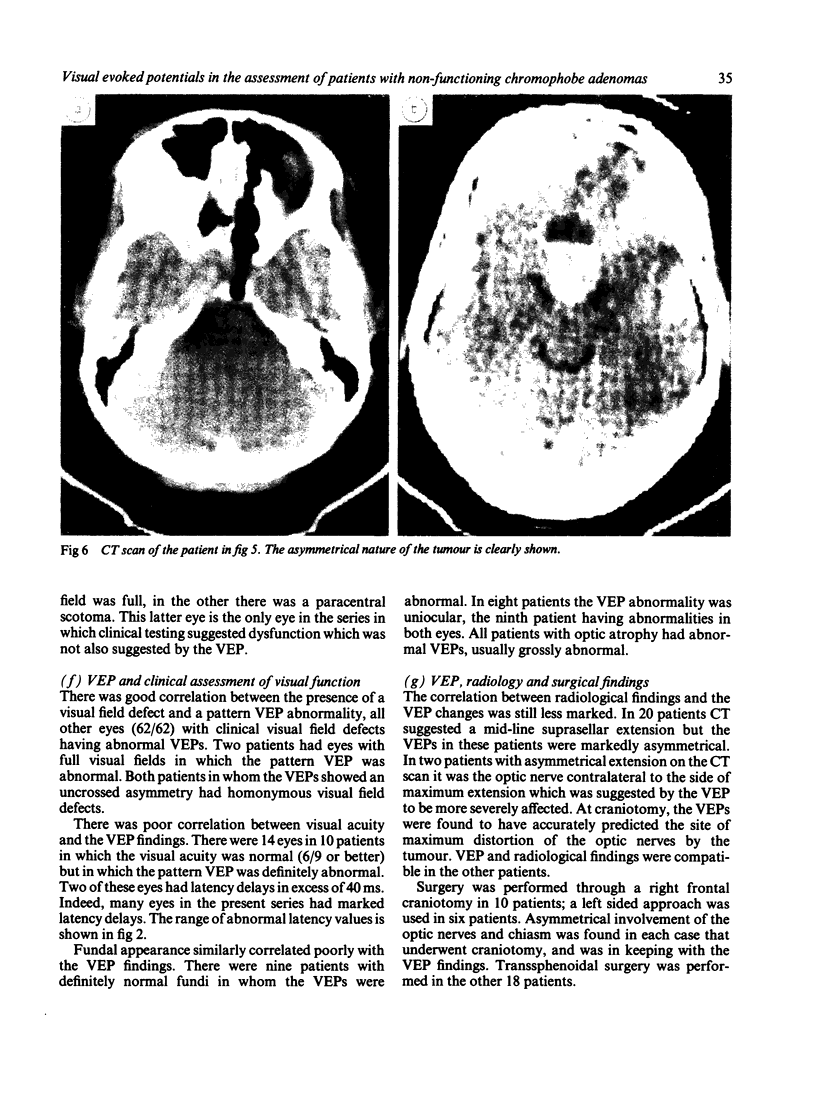
The results of visual evoked potential (VEP) examination in 34 patients with histologically confirmed chromophobe adenoma are described and discussed in relation to the clinical, radiological and surgical findings. The VEP is shown to be a reliable method of assessing the function of the intracranial visual pathways which is often more sensitive than conventional methods of examination.
Full text
PDF
Images in this article
Selected References
These references are in PubMed. This may not be the complete list of references from this article.
- Barett G., Blumhardt L., Halliday A. M., Halliday E., Kriss A. A paradox in the lateralisation of the visual evoked response. Nature. 1976 May 20;261(5557):253–255. doi: 10.1038/261253a0. [DOI] [PubMed] [Google Scholar]
- Camacho L. M., Wenzel W., Aschoff J. C. The pattern-reversal visual evoked potential in the clinical study of lesions of the optic chiasm and visual pathway. Adv Neurol. 1982;32:49–59. [PubMed] [Google Scholar]
- Garfield J., Neil-Dwyer G. Delay in diagnosis of optic nerve and chiasmal compression presenting with unilateral failing vision. Br Med J. 1975 Jan 4;1(5948):22–25. doi: 10.1136/bmj.1.5948.22. [DOI] [PMC free article] [PubMed] [Google Scholar]
- Gott O. S., Weiss M. H., Apuzzo M., Van Der Meulen J. P. Checkerboard visual evoked response in evaluation and management of pituitary tumors. Neurosurgery. 1979 Nov;5(5):553–558. doi: 10.1227/00006123-197911000-00002. [DOI] [PubMed] [Google Scholar]
- Haimovic I. C., Pedley T. A. Hemi-field pattern reversal visual evoked potentials. II. Lesions of the chiasm and posterior visual pathways. Electroencephalogr Clin Neurophysiol. 1982 Aug;54(2):121–131. doi: 10.1016/0013-4694(82)90154-7. [DOI] [PubMed] [Google Scholar]
- Halliday A. M., Halliday E., Kriss A., McDonald W. I., Mushin J. The pattern-evoked potential in compression of the anterior visual pathways. Brain. 1976 Jun;99(2):357–374. doi: 10.1093/brain/99.2.357. [DOI] [PubMed] [Google Scholar]
- Holder G. E. Pattern visual evoked potential in patients with posteriorly situated space-occupying lesions. Doc Ophthalmol. 1985 Feb;59(2):121–128. doi: 10.1007/BF00160608. [DOI] [PubMed] [Google Scholar]
- Holder G. E. Significance of abnormal pattern electroretinography in anterior visual pathway dysfunction. Br J Ophthalmol. 1987 Mar;71(3):166–171. doi: 10.1136/bjo.71.3.166. [DOI] [PMC free article] [PubMed] [Google Scholar]
- Holder G. E. The effects of chiasmal compression on the pattern visual evoked potential. Electroencephalogr Clin Neurophysiol. 1978 Aug;45(2):278–280. doi: 10.1016/0013-4694(78)90011-1. [DOI] [PubMed] [Google Scholar]
- Onofrj M., Bodis-Wollner I., Mylin L. Visual evoked potential diagnosis of field defects in patients with chiasmatic and retrochiasmatic lesions. J Neurol Neurosurg Psychiatry. 1982 Apr;45(4):294–302. doi: 10.1136/jnnp.45.4.294. [DOI] [PMC free article] [PubMed] [Google Scholar]
- Pruett R. C., Wepsic J. G. Delayed diagnosis of chiasmal compression. Am J Ophthalmol. 1973 Aug;76(2):229–236. doi: 10.1016/0002-9394(73)90166-9. [DOI] [PubMed] [Google Scholar]
- Pullan P. T., Carroll W. M., Chakera T. M., Khangure M. S., Vaughan R. J. Management of extra-sellar pituitary tumours with bromocriptine: comparison of prolactin secreting and non-functioning tumours using half-field visual evoked potentials and computerised tomography. Aust N Z J Med. 1985 Apr;15(2):203–208. doi: 10.1111/j.1445-5994.1985.tb04006.x. [DOI] [PubMed] [Google Scholar]
- Senelick R. C., Van Dyk H. J. Chromophobe adenoma masquerading as corticosteroid-responsive optic neuritis. Am J Ophthalmol. 1974 Sep;78(3):485–488. doi: 10.1016/0002-9394(74)90235-9. [DOI] [PubMed] [Google Scholar]
- Stark D. J., Lenton L. Electrophysiological assessment of compressive lesions of anterior visual pathways. Aust J Ophthalmol. 1981 May;9(2):135–141. [PubMed] [Google Scholar]
- Wilson P., Falconer M. A. Patterns of visual failure with pituitary tumours. Clinical and radiological correlations. Br J Ophthalmol. 1968 Feb;52(2):94–110. doi: 10.1136/bjo.52.2.94. [DOI] [PMC free article] [PubMed] [Google Scholar]
- Wray S. H. Neuro-ophthalmologic manifestations of pituitary and parasellar lesions. Clin Neurosurg. 1977;24:86–117. doi: 10.1093/neurosurgery/24.cn_suppl_1.86. [DOI] [PubMed] [Google Scholar]


