Abstract
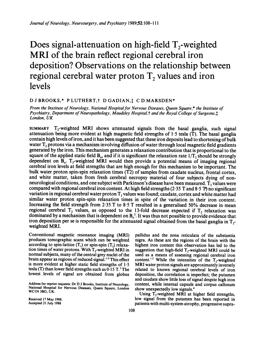
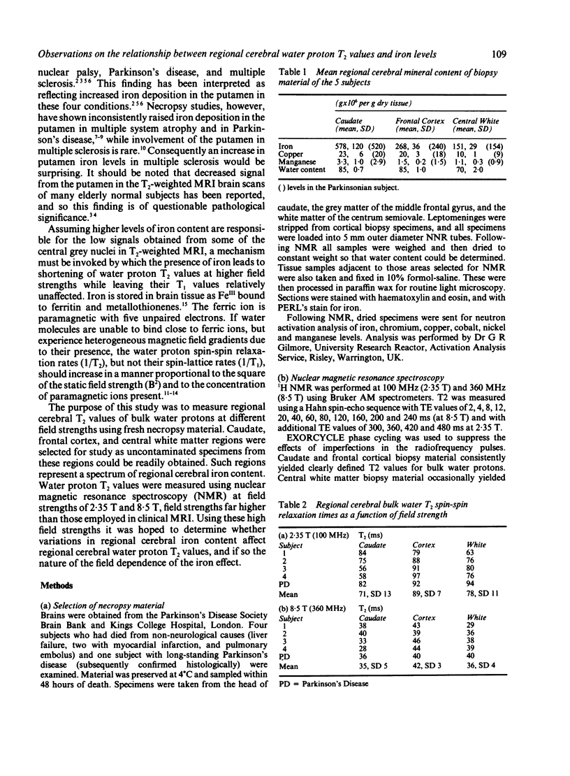
T2-weighted MRI shows attenuated signals from the basal ganglia, such signal attenuation being more evident at high magnetic field strengths of 1.5 tesla (T). The basal ganglia contain high levels of iron, and it has been suggested that these iron deposits lead to shortening of bulk water T2 protons via a mechanism involving diffusion of water through local magnetic field gradients generated by the iron. This mechanism generates a relaxation contribution that is proportional to the square of the applied static field B0, and if it is significant the relaxation rate 1/T2 should be strongly dependent on Bo. T2-weighted MRI would then provide a potential means of imaging regional cerebral iron levels at field strengths that are high enough for this mechanism to be important. The bulk water proton spin-spin relaxation times (T2) of samples from caudate nucleus, frontal cortex, and white matter, taken from fresh cerebral necropsy material of four subjects dying of non-neurological conditions, and one subject with Parkinson's disease have been measured. T2 values were compared with regional cerebral iron content. At high field strengths (2.35 T and 8.5 T) no significant variation in regional cerebral water proton T2 values was found; caudate, cortex and white matter had similar water proton spin-spin relaxation times in spite of the variation in their iron content. Increasing the field strength from 2.35 T to 8.5 T resulted in a generalised 50% decrease in mean regional cerebral T2 values, as opposed to the 13-fold decrease expected if T2 relaxation was dominated by a mechanism that is dependent on B02. It was thus not possible to provide evidence that iron deposition per se is responsible for the attenuated signal obtained from the basal ganglia in T2-weighted MRI.
Full text
PDF


Selected References
These references are in PubMed. This may not be the complete list of references from this article.
- BROWNELL B., HUGHES J. T. The distribution of plaques in the cerebrum in multiple sclerosis. J Neurol Neurosurg Psychiatry. 1962 Nov;25:315–320. doi: 10.1136/jnnp.25.4.315. [DOI] [PMC free article] [PubMed] [Google Scholar]
- Brindle K. M., Brown F. F., Campbell I. D., Grathwohl C., Kuchel P. W. Application of spin-echo nuclear magnetic resonance to whole-cell systems. Membrane transport. Biochem J. 1979 Apr 15;180(1):37–44. doi: 10.1042/bj1800037. [DOI] [PMC free article] [PubMed] [Google Scholar]
- Drayer B. P., Burger P., Hurwitz B., Dawson D., Cain J., Leong J., Herfkens R., Johnson G. A. Magnetic resonance imaging in multiple sclerosis: decreased signal in thalamus and putamen. Ann Neurol. 1987 Oct;22(4):546–550. doi: 10.1002/ana.410220418. [DOI] [PubMed] [Google Scholar]
- Drayer B. P., Olanow W., Burger P., Johnson G. A., Herfkens R., Riederer S. Parkinson plus syndrome: diagnosis using high field MR imaging of brain iron. Radiology. 1986 May;159(2):493–498. doi: 10.1148/radiology.159.2.3961182. [DOI] [PubMed] [Google Scholar]
- Edelman R. R., Johnson K., Buxton R., Shoukimas G., Rosen B. R., Davis K. R., Brady T. J. MR of hemorrhage: a new approach. AJNR Am J Neuroradiol. 1986 Sep-Oct;7(5):751–756. [PMC free article] [PubMed] [Google Scholar]
- Hill J. M., Switzer R. C., 3rd The regional distribution and cellular localization of iron in the rat brain. Neuroscience. 1984 Mar;11(3):595–603. doi: 10.1016/0306-4522(84)90046-0. [DOI] [PubMed] [Google Scholar]
- Pastakia B., Polinsky R., Di Chiro G., Simmons J. T., Brown R., Wener L. Multiple system atrophy (Shy-Drager syndrome): MR imaging. Radiology. 1986 May;159(2):499–502. doi: 10.1148/radiology.159.2.3961183. [DOI] [PubMed] [Google Scholar]
- Spokes E. G., Bannister R., Oppenheimer D. R. Multiple system atrophy with autonomic failure: clinical, histological and neurochemical observations on four cases. J Neurol Sci. 1979 Sep;43(1):59–82. doi: 10.1016/0022-510x(79)90073-x. [DOI] [PubMed] [Google Scholar]
- Thulborn K. R., Waterton J. C., Matthews P. M., Radda G. K. Oxygenation dependence of the transverse relaxation time of water protons in whole blood at high field. Biochim Biophys Acta. 1982 Feb 2;714(2):265–270. doi: 10.1016/0304-4165(82)90333-6. [DOI] [PubMed] [Google Scholar]
- Young I. R., Khenia S., Thomas D. G., Davis C. H., Gadian D. G., Cox I. J., Ross B. D., Bydder G. M. Clinical magnetic susceptibility mapping of the brain. J Comput Assist Tomogr. 1987 Jan-Feb;11(1):2–6. doi: 10.1097/00004728-198701000-00002. [DOI] [PubMed] [Google Scholar]


