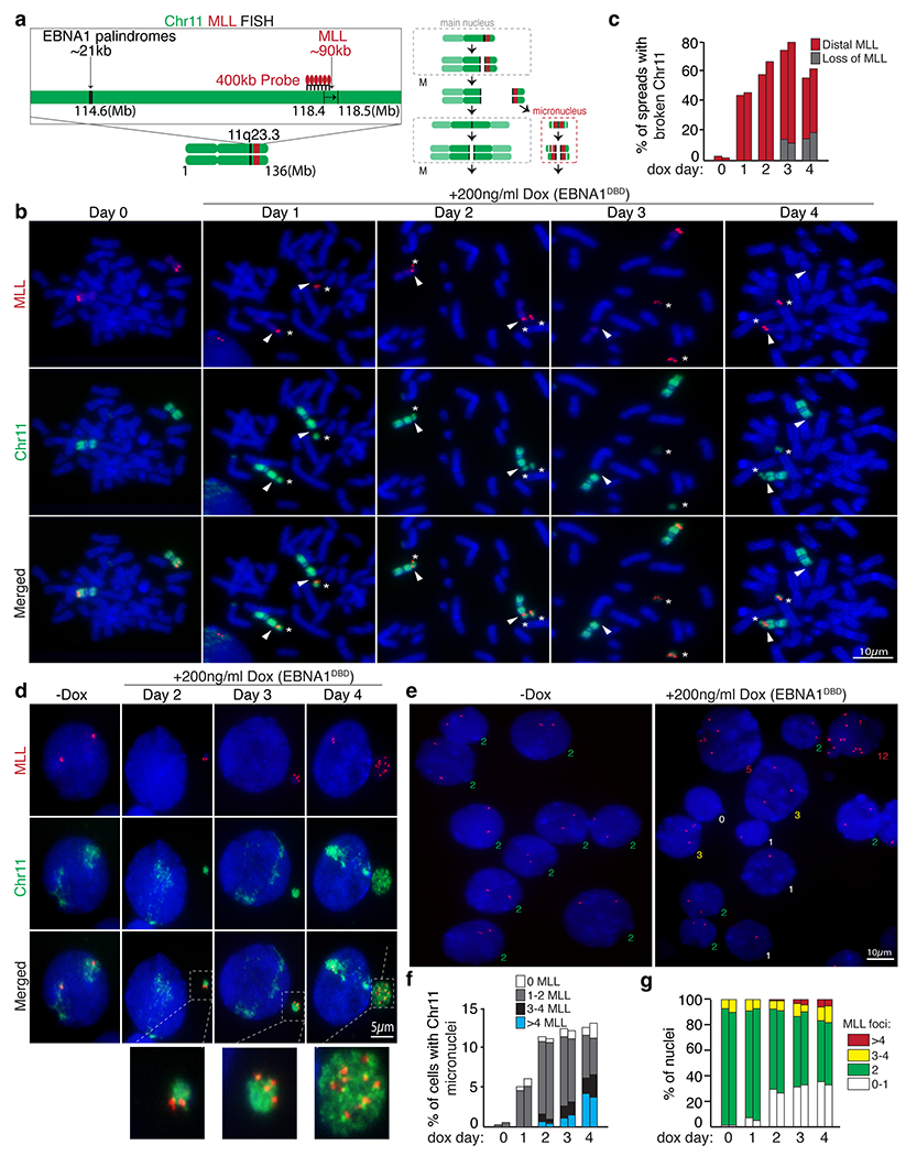
Extended Data Figure 9. EBNA1-induced breakage of chromosome 11 produces an MLL-containing distal fragment that undergoes micronucleation.

(a) Schematic of dual-colored FISH using whole chromosome 11 probe (green) and probe against MLL (red). (b) Representative FISH images of DLD1 cells showing intact chromosome 11’s (Day 0) and broken chromosome 11 fragments containing MLL distal to the site of breakage at day 1 to 4 post dox induction. (c) Data represent the percentage of spreads with broken chromosome 11 from two independent experiments. 71 mitotic spreads were quantified on Day 0, 49 on Day 1, 50 on Day2, 53 on Day 3, and 71 on Day 4. (d) Representative FISH images showing micronucleation of chromosome 11 fragments with or without MLL foci. (f) Stacked bars represent the percentage of cells with chromosome 11 micronuclei with the indicated number of MLL foci from two independent experiments. 400 cells were quantified on Day 0, 210 cells on Day 1, 235 on Day 2, 190 on Day 3, and 180 on Day 4. (e) Representative FISH images showing MLL foci in primary nuclei. (g) Stacked bars represent the percentage of primary nuclei with the indicated number of MLL foci. 950 cells were quantified on Day 0, 880 cells on Day 1, 980 on Day 2, 1100 on Day 3, and 950 on Day 4.
