

Abstract
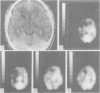
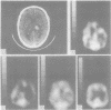
Cortical regional cerebral perfusion was assessed by N, N, N1-trimethyl-N1-(2)-hydroxy-3-methyl-5-(I-123) iodobenzyl-1, 3-propanediamine 2 HCl I-123 (HIPDM) and single photon emission computerised tomography (SPECT) in six aphasic and two neglect patients with unilateral subcortical vascular lesions. Assessments were carried out both in the acute phase and after a period ranging from 1 to 6 months after stroke onset. In all patients an almost complete spontaneous recovery occurred and was associated with a significant improvement of cortical perfusion. A relationship between severity of aphasia and degree of cortical hypoperfusion was found, in both the acute and the follow up assessments, in the aphasic subgroup.
Full text
PDF


Images in this article

Selected References
These references are in PubMed. This may not be the complete list of references from this article.
- Astrup J., Siesjö B. K., Symon L. Thresholds in cerebral ischemia - the ischemic penumbra. Stroke. 1981 Nov-Dec;12(6):723–725. doi: 10.1161/01.str.12.6.723. [DOI] [PubMed] [Google Scholar]
- Baron J. C., D'Antona R., Pantano P., Serdaru M., Samson Y., Bousser M. G. Effects of thalamic stroke on energy metabolism of the cerebral cortex. A positron tomography study in man. Brain. 1986 Dec;109(Pt 6):1243–1259. doi: 10.1093/brain/109.6.1243. [DOI] [PubMed] [Google Scholar]
- Bosley T. M., Dann R., Silver F. L., Alavi A., Kushner M., Chawluk J. B., Savino P. J., Sergott R. C., Schatz N. J., Reivich M. Recovery of vision after ischemic lesions: positron emission tomography. Ann Neurol. 1987 May;21(5):444–450. doi: 10.1002/ana.410210505. [DOI] [PubMed] [Google Scholar]
- Cappa S. F., Vignolo L. A. "Transcortical" features of aphasia following left thalamic hemorrhage. Cortex. 1979 Mar;15(1):121–130. doi: 10.1016/s0010-9452(79)80012-x. [DOI] [PubMed] [Google Scholar]
- Cappa S. F., Vignolo L. A. CT scan studies of aphasia. Hum Neurobiol. 1983;2(3):129–134. [PubMed] [Google Scholar]
- Cappa S., Cavallotti G., Vignolo L. A. Phonemic and lexical errors in fluent aphasia: correlation with lesion site. Neuropsychologia. 1981;19(2):171–177. doi: 10.1016/0028-3932(81)90102-0. [DOI] [PubMed] [Google Scholar]
- Crosson B. Role of the dominant thalamus in language: a review. Psychol Bull. 1984 Nov;96(3):491–517. [PubMed] [Google Scholar]
- Damasio A. R., Damasio H., Rizzo M., Varney N., Gersh F. Aphasia with nonhemorrhagic lesions in the basal ganglia and internal capsule. Arch Neurol. 1982 Jan;39(1):15–24. doi: 10.1001/archneur.1982.00510130017003. [DOI] [PubMed] [Google Scholar]
- Dauth G. W., Gilman S., Frey K. A., Penney J. B., Jr Basal ganglia glucose utilization after recent precentral ablation in the monkey. Ann Neurol. 1985 May;17(5):431–438. doi: 10.1002/ana.410170503. [DOI] [PubMed] [Google Scholar]
- Demeurisse G., Capon A., Verhas M. Prognostic value of computed tomography in aphasic stroke patients. Eur Neurol. 1985;24(2):134–139. doi: 10.1159/000115774. [DOI] [PubMed] [Google Scholar]
- Feeney D. M., Baron J. C. Diaschisis. Stroke. 1986 Sep-Oct;17(5):817–830. doi: 10.1161/01.str.17.5.817. [DOI] [PubMed] [Google Scholar]
- Ferro J. M., Kertesz A., Black S. E. Subcortical neglect: quantitation, anatomy, and recovery. Neurology. 1987 Sep;37(9):1487–1492. doi: 10.1212/wnl.37.9.1487. [DOI] [PubMed] [Google Scholar]
- Gilman S., Dauth G. W., Frey K. A., Penney J. B., Jr Experimental hemiplegia in the monkey: basal ganglia glucose activity during recovery. Ann Neurol. 1987 Sep;22(3):370–376. doi: 10.1002/ana.410220314. [DOI] [PubMed] [Google Scholar]
- Hier D. B., Mondlock J., Caplan L. R. Recovery of behavioral abnormalities after right hemisphere stroke. Neurology. 1983 Mar;33(3):345–350. doi: 10.1212/wnl.33.3.345. [DOI] [PubMed] [Google Scholar]
- Kertesz A., Harlock W., Coates R. Computer tomographic localization, lesion size, and prognosis in aphasia and nonverbal impairment. Brain Lang. 1979 Jul;8(1):34–50. doi: 10.1016/0093-934x(79)90038-5. [DOI] [PubMed] [Google Scholar]
- Knopman D. S., Rubens A. B., Selnes O. A., Klassen A. C., Meyer M. W. Mechanisms of recovery from aphasia: evidence from serial xenon 133 cerebral blood flow studies. Ann Neurol. 1984 Jun;15(6):530–535. doi: 10.1002/ana.410150604. [DOI] [PubMed] [Google Scholar]
- Lucignani G., Nehlig A., Blasberg R., Patlak C. S., Anderson L., Fieschi C., Fazio F., Sokoloff L. Metabolic and kinetic considerations in the use of [125I]HIPDM for quantitative measurement of regional cerebral blood flow. J Cereb Blood Flow Metab. 1985 Mar;5(1):86–96. doi: 10.1038/jcbfm.1985.12. [DOI] [PubMed] [Google Scholar]
- Metter E. J., Jackson C., Kempler D., Riege W. H., Hanson W. R., Mazziotta J. C., Phelps M. E. Left hemisphere intracerebral hemorrhages studied by ( (F-18)-fluorodeoxyglucose PET. Neurology. 1986 Sep;36(9):1155–1162. doi: 10.1212/wnl.36.9.1155. [DOI] [PubMed] [Google Scholar]
- Olsen T. S., Bruhn P., Oberg R. G. Cortical hypoperfusion as a possible cause of 'subcortical aphasia'. Brain. 1986 Jun;109(Pt 3):393–410. doi: 10.1093/brain/109.3.393. [DOI] [PubMed] [Google Scholar]
- Perani D., Vallar G., Cappa S., Messa C., Fazio F. Aphasia and neglect after subcortical stroke. A clinical/cerebral perfusion correlation study. Brain. 1987 Oct;110(Pt 5):1211–1229. doi: 10.1093/brain/110.5.1211. [DOI] [PubMed] [Google Scholar]
- Powers W. J., Raichle M. E. Positron emission tomography and its application to the study of cerebrovascular disease in man. Stroke. 1985 May-Jun;16(3):361–376. doi: 10.1161/01.str.16.3.361. [DOI] [PubMed] [Google Scholar]
- Yamaguchi F., Meyer J. S., Sakai F., Yamamoto J. Case reports of three dysphasic patients to illustrate rCMF responses during behavioral activation. Brain Lang. 1980 Jan;9(1):145–148. doi: 10.1016/0093-934x(80)90080-2. [DOI] [PubMed] [Google Scholar]