Abstract
Objectives:
To investigate changes in angle-specific knee extensor torque between limbs from 4-12 months post-anterior cruciate ligament reconstruction(ACLR) in Division I collegiate athletes at two different isokinetic velocities.
Design:
Cross-sectional study.
Setting:
Laboratory-based.
Participants:
Isokinetic knee flexion and extension assessments of 17 athletes (11 female) at 4, 8, and 12 months after ACLR with bone-patellar tendon-bone autograft were evaluated.
Main Outcome Measures:
Angle-specific curve analyses were performed using statistical parametric mapping for torque data obtained between 14-101° at 60°/s and 240°/s velocities.
Results:
At 60°/s, knee extensor torque of the operated limb increased between 4 and 8 months (18-101°,p<0.001), 4 and 12 months (28-101°,p<0.001), and 8 and 12 months post-surgery (62-70°,p=0.002, and 79-90°,p<0.001). Knee extensor torque was lower in the operated limb compared to the non-operated limb at 4 (47-97°,p<0.001) and 8 months (65-90°,p<0.001) for 60°/s, at 4 (21-89°,p<0.001) and 8 months (50-77°,p<0.001) for 240°/s, with no between-limb differences at 12 months post-ACLR for both velocities.
Conclusions:
Operated limb knee extensor torque increased throughout the majority of knee range of motion from 4 to 12 months post-ACLR at both isokinetic velocities, while non-operated limb torque only improved through a reduced arc of motion in greater knee flexion angles.
Keywords: ACL Injuries, Anterior Cruciate Ligament Reconstruction, College Athletes, Muscle Strength Dynamometer
Introduction
Isokinetic assessments of knee extensor and flexor torque production are commonly used clinically to guide rehabilitation efforts and assess return to sport readiness after anterior cruciate ligament reconstruction (ACLR) (1-5). Isokinetic dynamometers allow for various contraction types (eccentric, concentric, and isometric) and provide continuous torque assessment throughout the available joint range of motion (2). However, it is common practice clinically to characterize thigh muscle function by recording the peak knee extensor and flexor torque values generated during an isokinetic contraction, irrespective of the knee joint angle-specific torques. While this approach accurately captures thigh muscle strength deficits in patients post-ACLR (6, 7), it may be an incomplete measure of quadriceps muscle function. As peak torque values can occur at different joint angles between limbs, peak torque metrics may not reflect angle-specific muscle performance symmetry (8, 9). Considering that athletic tasks require effective joint torques through a large range of knee joint angles, identifying angle-specific asymmetries could facilitate more effective patient-specific rehabilitation efforts and/or improve return-to-sport assessments.
Curve analysis methods, such as statistical parametric mapping (SPM) (10, 11) and functional data analyses (12), have been applied to angle-specific isokinetic knee extensor torque data from athletes after ACLR (8, 9, 13). Knee extensor torque asymmetries of the operated limb were found throughout the joint range of motion (28-81°) up to ~6 months post-ACLR with deficits limited to more flexed knee angles (51-80°) by ~9 months post-ACLR (9). This reflects a change in quadriceps performance during a crucial time period in post-ACLR recovery that may not be effectively captured by traditional peak torque values. In the angle-specific torque analysis performed between 8-10 months after ACLR, bone-patellar tendon-bone and hamstring tendon grafts were compared and extensor inter-limb asymmetry was found to be higher between 30°−85° in those with bone-patellar tendon-bone graft (13). Evaluating between-limb asymmetries in torque-angle curves throughout the post-ACLR rehabilitation period can be useful to monitor the patterns of strength recovery in the operated limb and inform optimal exercise selection to target deficits.
Previous studies have performed angle-specific knee flexor and extensor strength analysis post-ACLR, but factors such as the influence of contraction velocity, and the effects in high-level athletes across a variety of sports and up to 1 year post-surgery have not been explored. Longitudinally, only 60°/s angle-specific isokinetic torques have been analyzed using SPM in individuals out to 9 months post-ACLR (9), despite faster velocities (e.g., 180, 240, or 300°/s) commonly included in clinical assessments (7). Additionally, angle-specific analyses of quadriceps performance in high level male and female athletes post-ACLR across a variety of collegiate sports has not been explored (8, 12, 14). Collegiate athletes are well monitored, high level performers with unrestricted access to sports medicine facilities and care. Due to these factors along with the external pressures to return to sport and the need to achieve a high level of performance upon returning to sport, collegiate athletes may show discrepancies in the rate of recovery compared to a more recreational population. As peak torque deficits are routinely identified ≥ 1 year post-surgery (6), better understanding of longitudinal changes in angle-specific torque production at multiple velocities may aid clinicians in exercise selection and prescription.
The primary purpose of this exploratory study was to investigate changes in angle-specific isokinetic knee extensor torque between limbs from 4-12 months post-ACLR in Division I collegiate athletes at two different test velocities. We hypothesized that: 1) knee extensor torque asymmetries would be observed throughout the entire evaluated knee range of motion at 4 months post-surgery, with asymmetries only detected at > 60 deg of knee flexion at 8 and 12 months post-surgery; 2) operated limb extensor torques would increase between 4 and 8 months post-surgery and between 8 and 12 months post-surgery while non-operated limb extensor torques would increase only between 4 and 12 months post-surgery; 3) there would be similar changes in the torque-angle curves across test velocities. These hypotheses were based on 1) the results of previous investigations reporting quadriceps strength results of both limbs within the first year post-ACLR (6, 9) and 2) pilot studies within our laboratory. A secondary purpose was to assess changes in knee flexor torque-angle curves between limbs and over time following ACLR. A tertiary purpose was to perform sex-specific analysis of torque-angle curves to provide a preliminary assessment of the effect of sex on angle-specific knee joint torques post-ACLR.
Methods
Participants
Data for this study were identified through an analysis of 10 years (2012-21) of routinely, prospectively collected data from the Badger Athletic Performance database. This database contains performance and healthcare data from NCAA Division I athletes at the University of Wisconsin-Madison. The University’s Health Sciences Institutional Review Board approved this records review. Records were included if the athlete: 1) underwent a primary ACLR using a bone-patellar tendon-bone graft; 2) did not undergo a secondary or revision surgery during the 12-month follow-up period; 3) completed isokinetic testing at approximately 4, 8, and 12 months post-ACLR; and 4) completed all isokinetic tests without a significant limitation in range of motion (between 14° and 101° of knee flexion). We did not exclude athletes with concomitant meniscal tears or additional knee ligament injuries. All athletes followed a standardized rehabilitation protocol.
Data Collection and Analysis
Strength testing was performed using an isokinetic dynamometer (Biodex System 4, Biodex Medical Systems, Shirley, NY) with the non-operated limb tested first. Participants were seated with the seat angle at 85°, the knee axis of rotation aligned with the dynamometer shaft, and the lower edge of the resistance pad placed immediately superior to the medial malleolus (15). Concentric knee extension/flexion isokinetic testing was performed at 60°/s and 240°/s from the maximally comfortable knee flexion angle through the maximally comfortable knee extension angle. A progressive warm-up of 50%, 75%, and 90% of maximal effort was performed to familiarize the athlete to the test at each speed. Athletes were then instructed to kick and pull as hard as possible through the entire range of motion for 5 consecutive repetitions at 60°/s and 12 consecutive repetitions at 240°/s. Torque and angle signals were sampled at 100 Hz (Biodex System 4, Biodex Medical Systems, Shirley, NY).
Across all repetitons, limbs, time points, and athletes, a common torque-angle range of motion was identified (14° – 101°) and used for the subsesquent analyses. As the inclusion criteria based on range of motion does not consider potential variations in velocity at the end ranges, velocities less than 58°/s for all repetitions at 60°/s were identified, along with velocities less than 220°/s for all repetitions at 240°/s. Torque values were normalized to the athlete’s mass (kg) and averaged across all repetitions on a point-by-point basis, resulting in one torque-angle curve at each test velocity for each limb, time point, and athlete. Finally, each average torque-angle curve was registered to 101 data points using a cubic interpolation. All data processing was conducted in Matlab (R2021b, The Mathworks Inc, Natick, MA). Descriptive statistics, including means and standard deviations of global peak torque, angle of peak torque, and limb symmetry indices (defined as: (Surgical Limb ÷ Non-Surgical Limb) × 100) were computed.
Statistical Analysis
Statistical parametric mapping (SPM) two-factor (limb x time) repeated measures ANOVAs were used to compare the knee flexor and extensor torque-angle curves with statistical significance set at p < 0.05 (10, 16). Paired t-tests for pairwise comparisons of limb and time points were performed with significance set using a Bonferroni correction (p< 0.0056). All SPM analyses were implemented using the open-source spm1d package (http://spm1d.org/; Pataky, 2012) in Matlab (R2021b, The Mathworks Inc, Natick, MA). All angle-specific curve analyses across both limbs and velocities were repeated with separate assessement of male and female athletes
Results
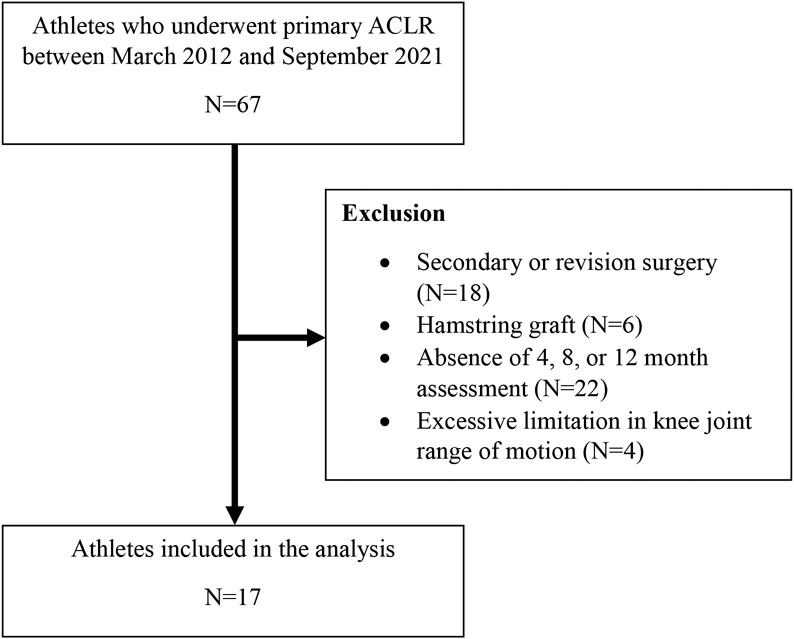
Of the 67 athletes who underwent ACLR surgery between 2010 and 2021, 17 athletes met the inclusion criteria for this study (Figure 1). Athletes participated in soccer (7), spirit (3), football (2), basketball (2), track and field (2), and softball (1). Participant characteristics are presented in Table 1. Eight athletes underwent ACLR; 5, ACLR + meniscectomy; 3, ACLR + meniscal repair; 1, ACLR + LCL reconstruction. Within the range of motion of interest for the 60°/s isokinetic test, only 6 extension or flexion repetitions across all athletes, limbs, and time had velocities below 58°/s (0.6%). All athletes had velocities greater than 58°/s between 17° and 99° of knee flexion. Within the range of motion of interest for the 240°/s isokinetic test, 413 extension or flexion repetitions across all athletes, limbs, and time had velocities below 220°/s (16.9%). All athletes achieved velocities greater than 220°/s between 17° and 99°.
Figure 1.
Flowchart Diagram
Table 1.
Participant characteristics
| Participant Information (N=17)* | |
|---|---|
| Age (years) | 22.1 (1.4) |
| Height (cm) | 174.9 (10.1) |
| Mass (kg) | 78.7 (23.4) |
| Body mass index (kg/m2) | 25.3 (4.9) |
| Females, n (% of total participants) | 11 (65%) |
| Time points post-operatively | |
| 4 Month | 4.5 ± 0.9 |
| 8 Month | 8.3 ± 0.9 |
| 12 Month | 12.0 ± 0.7 |
Values reported as mean and standard deviation unless indicated.
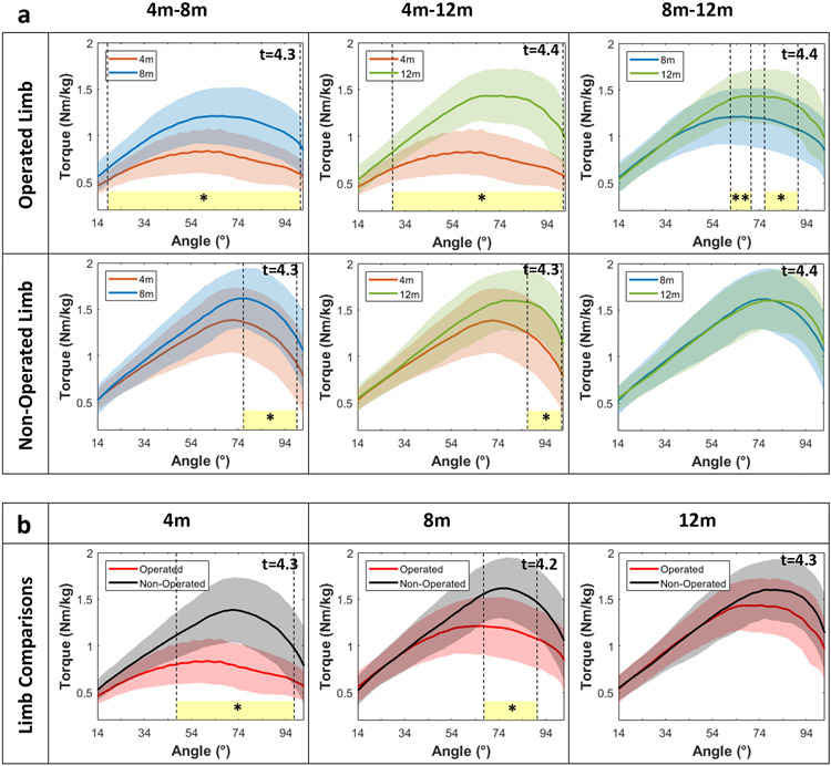
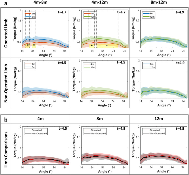
At 60°/s, an interaction between time point and limb was detected for knee extensor torque between 25° and 91° (F-statistic threshold, 5.691; p<0.001), with pairwise comparisons shown in Figure 2. Knee extensor torque of the operated limb increased between 4 and 8 months (18-101°, p<0.001), 4 and 12 months (28-101°, p<0.001), and 8 and 12 months (62-70°, p=0.002 and 79-90°, p<0.001)(Figure 2a). For the non-operated limb, knee extensor torque increased between 4 and 8 months (76-99°, p<0.001) and between 4 and 12 months (87-101°, p<0.001)(Figure 2a). Knee extensor torque was lower in the operated limb compared to the non-operated limb at 4 (47-97°, p<0.001) and 8 months (65-90°, p<0.001), with no between-limb differences at 12 months (Figure 2b). Peak torque, angle of peak torque, and limb symmetry indices at 60°/s for each limb and from 4-12 months post-surgery are presented in table 2.
Figure 2. Knee extensor isokinetic torque-angle curves at 60°/s for each limb and over time.
m:months, t:critical threshold for SPM paired t-test, yellow-shaded area:statistically significant different zone, *:p<.001, **:p=.002
Table 2.
Peak torque values, angle of peak torque, and limb symmetry indices at 60°/s for each limb at 4, 8, and 12 months post-surgery. Means ± standard deviations are reported.
| 4 Months (n=17) | 8 Months (n=17) | 12 Months (n=17) | |||||
|---|---|---|---|---|---|---|---|
| Peak Torque (Nm/kg) |
Angle of Peak Torque (°) |
Peak Torque (Nm/kg) |
Angle of Peak Torque (°) |
Peak Torque (Nm/kg) |
Angle of Peak Torque (°) |
||
| Knee Extensors | Operated Limb | 0.88 ± 0.28 | 57.5 ± 14.3 | 1.28 ±.31 | 65.1 ± 14.2 | 1.51 ± 0.28 | 73.3 ± 11.0 |
| Non-Operated Limb | 1.46 ± 0.34 | 68.7 ± 10.2 | 1.68 ± 0.35 | 74.7 ± 7.7 | 1.71 ± 0.32 | 78.8 ± 11.5 | |
| Limb Symmetry Index (%) | 61 ± 19 | 77 ± 18 | 90 ± 16 | ||||
| Knee Flexors | Operated Limb | 0.69 ± 0.15 | 42.1 ± 15.3 | 0.91 ± 0.18 | 29.4 ± 9.9 | 0.94 ± 0.17 | 29.1 ± 17.0 |
| Non-Operated Limb | 0.73 ± 0.14 | 43.4 ± 18.2 | 0.87 ± 0.18 | 36.5 ± 23.1 | 0.89 ± 0.15 | 33.3 ± 19.2 | |
| Limb Symmetry Index (%) | 95 ± 19 | 106 ± 19 | 107 ± 20 | ||||
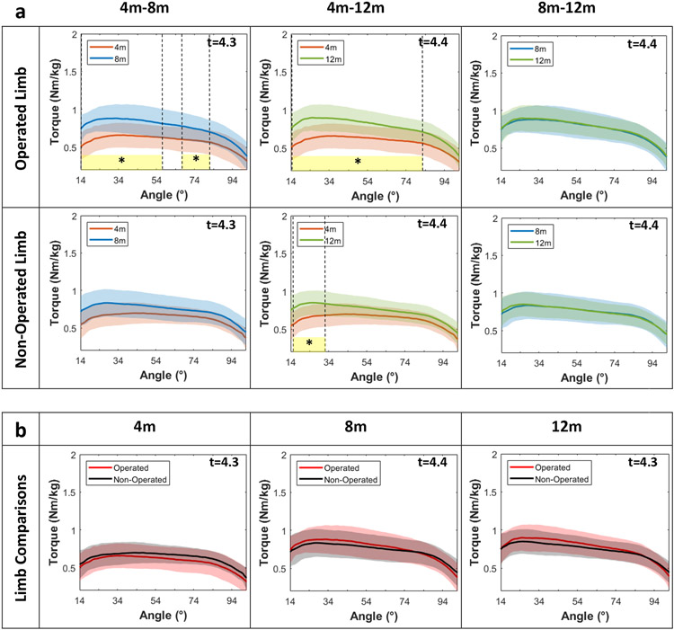
An interaction between time point and limb was present for knee flexor torque at 60°/s between 33° and 77° (F-statistic threshold, 5.657; p<0.001), with pairwise comparisons shown in Figure 3. Knee flexor torques of the operated limb increased between 4 and 8 months (14-57°, 68-82°, both p<0.001) and between 4 and 12 months (14-82°, p<0.001), with no differences between 8 and 12 months (Figure 3a). For the non-operated limb, knee flexor torque increased between 4 and 12 months (15-32°, p<0.001) (Figure 3a). No between-limb differences for knee flexor torque at 60°/s were detected at any time point (Figure 3b).
Figure 3. Knee flexor isokinetic torque-angle curves at 60°/s for each limb and over time.
m:months, t:critical threshold for SPM paired t-test, yellow-shaded area:statistically significant different zone, *:p<.001
At 240°/s, an interaction between time point and limbs was present for knee extensor torque between 18° and 90° (F-statistic threshold, 6.026; p<0.001), with pairwise comparisons shown in Figure 4. Knee extensor torques of the operated limb increased between 4 and 8 months (40-99°, p<0.001) and 4 and 12 months (37-97°, p<0.001) (Figure 4a). For the non-operated limb, knee extensor torque increased between 4 and 12 months (88-97°, p<0.001) (Figure 4a). Knee extensor torque was lower in the operated limb compared to the non-operated limb at 4 (21-89°, p<0.001) and 8 months (50-77°, p<0.001), with no between-limb differences at 12 months (Figure 4b). Peak torque, angle of peak torque, and limb symmetry indices at 240°/s for each limb and from 4-12 months post-surgery are presented in table 3.
Figure 4. Knee extensor isokinetic torque-angle curves at 240°/s between limbs and over time.
m:months, t:critical threshold for SPM paired t-test, yellow-shaded area:statistically significant different zone, *:p<.001
Table 3.
Peak torque values, angle of peak torque, and limb symmetry indices at 240°/s for each limb at 4, 8, and 12 months post-surgery. Means ± standard deviations are reported.
| 4 Months (n=17) | 8 Months (n=17) | 12 Months (n=17) | |||||
|---|---|---|---|---|---|---|---|
| Peak Torque (Nm/kg) |
Angle of Peak Torque (°) |
Peak Torque (Nm/kg) |
Angle of Peak Torque (°) |
Peak Torque (Nm/kg) |
Angle of Peak Torque (°) |
||
| Knee Extensors | Operated Limb | 0.63 ± 0.20 | 61.8 ± 9.8 | 0.85 ±0.15 | 70.7 ± 14.3 | 0.96 ± 0.15 | 69.2 ± 12.2 |
| Non-Operated Limb | 0.92 ± 0.18 | 68.0 ± 10.7 | 1.03 ± 0.16 | 72.3 ± 12.4 | 1.05 ± 0.19 | 69.6 ± 12.8 | |
| Limb Symmetry Index (%) | 31 ± 14 | 83 ± 15 | 94 ± 15 | ||||
| Knee Flexors | Operated Limb | 0.52 ± 0.14 | 34.1 ± 20.2 | 0.69 ± 0.12 | 32.0 ± 10.9 | 0.72 ± 0.11 | 32.7 ± 20.2 |
| Non-Operated Limb | 0.55 ± 0.15 | 54.2 ± 30.8 | 0.65 ± 0.15 | 25.4 ± 9.0 | 0.67 ± 0.14 | 29.7 ± 8.5 | |
| Limb Symmetry Index (%) | 95 ± 18 | 109 ± 27 | 109 ± 20 | ||||
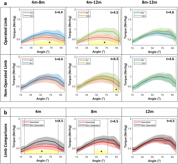
There was no interaction between time point and limbs for knee flexor torque at 240°/s (F-statistic threshold, 6.744; p>0.05), with a main effect for time points (15-85°, p<0.001). Pairwise comparisons are shown in Figure 5. Knee flexor torques of the operated limb increased between 4 and 8 months (20-28°, 33-40°, both p<0.001), as well as 4 and 12 months (28-38°, 42-82°, both p<0.001) (Figure 5a). There were no differences in knee flexor torque for the non-operated limb (Figure 5a), and between limbs at any time point (Figure 5b).
Figure 5. Knee flexor isokinetic torque-angle curves at 240°/s between limbs and over time.
m:months, t:critical threshold for SPM paired t-test, yellow-shaded area:statistically significant different zone, *:p<.001
An exploratory analysis of sex-specific torque-angle relationships was completed, with pairwise comparisons for both the knee extensors and flexors at both speeds shown in the Supplementary material (Figures S1 - S8).
Discussion
The primary purpose of this preliminary investigation was to study changes in angle-specific isokinetic knee extensor torque within and between limbs from 4-12 months following ACLR in Division I collegiate athletes. Consistent with our first hypothesis, the knee extensor torque of the operated limb increased over time across much of the range of motion at both test velocities; however, by 12 months post-ACLR, no significant difference in torque-angle curves between limbs were detected. Consistent with our second hypothesis, operated limb torque increased between both 4-8 and 8-12 months post-surgery, while the non-operated limb demonstrated an increase in knee extensor torque from 4-12 months post-surgery. However, we also observed a non-operative limb torque increase from 4-8 months post-surgery. Lastly, operated limb knee extensor torque at 240°/s did not change beyond 8 months post-surgery, which was contradictory to our third hypothesis. Findings from this study add to the growing body of literature on angle-specific torque analyses by describing the post-ACLR recovery in knee extensor and flexor torque production at both 60°/s and 240°/s in high-level athletes out to 1 year post-surgery.
In the operated limb, we observed improvements in knee extensor torque throughout the majority of the range of motion (18°−100°), which is consistent with prior literature measured between 4 and 8 months (28°−81°) (9). We also observed increased knee extensor torques of the non-operated limb, primarily at greater knee flexion angles (>76°) (9). While in contrast to the findings of previous angle-specific torque analyses post-ACLR (9), this is not a suprising result as bilateral deficits in quadriceps neuromuscular function are common following ACL injury and surgery (17). The divergence in findings may be attributed to variations in pre or post-operative rehabililtation programs, graft types, or subject-specific characteristics (18).
Between-limb comparison of knee extensor torques at 60 deg/s revealed asymmetries from 43°−98° of knee flexion at 4 months and 63°−92° at 8 months. Our observation of quadriceps strength asymmetries in greater knee flexion ranges of motion is consistent with prior literature (9) and has potentially important clinical implications. After ACLR, people perform weightbearing tasks with reduced knee flexion angles (19-22). This is a compensatory strategy at least in part due to quadriceps weakness (23-27). As such, patients post-ACLR may struggle to attain greater knee flexion angles when performing common closed-chain quadriceps strengthening exercises such as squats, step ups, and lunges. This could be a limiting factor to restoring quadriceps strength in this population, as resistance training in long quadriceps muscle lengths may be important for stimulating muscular hypertrophy (28) and facilitating greater strength gains (29-31). Rehabilitation strategies emphasizing deeper knee flexion angles during closed-chain exercises and angle-specific, open-chain knee extensor training in greater knee flexion angles may be warranted for patients post-ACLR with persistent quadriceps strength asymmetries in > 60 ° of knee flexion.
By 12 months post-ACLR, no significant between-limb differences were observed in knee extensor torque at any angle. This is inconsistent with prior literature which has demonstrated persistent strength deficits beyond 1 year post-ACLR, albeit curve anlyses were not utilized (6, 14). The lack of significant between-limb differences at 12 months may in part be due to the conservative statistical approach employed, in which we used a Bonferroni adjusted p-value (p< 0.0056). Although this protects against a type 1 statistical error, it reduces our ability to detect smaller effects which may be clinically relevant.
Knee extensor torques assessed at 240°/s demonstrated similar changes as 60°/s for both the surgical limb and between-limb comparisons except in the surgical limb from 8 to 12 months post-operatively, in which no significant changes were detected. Isokinetic assessment of knee extensor and flexor torque is often performed across multiple speeds, with the implication that faster speeds may provide valuable information regarding the ability to rapidly generate knee torques. However, isokinetic assessments of torque rates are problematic (32) and our findings suggest that assessments of thigh muscle strength at 60°/s may be more discrimative than assessments at 240°/s. Thus, from a clinical perspective, assessing isokinetic knee flexor and extensor torques at both speeds may be redundant and inefficient.
Knee flexor torques of the operated limb increased from 4 to 8 months across much of the range of motion for both test velocities, with minimal changes evident in the non-operated limb. No between-limb differences were present, which is in contrast to prior studies which found reduced knee flexor torques in the operated limb compared to the non-operated limb across at least a portion of the range of motion (8, 9). The participants in prior studies were at a comparable time point post-ACLR; however, 30% (9) and 100% (8) of subjects included in these studies received a hamstring graft which likely explains the difference in findings from the present study in which all participants underwent reconstruction with a bone-patellar tendon-bone graft (33).
An exploratory analysis of sex-specific torque-angle curves was completed due to the previously observed differences in quadriceps strength-to-body weight ratios in males and females post-ACLR (34). Torque-angle patterns were similar across males and females, although minimal statistical differences were detected in male athletes, most likely due to the low sample size (n=6). Future investigations of angle-specific analysis by sex with larger sample sizes are required to better determine if knee extensor and flexor torque-angle relationships differ by sex post-ACLR.
Although significant differences were found in many of the comparisons, the small sample size may have influenced the precision of identifying the exact arcs of motion where torque differences were detected. Further, the data for this study was linearly registered for the SPM analysis, which may result in temporal shifts of the peaks. Other strategies, such as a nonlinear registration paired with SPM analysis of the timing and amplitude trajectories seperately may yield more precise estimates (35). While we are confident in the general angles that were identified, the precision of the defined regions should be interpreted with caution. Additionally, significant between-athlete variability in torque-angle curves was observed in this cohort. Although all athletes followed a standard post-ACLR protocol, minor differences in treatment strategies of individual athletes (either by sport or over time) were possible and could have contributed to individual variations in results. Another potential factor that could contribute to the between-athlete variability we observed are the associated injuries (e.g. meniscus tears) and/or surgeries (e.g. meniscus repair) experienced by some of the athletes included in this study. Therefore, from a clinical perspective it is important to assess torque-angle curve differences on an individual basis and tailor exercise prescription accordingly. While joint angles were matched for all trials and subjects (14-101°), this does not correspond to the full range of motion that all athletes completed. Many were tested beyond this range of motion, and therefore, may have been within different portions of the acceleration and/or deceleration phase inherent to isokinetic testing. This may have affected the torque data at the outerbounds of the range of motion assessed, particularly at 240°/s. Thus, the outerbounds should be interpreted with caution. However, all athletes achieved isovelocity between 17-99° and 20-94° for the 60°/s and 240°/s tests, respectively. Additionally, this study included only collegiate athletes with bone-patellar tendon-bone autografts, and thus, the generalizability of these findings to other populations may be limited.
Conclusion
Angle-specific analyses of the operated limb knee extensor torques identified increases across much of the range of motion from 4 to 12 months post-ACLR at both testing velocities. Between-limb differences in knee extensor angle-specific torques were present at 4 and 8 months. Isokinetic testing of knee extension torques at multiple speeds post-ACLR may be redundant. The information in our study may provide a basis for clinically assessing angle-specific torque changes in athletes after ACLR from 4-12 months post-operatively.
Supplementary Material
References
- 1.Czuppon S, Racette BA, Klein SE, Harris-Hayes M. Variables associated with return to sport following anterior cruciate ligament reconstruction: a systematic review. British journal of sports medicine. 2014;48(5):356–64. [DOI] [PMC free article] [PubMed] [Google Scholar]
- 2.Maestroni L, Read P, Turner A, Korakakis V, Papadopoulos K. Strength, rate of force development, power and reactive strength in adult male athletic populations post anterior cruciate ligament reconstruction-A systematic review and meta-analysis. Physical Therapy in Sport. 2021;47:91–104. [DOI] [PubMed] [Google Scholar]
- 3.Perriman A, Leahy E, Semciw AI. The effect of open-versus closed-kinetic-chain exercises on anterior tibial laxity, strength, and function following anterior cruciate ligament reconstruction: a systematic review and meta-analysis. journal of orthopaedic & sports physical therapy. 2018;48(7):552–66. [DOI] [PubMed] [Google Scholar]
- 4.Shelbourne KD, Gray T, Haro M. Incidence of subsequent injury to either knee within 5 years after anterior cruciate ligament reconstruction with patellar tendon autograft. The American journal of sports medicine. 2009;37(2):246–51. [DOI] [PubMed] [Google Scholar]
- 5.Werner JL, Burland JP, Mattacola CG, Toonstra J, English RA, Howard JS. Decision to return to sport participation after anterior cruciate ligament reconstruction, part II: self-reported and functional performance outcomes. Journal of athletic training. 2018;53(5):464–74. [DOI] [PMC free article] [PubMed] [Google Scholar]
- 6.Brown C, Marinko L, LaValley MP, Kumar D. Quadriceps strength after anterior cruciate ligament reconstruction compared with uninjured matched controls: a systematic review and meta-analysis. Orthopaedic Journal of Sports Medicine. 2021;9(4):2325967121991534. [DOI] [PMC free article] [PubMed] [Google Scholar]
- 7.Wilk KE, Arrigo CA. Rehabilitation principles of the anterior cruciate ligament reconstructed knee: twelve steps for successful progression and return to play. Clinics in sports medicine. 2017;36(1):189–232. [DOI] [PubMed] [Google Scholar]
- 8.Baumgart C, Welling W, Hoppe MW, Freiwald J, Gokeler A. Angle-specific analysis of isokinetic quadriceps and hamstring torques and ratios in patients after ACL-reconstruction. BMC Sports Science, Medicine and Rehabilitation. 2018;10(1):1–8. [DOI] [PMC free article] [PubMed] [Google Scholar]
- 9.Read PJ, Trama R, Racinais S, McAuliffe S, Klauznicer J, Alhammoud M. Angle specific analysis of hamstrings and quadriceps isokinetic torque identify residual deficits in soccer players following ACL reconstruction: a longitudinal investigation. Journal of Sports Sciences. 2022:1–7. [DOI] [PubMed] [Google Scholar]
- 10.Pataky TC, Vanrenterghem J, Robinson MA. Zero-vs. one-dimensional, parametric vs. non-parametric, and confidence interval vs. hypothesis testing procedures in one-dimensional biomechanical trajectory analysis. Journal of biomechanics. 2015;48(7):1277–85. [DOI] [PubMed] [Google Scholar]
- 11.Warmenhoven J, Harrison A, Robinson MA, Vanrenterghem J, Bargary N, Smith R, et al. A force profile analysis comparison between functional data analysis, statistical parametric mapping and statistical non-parametric mapping in on-water single sculling. Journal of Science and Medicine in Sport. 2018;21(10):1100–5. [DOI] [PubMed] [Google Scholar]
- 12.Tengman E, Schelin L, Häger C. Angle-specific torque profiles of concentric and eccentric thigh muscle strength 20 years after anterior cruciate ligament injury. Sports Biomechanics. 2022:1–17. [DOI] [PubMed] [Google Scholar]
- 13.Hart LM, Izri E, King E, Daniels KA. Angle-specific analysis of knee strength deficits after ACL reconstruction with patellar and hamstring tendon autografts. Scandinavian Journal of Medicine & Science in Sports. 2022. [DOI] [PubMed] [Google Scholar]
- 14.Ebert JR, Edwards P, Joss B, Annear P, Radic R, D'Alessandro P. Isokinetic torque analysis demonstrates deficits in knee flexor and extensor torque in patients at 9–12 months after anterior cruciate ligament reconstruction, despite peak torque symmetry. The Knee. 2021;32:9–18. [DOI] [PubMed] [Google Scholar]
- 15.Hamilton RT, Shultz SJ, Schmitz RJ, Perrin DH. Triple-hop distance as a valid predictor of lower limb strength and power. Journal of athletic training. 2008;43(2):144–51. [DOI] [PMC free article] [PubMed] [Google Scholar]
- 16.Friston KJ, Holmes AP, Poline J, Grasby P, Williams S, Frackowiak RS, et al. Analysis of fMRI time-series revisited. Neuroimage. 1995;2(1):45–53. [DOI] [PubMed] [Google Scholar]
- 17.Pamukoff DN, Montgomery MM, Choe KH, Moffit TJ, Garcia SA, Vakula MN. Bilateral alterations in running mechanics and quadriceps function following unilateral anterior cruciate ligament reconstruction. Journal of Orthopaedic & Sports Physical Therapy. 2018;48(12):960–7. [DOI] [PubMed] [Google Scholar]
- 18.Ebert JR, Edwards P, Yi L, Joss B, Ackland T, Carey-Smith R, et al. Strength and functional symmetry is associated with post-operative rehabilitation in patients following anterior cruciate ligament reconstruction. Knee Surgery, Sports Traumatology, Arthroscopy. 2018;26(8):2353–61. [DOI] [PubMed] [Google Scholar]
- 19.Knurr KA, Kliethermes SA, Stiffler-Joachim MR, Cobian DG, Baer GS, Heiderscheit BC. Running biomechanics before injury and 1 year after anterior cruciate ligament reconstruction in Division I collegiate athletes. The American journal of sports medicine. 2021;49(10):2607–14. [DOI] [PMC free article] [PubMed] [Google Scholar]
- 20.Sigward SM, Lin P, Pratt K. Knee loading asymmetries during gait and running in early rehabilitation following anterior cruciate ligament reconstruction: a longitudinal study. Clinical biomechanics. 2016;32:249–54. [DOI] [PubMed] [Google Scholar]
- 21.King E, Richter C, Franklyn-Miller A, Wadey R, Moran R, Strike S. Back to normal symmetry? Biomechanical variables remain more asymmetrical than normal during jump and change-of-direction testing 9 months after anterior cruciate ligament reconstruction. The American Journal of Sports Medicine. 2019;47(5):1175–85. [DOI] [PubMed] [Google Scholar]
- 22.Hughes G, Musco P, Caine S, Howe L. Lower limb asymmetry after anterior cruciate ligament reconstruction in adolescent athletes: a systematic review and meta-analysis. Journal of athletic training. 2020;55(8):811. [DOI] [PMC free article] [PubMed] [Google Scholar]
- 23.Losciale JM, Ithurburn MP, Paterno MV, Schmitt LC. Passing return-to-sport criteria and landing biomechanics in young athletes following anterior cruciate ligament reconstruction. Journal of Orthopaedic Research®. 2022;40(1):208–18. [DOI] [PMC free article] [PubMed] [Google Scholar]
- 24.Palmieri-Smith RM, Lepley LK. Quadriceps strength asymmetry after anterior cruciate ligament reconstruction alters knee joint biomechanics and functional performance at time of return to activity. The American journal of sports medicine. 2015;43(7):1662–9. [DOI] [PMC free article] [PubMed] [Google Scholar]
- 25.Palmieri-Smith RM, Curran MT, Garcia SA, Krishnan C. Factors that predict sagittal plane knee biomechanical symmetry after anterior cruciate ligament reconstruction: A decision tree analysis. Sports Health. 2022;14(2):167–75. [DOI] [PMC free article] [PubMed] [Google Scholar]
- 26.Lisee C, Birchmeier T, Yan A, Kuenze C. Associations between isometric quadriceps strength characteristics, knee flexion angles, and knee extension moments during single leg step down and landing tasks after anterior cruciate ligament reconstruction. Clinical biomechanics. 2019;70:231–6. [DOI] [PubMed] [Google Scholar]
- 27.Lewek M, Rudolph K, Axe M, Snyder-Mackler L. The effect of insufficient quadriceps strength on gait after anterior cruciate ligament reconstruction. Clinical biomechanics. 2002;17(1):56–63. [DOI] [PubMed] [Google Scholar]
- 28.Noorkõiv M, Nosaka K, BLAZEVICH A. Neuromuscular adaptations associated with knee joint angle-specific force change. 2014. [DOI] [PubMed] [Google Scholar]
- 29.Noorkõiv M, Nosaka K, Blazevich AJ. Effects of isometric quadriceps strength training at different muscle lengths on dynamic torque production. Journal of sports sciences. 2015;33(18):1952–61. [DOI] [PubMed] [Google Scholar]
- 30.Alegre LM, Ferri-Morales A, Rodriguez-Casares R, Aguado X. Effects of isometric training on the knee extensor moment–angle relationship and vastus lateralis muscle architecture. European journal of applied physiology. 2014;114(11):2437–46. [DOI] [PubMed] [Google Scholar]
- 31.Pedrosa GF, Lima FV, Schoenfeld BJ, Lacerda LT, Simões MG, Pereira MR, et al. Partial range of motion training elicits favorable improvements in muscular adaptations when carried out at long muscle lengths. European Journal of Sport Science. 2021:1–11. [DOI] [PubMed] [Google Scholar]
- 32.Maffiuletti NA, Aagaard P, Blazevich AJ, Folland J, Tillin N, Duchateau J. Rate of force development: physiological and methodological considerations. European journal of applied physiology. 2016;116(6):1091–116. [DOI] [PMC free article] [PubMed] [Google Scholar]
- 33.Hughes JD, Burnham JM, Hirsh A, Musahl V, Fu FH, Irrgang JJ, et al. Comparison of short-term biodex results after anatomic anterior cruciate ligament reconstruction among 3 autografts. Orthopaedic Journal of Sports Medicine. 2019;7(5):2325967119847630. [DOI] [PMC free article] [PubMed] [Google Scholar]
- 34.Schwery NA, Kiely MT, Larson CM, Wulf CA, Heikes CS, Hess RW, et al. Quadriceps strength following anterior cruciate ligament reconstruction: normative values based on sex, graft type and meniscal status at 3, 6 & 9 months. International Journal of Sports Physical Therapy. 2022;17(3):434. [DOI] [PMC free article] [PubMed] [Google Scholar]
- 35.Pataky TC, Robinson MA, Vanrenterghem J, Donnelly CJ. Simultaneously assessing amplitude and temporal effects in biomechanical trajectories using nonlinear registration and statistical nonparametric mapping. Journal of Biomechanics. 2022;136:111049. [DOI] [PubMed] [Google Scholar]
Associated Data
This section collects any data citations, data availability statements, or supplementary materials included in this article.


