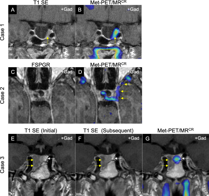
Fig. 1.
Illustrative cases demonstrating potential utility of molecular (PET) imaging in refractory pituitary adenomas. A-B, Case 1: Middle-aged man with acromegaly in whom initial imaging demonstrated a largely empty sella (IGF-1 3.6 ×ULN). Following primary medical therapy with maximum dose first generation somatostatin receptor ligand, the patient remained symptomatic with continued IGF-1 elevation (IGF-1 2.6 ×ULN). A: Post-contrast coronal T1 SE MRI showing a largely empty sella, but with an area suspicious for residual tumor adjacent to the left cavernous sinus (yellow arrow). B: Met-PET/MRCR reveals focal tracer uptake at this site only (yellow arrow). At subsequent transsphenoidal surgery this tissue was resected, and histology confirmed a sparsely granulated somatotroph adenoma. Postoperatively, IGF-1 fully normalized, and the patient had complete resolution of symptoms. C–D, Case 2: Young woman with persistent Cushing disease following initial presentation with pituitary apoplexy in a 2.5 cm macroadenoma. C: FSPGR (volumetric) MRI in axial plane showing indeterminate appearances. D: Met-PET/MRCR reveals significant parasellar disease not initially suspected on MRI, with tracer uptake extending along the lateral wall of the cavernous sinus towards the orbital apex (yellow arrows). E–F, Case 3: Middle-aged man with macroprolactinoma [presenting serum prolactin 552 ng/mL (RR 4–15)] in whom serial MRI showed only minimal reduction in tumor size following cabergoline therapy, despite complete normalization of serum prolactin (6.6 ng/mL). E: Post-contrast coronal T1 SE MRI at presentation demonstrating a predominantly right sided macroadenoma with infrasellar extension (yellow arrow). F: Repeat MRI at 24 months showing minimal tumor shrinkage (yellow arrow). G: Met-PET/MRCR reveals only physiological tracer uptake by the normal gland in the left side of the sella (white arrow) and confirming absence of tumor activity.
Key: FSPGR, fast spoiled gradient recalled echo; Met-PET/MRCR, 11 C-methionine PET/CT coregistered with FSPGR MRI; MRI, magnetic resonance imaging; SE, spin echo; T1, T1-weighted MRI.

