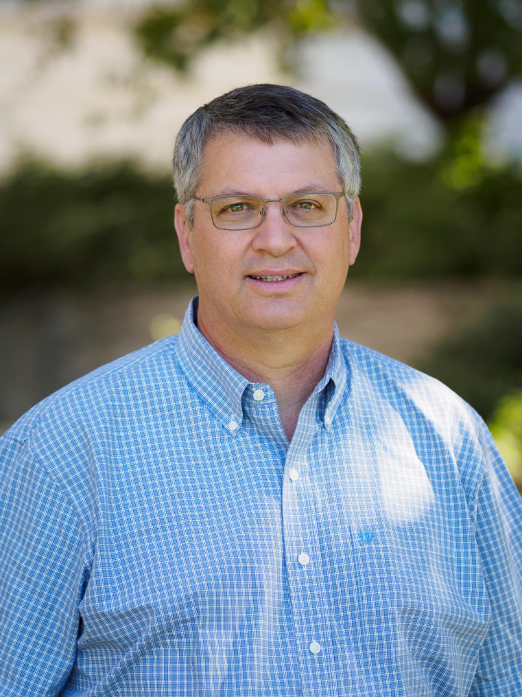
David Drubin has spent his career piecing together the internal workings of eukaryotic cells. Drubin, a cell biologist at the University of California, Berkeley, has carried out impactful work on the hidden players that keep healthy cells humming. His graduate work describing the biological function of the protein tau later allowed other researchers to understand the protein’s role in Alzheimer’s disease (AD). Drubin went on to identify the key regulators that dictate the assembly and organization of the cytoskeletal protein actin and its role in endocytosis, a process cells use to internalize molecules from a cell’s external environment. Along the way, he has described a complex pathway involving 60 proteins that control endocytosis. Elected to the National Academy of Sciences in 2022, Drubin reports in his inaugural article (IA) (1) efforts to reengineer part of the endocytosis pathway outside the cell as a way to understand each step of this life-sustaining process.
Image credit: Mark Joseph Hanson (photographer).
A Clear Path
Born in Tarrytown, New York, Drubin moved often as his electrical engineer father pursued jobs in the aerospace industry. From New York, the family moved to Massachusetts, Michigan, and California. Drubin recalls always gravitating toward science. The space race of his youth and a summer science program at the University of Michigan when he was eight years old cemented his interest in science. Through the Michigan program, Drubin recalls, “We went to Ohio to do fossil hunting; we went into bogs, where we stood waist-deep in mud to take core samples to look at things that were thousands of years old; we watched bats at night on the lake. It was an amazing program.”
Despite excellent high-school science teachers, when Drubin began college, he considered majoring in English. “I expressed myself well in writing and thought for a while I would go into environmental law,” says Drubin, who did his undergraduate work at Berkeley, where he would eventually spend his entire professional career. A few chance conversations steered him toward biochemistry. By the summer between his junior and senior years, he was vacillating between research and medical school. To hedge his bets, he asked Berkeley biochemist Michael Chamberlin about working in his lab, while at the same time applying for an American Heart Association fellowship, which was rumored to ensure acceptance into medical school. He got offers from both. “When faced with the decision, I thought the basic research sounded much more interesting,” he recalls. “It was a key junction in my life. I had two paths, and it was very clear to me that I should take the research path.”
Drubin spent the rest of his time at Berkeley working in Chamberlin’s lab. Eight years later, he returned to Berkeley as an assistant professor, eventually occupying Chamberlin’s former lab space. As an undergraduate, he worked with a graduate student, Janey Wiggs, now an ophthalmologist at Harvard Medical School, analyzing the enzymology of Bacillus subtilis RNA polymerase. “It was really influential because Mike is an extremely rigorous scientist,” says Drubin. “He taught me how to do very precise research, and Wiggs taught me how to be systematic, thorough, and quantitative.”
From 2D to 3D
Developing precise research skills served Drubin well in the graduate school at the University of California, San Francisco. He chose UCSF for its excellent reputation and because it was among the pioneering centers working on gene transcription in eukaryotic cells. His interest in transcription was quickly superseded, however, after he heard cell biologist Marc Kirschner’s talk about cell biology. “I loved how he described the special organization of cells, how everything is regulated, and how processes occur in three dimensions,” recalls Drubin. “Transcription was literally 2D. Cell biology looks at a whole 3D cell. It seemed so exciting, so I decided to do my PhD with Kirschner.”
For his graduate thesis, Drubin studied tau, or tubulin-associated unit, a protein discovered in Kirschner’s lab. Tau regulates microtubule assembly in cells—part of a cell’s cytoskeleton—and would, much later, be discovered as a major factor in AD. Drubin established tau’s fundamental function in living cells by cloning its gene and using biochemistry and cell biology (2–4). “People cite those early papers a lot because they’re some of the foundational papers on tau,” says Drubin. “But it could have just as easily been an obscure protein. That’s why fundamental research is so important. You need to know what something does in normal physiology to understand what goes wrong in disease.”
Another chance encounter at UCSF shaped the next phase of Drubin’s career. He attended a lecture on using genetics to study cells’ cytoskeleton by Massachusetts Institute of Technology geneticist David Botstein. “He’s an amazing speaker, and I was totally sold on his approach using genetics in yeast cells,” says Drubin. “I had reached the limits of the technology I was using, and genetics looked like it provided much more opportunity to examine what was happening in living cells. With genetics you can knock out genes, impair gene function, examine gene mutations, and begin to focus in on a protein’s function.”
Botstein accepted Drubin as a postdoctoral associate. Drubin planned to continue studying microtubules but, when he arrived at MIT, Botstein told him there were enough people working on microtubules in his lab but no one working on actin, another protein active in the eukaryotic cell cytoskeleton. “It was a great opportunity,” says Drubin, who still works on actin and related processes. “It was wide-open for me to explore.”
In Botstein’s lab, Drubin was the sole biochemist, surrounded by geneticists; everyone had something to teach him. He worked on live yeast cells, parsing the proteins that help regulate actin (5–7). It was there that he also met his future wife, geneticist Georjana Barnes, another Botstein postdoctoral associate. Fortuitously, Botstein moved his lab to Genentech in San Francisco around the time Berkeley offered Drubin a job. So, Drubin and Barnes moved together to California. Being back at Berkeley was Drubin’s dream. “I had other offers, but I loved Berkeley,” he says. “I just couldn’t believe they offered me the job. It was one of the best days of my life.”
Barnes stayed with Botstein, first at Genentech, then at Stanford University, until Berkeley hired her in 1991. There, she and Drubin share a lab. “It’s been the most wonderful adventure,” says Drubin. “She works on microtubules in yeast, so we’re very complementary. It’s been a blast to run the lab together.”
Parsing Endocytosis
Drubin continues to find actin, microtubules, and other aspects of eukaryotic cells’ cytoskeleton endlessly exciting. “Polymers of actin and tubulin assemble and disassemble very rapidly in a cell,” he explains. “It’s incredibly dynamic.”
Generally, Drubin starts a line of research in yeast before moving his experiments to human stem cells. “For the most part, the process is the same and the machinery is the same in yeast and human cells,” says Drubin. “Making comparisons across huge evolutionary distances like that is a way to distill out what is fundamental because things that are common tend to be fundamental and things that are different tend to indicate how a process gets tweaked for specific demands of a specific type of cell.”
Beginning at MIT and continuing at Berkeley, Drubin has methodically pieced together actin’s role in the cytoskeleton. His lab has identified key regulators that dictate actin’s assembly and organization (8–10) and implicated actin as a key player in endocytosis (11). Along with actin, they discovered many of the approximately 60 proteins involved in a complex endocytosis pathway (12).
His work on endocytosis also reveals general principles about how cells translate chemical energy into mechanical force, including the dynamic interplay and feedback between force generation, force sensing, and the geometry of cell membranes (13–15). “The novel notion is that, on a subcellular scale, biochemical reactions and the geometry of cell membranes dynamically influence each other, with the activities of proteins that exert forces to shape membranes, and proteins that sense the resulting membrane curvature, being influenced by the curvature itself,” he says.
Most cell biologists working with human cells use cancer cell lines that harbor mutations. Drubin uses human pluripotent stem cells in an attempt to study normal cells, hewing to his belief that understanding cellular abnormalities requires a grasp of the normal biology of healthy cells. “Generally, we have tried to develop in human cells the cell biology approaches that were previously employed only in genetically tractable organisms like yeast,” says Drubin, who used genome editing to make human cells that express fluorescent proteins to track the dynamic localizations of the proteins expressed at native levels (16–18).
Drubin’s IA (1) continues his work on endocytosis, piecing together how each of the 60 proteins involved fits into the process. To explain the work, Drubin quotes Richard Feynman, who said, “What I cannot create, I cannot understand.” In that vein, his team dismantled the crucial final piece of endocytosis—where actin pulls vesicles off the cell membrane—and tried to recreate it outside the cell in a test tube. “We reconstituted the final stages, where you actually produce this package of molecules that you take up from outside the cell,” he says.
To mimic a cell’s membrane, the group put an artificial membrane around little microspheres. Next, they broke open yeast cells and mixed the extract with the microspheres. “Sure enough, some of the endocytic machinery assembled on the membrane, and the actin organized itself properly, and it pulled off vesicles,” explains Drubin. Showing that a burst actin filament assembly is sufficient to pull off bits of membrane into vesicles suggests that the process might be primordial and that it later became more elaborate as eukaryotic cells evolved, says Drubin. “Of course, you can never prove how something evolved,” he admits. “You can just propose models.”
His group has already begun similar studies to examine the steps upstream of vesicle formation. The ultimate goal is to fully reconstitute the entire process of endocytosis. Meanwhile, Drubin is turning toward a new model that will allow him to examine cells on a subcellular level inside a multicellular living organism. Microscopy advances now allow researchers to noninvasively image cells inside animals such as the translucent zebrafish. The Berkeley Advanced Bioimaging Center gives Drubin access to one such microscope, and his Berkeley colleague Ian Swinburne maintains tanks of zebrafish. “We’re starting to use genome editing in zebrafish and the advanced microscopes to see how these processes are different in a living animal,” says Drubin. “It’s lot of fun for me, working in a brand-new area.”
Footnotes
This is a Profile of a member of the National Academy of Sciences to accompany the member’s Inaugural Article, e2302622120, in vol. 120, issue 22.
References
- 1.Stoops E. H., Ferrin M. A., Jorgens D. M., Drubin D. G., Self-organizing actin networks drive sequential endocytic protein recruitment and vesicle release on synthetic lipid bilayers. Proc. Natl. Acad. Sci. U.S.A. 120, e2302622120 (2023). [DOI] [PMC free article] [PubMed] [Google Scholar]
- 2.Drubin D. G., Kirschner M. W., Tau protein function in living cells. J. Cell Biol. 103, 2739–2746 (1986). [DOI] [PMC free article] [PubMed] [Google Scholar]
- 3.Drubin D. G., Feinstein S. C., Shooter E. M., Kirschner M. W., Nerve growth factor-induced neurite outgrowth in PC12 cells involves the coordinate induction of microtubule assembly and assembly-promoting factors. J. Cell Biol. 101, 1799–1807 (1985). [DOI] [PMC free article] [PubMed] [Google Scholar]
- 4.Drubin D. G., Caput D., Kirschner M. W., Studies on the expression of the microtubule-associated protein, tau, during mouse brain development, with newly isolated complementary DNA probes. J. Cell Biol. 98, 1090–1097 (1984). [DOI] [PMC free article] [PubMed] [Google Scholar]
- 5.Adams A. E. M., Botstein D., Drubin D. G., Yeast fimbrin is required in vivo for actin organization and morphogenesis. Nature 354, 404–408 (1991). [DOI] [PubMed] [Google Scholar]
- 6.Drubin D. G., Zhu Z., Mulholland J., Botstein D., Homology of a yeast actin-binding protein to signal transduction proteins and myosin-I. Nature 343, 288–290 (1990). [DOI] [PubMed] [Google Scholar]
- 7.Drubin D. G., Miller K., Botstein D., Yeast actin-binding proteins: evidence for a role in morphogenesis. J. Cell Biol. 107, 2551–2561 (1988). [DOI] [PMC free article] [PubMed] [Google Scholar]
- 8.Lappalainen P., Drubin D. G., Cofilin promotes rapid actin filament turnover in vivo. Nature 388, 78–82 (1997). [DOI] [PubMed] [Google Scholar]
- 9.Moon A. L., Janmey P. A., Louie K. A., Drubin D. G., Cofilin is an essential component of the Saccharomyces cerevisiae membrane cytoskeleton. J. Cell Biol. 120, 421–435 (1993). [DOI] [PMC free article] [PubMed] [Google Scholar]
- 10.Holtzman D. A., Yang S., Drubin D. G., Synthetic-lethal interactions identify two novel genes, SLA1 and SLA2, that control membrane cytoskeleton assembly in Saccharomyces cerevisiae. J. Cell Biol. 122, 635–644 (1993). [DOI] [PMC free article] [PubMed] [Google Scholar]
- 11.Kaksonen M., Sun Y., Drubin D. G., A pathway for association of receptors, adaptors and actin during endocytic internalization. Cell 115, 475–487 (2003). [DOI] [PubMed] [Google Scholar]
- 12.Kaksonen M., Toret C. P., Drubin D. G., A modular design for the clathrin- and actin-mediated endocytosis machinery. Cell. 123, 305–320 (2005). [DOI] [PubMed] [Google Scholar]
- 13.Cail R. C., Shirazinejad C. R., Drubin D. G., Induced Membrane curvature bypasses clathrin’s essential endocytic function. J. Cell Biol. 221, e202109013 (2022), 10.1083/jcb.202109013. [DOI] [PMC free article] [PubMed] [Google Scholar]
- 14.Akamatsu M., et al. , Principles of self-organization and load adaptation by the actin cytoskeleton during clathrin-mediated endocytosis. eLife 9, e49840 (2020), 10.7554/eLife.49840. [DOI] [PMC free article] [PubMed] [Google Scholar]
- 15.Kaplan K., et al. , Adaptive actin organization counteracts elevated membrane tension to ensure robust endocytosis. Mol. Biol. Cell. 7, mbcE21110589 (2022), 10.1091/mbc.E21-11-0589. [DOI] [Google Scholar]
- 16.Doyon J. B., et al. , Rapid and efficient clathrin-mediated endocytosis revealed in genome-edited mammalian cells. Nat. Cell Biol. 13, 331–337 (2011). [DOI] [PMC free article] [PubMed] [Google Scholar]
- 17.Dambournet D., et al. , Genome-edited human stem cells expressing fluorescently labeled endocytic markers allow quantitative analysis of clathrin-mediated endocytosis during differentiation. J. Cell Biol. 217, 3301–3311 (2018), 10.1083/jcb.201710084. [DOI] [PMC free article] [PubMed] [Google Scholar]
- 18.Drubin D. G., Hyman A. A., Stem cells: The new “model organism”. Mol. Biol. Cell. 28, 1409–1411 (2017), 10.1091/mbc.E17-03-0183. [DOI] [PMC free article] [PubMed] [Google Scholar]


