Abstract
Sinonasal teratoid carcinosarcoma or teratocarcinoma is an extremely rare aggressive tumour. It usually arises in the nasal cavity and paranasal sinuses. In this study, the authors described magnetic resonance imaging and computed tomography findings from a patient with sinonasal teratocarcinoma. Computed tomography of the sinonasal teratoid carcinosarcoma can mimic paranasal fungal infections. Magnetic resonance imaging is a very useful tool for making a differential diagnosis between the sinonasal teratoid carcinosarcoma and paranasal sinusitis.
Keywords: Magnetic resonance imaging, Malignant teratocarcinosarcoma
Background
Malignant teratocarcinosarcoma of the sinonasal cavity is an extremely rare neoplasm.1,2 It is a locally aggressive neoplasm.1,2 Patients frequently present with epistaxis and nasal obstruction.1 We describe the findings from magnetic resonance imaging (MRI) of a rare case of sinonasal teratocarcinosarcoma.
Case history
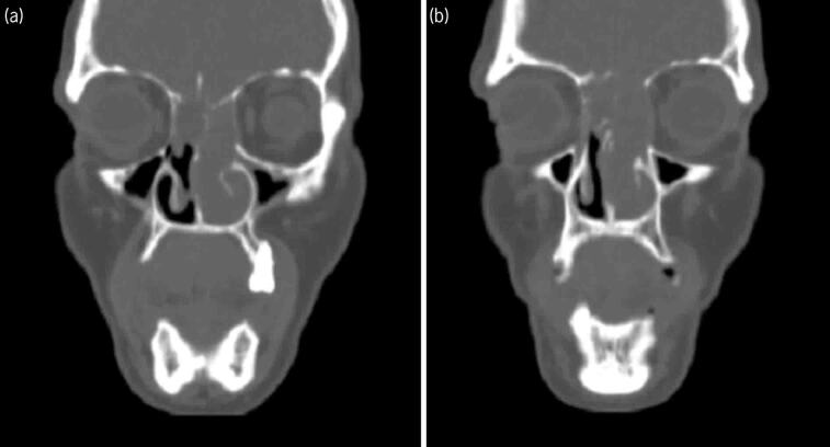
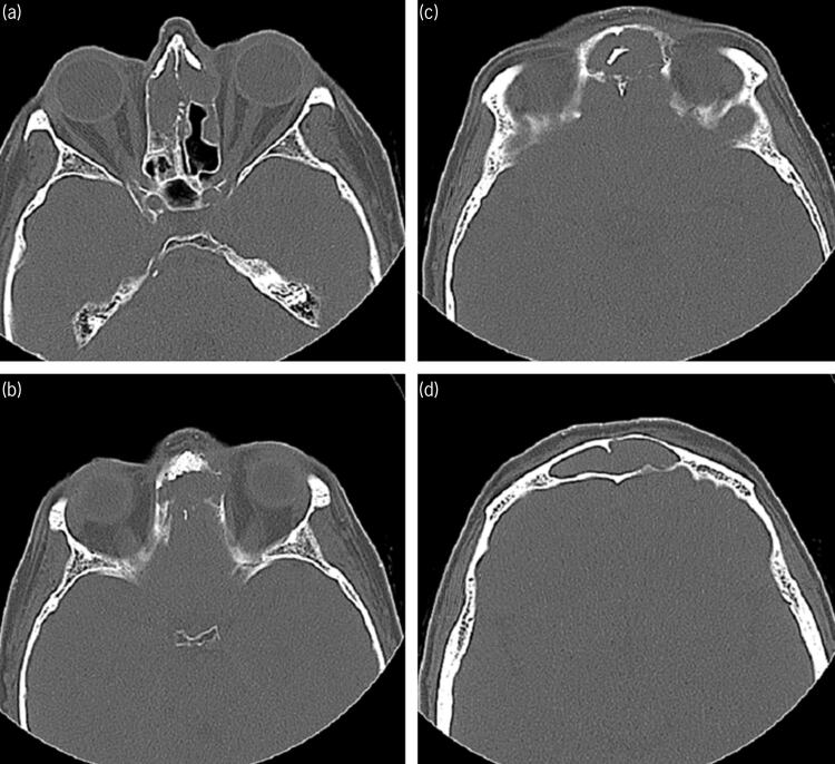
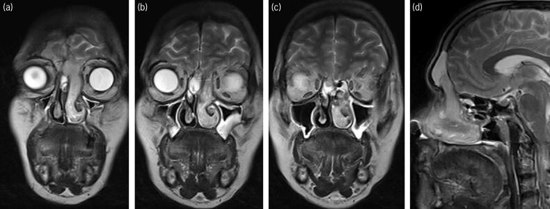
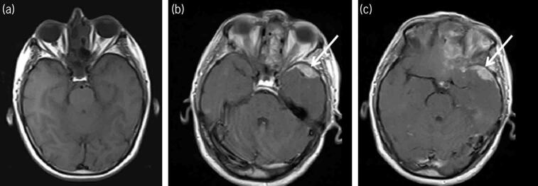
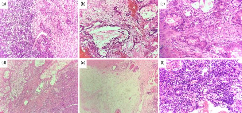
A 52-year-old woman presented to our clinic with a history of progressive maxillofacial swelling and blurred vision in both eyes. She had previously been treated for sinusitis about one week earlier and had a history of purulent nasal discharge. There was no history of neurological disease. On examination, her body temperature was 38.1 degrees C. All her blood investigations and blood pressure were within the normal ranges. Physical examination revealed maxillofacial swelling. The visual acuity of both eyes was 25/30 and there was no evidence of a relative afferent pupillary defect. Fundoscopic examination of both eyes showed bilaterally minimal papilla oedema. A neurological examination was normal. As the maxillofacial features suggested the possibility of sinonasal cavity lesion, she underwent computed tomography (CT) of the paranasal sinuses. Coronal and axial unenhanced CT images showed non-specific soft-tissue attenuation filling the ethmoid air cells, frontal sinus and left nasal cavity (Figure 1). The imaging also revealed a naso-ethmoidal soft-tissue mass which had destroyed the left medial orbital wall and ethmoid roof (Figure 2). These findings suggested several differential diagnoses, such as chronic invasive fungal sinusitis, invasive granulomatous disease and neoplasm of the paranasal sinuses. Further diagnostic work-up with pre-contrast and contrast-enhanced cerebral and maxillofacial MRI was undertaken to make a differential diagnosis. Coronal and sagittal T2-weighted images showed relatively T2 hyperintense left fronto-ethmoid soft-tissue mass extending into the anterior cranial fossa (Figure 3). Non-contrast and contrast-enhanced T1 weighted MRI revealed a contrast-enhanced sinonasal malignancy and its intracranial extension (Figure 4). Given the pathological findings, the lesion was determined to be a sinonasal teratocarcinosarcoma (Figure 5). The patient was recommended to receive radiotherapy. She decided to withdraw her treatment and died three months later.
Figure 1 .
(a, b) Coronal computed tomography showing non-specific soft-tissue attenuation filling the ethmoid air cells, frontal sinus and left nasal cavity
Figure 2 .
(a–d) Axial computed tomography (CT) revealing a naso-ethmoidal soft-tissue mass destroying the left medial orbital wall and ethmoid roof. CT (b, c) also reveals erosion of the posterior wall of the frontal sinus and the septum with spread across the midline
Figure 3 .
Coronal (a–c) and sagittal (d) T2-weighted images show relatively hyperintense left frontonaso-ethmoid soft-tissue mass extending into the anterior cranial fossa
Figure 4 .
Pre- (a) and post-contrast (b, c) T1 weighted magnetic resonance imaging revealing a contrast-enhanced sinonasal neoplasm and its intracranial extension (arrow)
Figure 5 .
(a) Haematoxylin and eosin (H&E) stain (magnification × 200) showing benign epithelial structures (right side) adjacent to primitive neuroectodermal/blastemal component (left side). (b, c) H&E (magnification × 400) revealing carcinomatous component with glandular pattern accompanied by adenocarcinoma. (d) H&E (magnification × 200) showing sarcomatous (right side) and carcinomatous (left side) components. (e) H&E (magnification × 200) showing myxoid stromal tissue adjacent to benign epithelial structures. (f) H&E (magnification × 400) revealing primitive neuroectodermal/blastemal component with rosette formation.
Discussion
Malignant teratocarcinosarcoma of the sinonasal cavity is a very rare tumour.1,2 The lesion frequently originates in the ethmoid sinus or maxillary antrum.1 Patients frequently present with epistaxis and nasal obstruction.1 If the disease spreads to the intracranial area, patients can rarely present neurological symptoms such as diplopia, headache and confusion. Teratocarcinosarcoma is a locally aggressive neoplasm.1,2
On radiological imaging of the malignant teratocarcinoma, paranasal CT is the tool of choice for the detection of osseous destruction and erosion. Intracranial involvement is better depicted on MRI. Chronic invasive fungal disease of the paranasal sinuses may have the radiological appearance of an aggressive sinonasal neoplasm.3 On CT imaging, prominent soft tissue density in the sinonasal cavity with associated sinus wall erosion and destruction is commonly seen.
In conclusion, fungal disease of the paranasal sinuses can rarely mimic sinonasal neoplasms. Conventional and contrast-enhanced MRI plays an important role in the differential diagnosis between the chronic invasive fungal disease of the paranasal sinuses and sinonasal teratocarcinosarcoma.
References
- 1.Chakraborty S, Chowdhury AR, Bandyopadhyay G. Sinonasal teratocarcinosarcoma: case report of an unusual neoplasm. J Oral Maxillofac Pathol 2016; 20: 147–150. 10.4103/0973-029X.180979 [DOI] [PMC free article] [PubMed] [Google Scholar]
- 2.Mohanty S, Somu L, Gopinath M. Sino nasal teratocarcinosarcoma-an interesting clinical entity. Indian J Surg 2013; 75: 141–142. 10.1007/s12262-012-0510-z [DOI] [PMC free article] [PubMed] [Google Scholar]
- 3.Mossa-Basha M, Ilica AT, Maluf Fet al. The many faces of fungal disease of the paranasal sinuses: CT and MRI findings. Diagn Interv Radiol 2013; 19: 195–200. 10.5152/dir.2012.003 [DOI] [PubMed] [Google Scholar]


