Abstract
Laparoscopic cholecystectomy is the standard of care for the surgical management of symptomatic gallstone disease. Gallstone spillage at laparoscopic cholecystectomy is common, with a reported incidence of 0.2–20%. In the majority of cases there are no complications associated with this spillage, but a series of studies report patients with complications of free peritoneal gallstones. We present a case of migration of gallstone to the lung resulting in an inflammatory mass in the right middle lobe as a complication of spillage at laparoscopic cholecystectomy.
Keywords: Intrathoracic gallstone, Gallstone ectopia, Solitary lung nodule, Laparoscopic cholecystectomy, Gallstone migration
Introduction
Laparoscopic cholecystectomy is the standard of care for symptomatic gallstone disease. Gallstone spillage at laparoscopic cholecystectomy is common, with a reported incidence of 0.2–20%.1 In the majority of cases there are no complications associated with this spillage, but a series of studies report patients with complications of free peritoneal gallstones, sometimes many years later.1,2 Typical complications include intraperitoneal abscess formation, wound sinus and fistula; less common complications include small bowel obstruction, small bowel fistula, colonic fistula, diaphragmatic irritation and stone erosion through the flank. Intrathoracic complications include haemoptysis and, less commonly, cholelithoptysis, empyema and intrathoracic abscess.1–3
Migration of gallstones into the thoracic cavity is rare, with very few reported cases.1–3 Cases of gallstone migration in the context of hepatopulmonary abscess have been described. In the context of spilled gallstones at cholecystectomy, often there is a preceding subphrenic abscess that involves the right lower lobe of the lung.3
We present a case of a gallstone inflammatory mass in the right middle lobe as a complication of spillage at laparoscopic cholecystectomy that migrated to the chest.
Case history
A 79-year-old man was referred for thoracic surgery with a right middle lobe nodule. Three months earlier he presented with acute cholangitis and underwent urgent laparoscopic cholecystectomy. Preoperative computed tomography (CT) imaging revealed a 2.5cm gallstone within an enlarged and inflamed gallbladder (Figure 1). The patient made a good recovery.
Figure 1 .
Computed tomography scan of the abdomen, showing an inflamed gallbladder with a 2.5cm gallstone before cholecystectomy
Histopathological examination of the surgical specimen confirmed acute severe inflammation of the gallbladder with abscess formation. The gallstone was not present and presumably had been spilled during surgery.
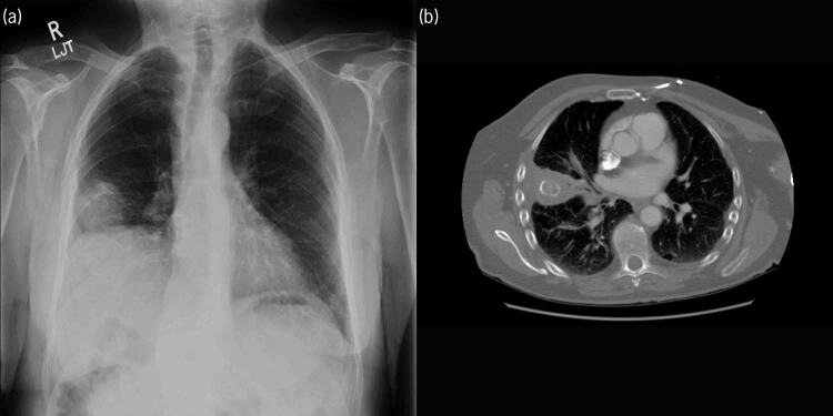
A month after discharge, the patient presented with a cough, streaky haemoptysis and shortness of breath. He was diagnosed with a lower respiratory tract infection and treated with antibiotics. His symptoms did not resolve, and after three further courses of antibiotics he was referred for a chest radiograph. The chest radiograph demonstrated a right lower zone opacity (Figure 2a).
Figure 2 .
(a) Chest x-ray showing opacity in the right middle zone. (b) Computed tomography scan showing the gallstone in the right middle lobe.
With concern over the possibility of a pulmonary malignancy, a CT scan was performed. This demonstrated focal consolidation of the subpleural region of the right middle lobe surrounding an ovoid well-circumscribed lesion with a laminated pattern of calcification (Figure 2b). There were enlarged hilar and mediastinal lymph nodes.
The appearance of the nodule was very similar to that of the gallstone on previous imaging. It was thought this may represent migration of the spilled gallstone through the diaphragm and into the middle lobe, resulting in the development of an inflammatory pseudo-tumour.
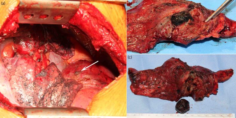
The case was discussed in the multidisciplinary team and referred to surgery. The patient underwent a right thoracotomy and middle lobectomy. Intraoperatively, the right lower lobe was adherent to the diaphragm and chest wall. There was a site of dense fibrosis on the diaphragm, which may identify the site of entry point to the chest (Figure 3a). An enlarged lymph node was excised for histology (station R11). The middle lobe was excised and opened on the back table. A 2cm gallstone was found within the lobe, completely surrounded by lung parenchyma (Figure 3b,c). Histopathological examination confirmed presence of the gallstone close to the middle lobe bronchus. The lung tissue showed evidence of chronic inflammation, with no evidence of malignancy in the lung parenchyma or lymph node.
Figure 3 .
(a) Intraoperative photograph showing healed defect in the diaphragm (arrow). (b) and (c) excised lobe, showing gallstone.
The patient made a good postoperative recovery and was discharged on the third postoperative day.
Discussion
Laparoscopic cholecystectomy is now the standard of care for the surgical management of cholelithiasis. Gallbladder perforation occurs in 10–32% of patients who have laparoscopic cholecystectomies. Gallstone spillage is estimated to occur in 0.2–20% of cases, with very few causing complications. A case review of 1130 consecutive laparoscopic cholecystectomies performed at 2 different institutions demonstrated a 0.3% complication rate from intraoperative stone spillage at 13-year follow-up.4
The first intrathoracic gallstone was reported by Schwengler and colleagues in 1975. Qiang and colleagues reviewed 15 cases of gallstones in the lung, including their own case, in 2014. In their review, symptoms included haemoptysis, chest infection and lung abscess. Gallstones were found in the right lower lobe in 14 of 15 cases and in the right middle lobe in 1 case.2
The time interval between clinical presentation (which includes haemoptysis or, less commonly, cholelithoptysis, empyema or intrathoracic abscess) and surgery ranges from 2 to 60 months (mean 12 months). By far the most common thoracic site is the right lower lobe, which is explained by the subphrenic abscess after complicated cholecystectomy with direct transdiaphragmatic erosion owing to a local inflammatory reaction.4
A study by Guest explored the porous diaphragm syndrome responsible for conditions such as the predominantly right-sided peritoneal dialysis-related hydrothorax, bilious right-sided effusion with perforated gastric or duodenal ulcer and Meigs syndrome.5 Only one case of middle lobe involvement has been reported.3
Some authors suggest removing gallstones to the greatest extent during cholelithiasis surgery to prevent stones leaving the intraperitoneal cavity and causing intraperitoneal and thoracic cavity complications. Other authors recommend using a retrieval bag to minimise the spillage of gallstones during surgery.4
We add our case to the literature as another gallstone migration to the lung. In our case, the stone went into the right middle lobe. We found a healed defect in the diaphragm surrounded by dense fibrosis. Our patient presented with chest infection resistant to medical treatment.
Conclusion
Gallstone migration to the chest is a rare condition that clinicians should be aware of. We recommend reporting missing gallstones during biliary surgery to the patient and their general practitioner for follow-up as the least accepted standard of practice. We recommend that patients with previous biliary surgery presenting with unexplained chest symptoms that do not respond to medical treatment should have chest CT imaging to exclude intrathoracic gallstone migration.
References
- 1.Fontaine JP, Issa RA, Yantiss RK, Podbielski FJ. Intrathoracic gallstones: a case report and literature review. JSLS 2006; 10: 375–378. [PMC free article] [PubMed] [Google Scholar]
- 2.Qiang Z, Xinli W, Chengxin Y, Yushu M, Peifeng L. Gallstone ectopia in the lungs: case report and literature review. Int J Clin Exp Med 2014; 7: 4530–4533. [PMC free article] [PubMed] [Google Scholar]
- 3.Binmahfouz AS, Steinke K. The wanderlust of a gallstone: a case report of intrathoracic migration of a gallstone post complicated cholecystectomy mimicking lung cancer. BJR Case Rep 2016; 2: 20150430. 10.1259/bjrcr.20150430 [DOI] [PMC free article] [PubMed] [Google Scholar]
- 4.Horton M, Florence MG. Unusual abscess patterns following dropped gallstones during laparoscopic cholecystectomy. Am J Surg 1998; 175: 3. 10.1016/S0002-9610(98)00048-8 [DOI] [PubMed] [Google Scholar]
- 5.Guest S. The curious right-sided predominance of peritoneal dialysis-related hydrothorax. Clin Kidney J 2015; 8: 212–214. 10.1093/ckj/sfu141 [DOI] [PMC free article] [PubMed] [Google Scholar]


