Abstract
Small bowel volvulus is a rare but life-threatening emergency. Volvulus of the duodenum is even rarer without the presence of predisposing factors. The clinical presentation is vague, including abdominal pain, nausea and vomiting, prompt diagnosis of volvulus therefore relies heavily on radiographs. The treatment options lie between conservative or surgical management, where the decision is influenced by the patient and their presentation. This case is of a 100-year-old female with an extensive surgical and medical background presenting with signs of small bowel obstruction. With the help of imaging, a rare case of duodenal volvulus was diagnosed but managed conservatively due to the patient's background, age and personal wishes.
Keywords: Duodenal, Volvulus
Background
Volvulus can be defined as rotation of the bowel around itself or its mesentery, compromising its lumen and vascular supply.1 Small bowel volvulus is the cause of 1% of cases of small bowel obstruction2 and colonic volvulus is the cause of 5% of cases of large bowel obstruction.3
Volvulus most commonly affects parts of the bowel that are mobile. Small bowel volvulus is uncommon; however, abnormal mobility or presence of adhesions can predispose the small bowel to rotate. Incidence of duodenal volvulus is very rare because the duodenum is retroperitoneal and fixed along most of its length. With age, there is progressive downward migration of the duodenum due to laxity of support and loss of intra-abdominal fat.1,4
Complications of volvulus stem from compromise in vascular supply, causing ischaemia, necrosis and perforation. Surgical management is a more appropriate approach if these complications are present.
Case history
This case is of a 100-year-old female who presented with one-day history of abdominal pain associated with nausea, bilious vomiting, not passing flatus and reduced stoma output. Her past surgical history included Crohn’s with pan-proctocolectomy with ileostomy formation in 1988 and previous adhesional small bowel obstruction. Past medical history included gastric polyps, glaucoma, eczema and hiatus hernia. The patient had multiple hospital admissions over the past 15 years due to adhesional small bowel obstruction which were all managed conservatively.
Physical examination showed a haemodynamically stable patient with soft, distended abdomen and right upper quadrant tenderness on palpation. On inspection, the stoma bag was empty. No concerns arose from examination of the respiratory and cardiovascular systems.
Investigations
Laboratory work showed normal inflammatory markers: white cell count of 6.9×10⁹/l (reference range 3.7–11×109/l); C-reactive protein of <4mg/l (reference range 0–10mg/l); and normal lactate of 1.98 on arterial blood gas. Arterial blood gas also showed metabolic acidosis with no respiratory compensation.
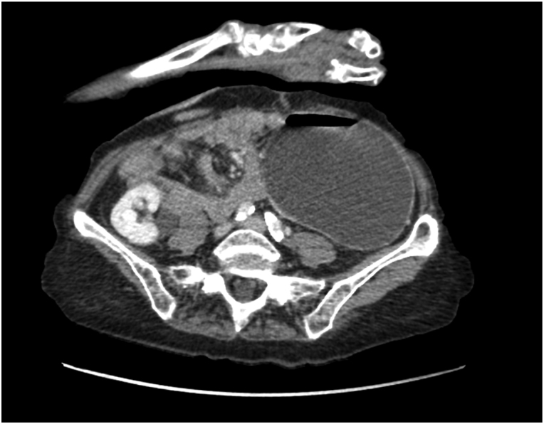
Abdomen and pelvic computerised tomography (CT) with contrast showed dilatation of the stomach (Figure 1) with collapse of the duodenum and small bowel. The transition point at the level of pylorus was in an inferior anatomical position and lay adjacent to third part of duodenum (Figure 2). The distal end of the loop ileostomy lay posterior to the transition point and demonstrated whirling (Figure 3). CT also highlighted the presence of aortic stenosis that was further investigated by echocardiogram which confirmed mild aortic stenosis with mild aortic regurgitation and mitral regurgitation.
Figure 1 .
Coronal view of the contrast CT abdomen and pelvis demonstrating dilated stomach
Figure 2 .
Axial view of the contrast CT abdomen and pelvis demonstrating the transition point at the level of the pylorus was in an inferior anatomical position and lays adjacent to the third part of the duodenum
Figure 3 .
Axial view of the contrast CT abdomen and pelvis demonstrating classic whirlpool sign of volvulus
Oesophago-gastro-duodenoscopy showed oesophagitis, large distended stomach and a distended first and second part of the duodenum. The gastroscope could not pass past the second part of the duodenum and was switched to a colonoscope. Omnipaque, the contrast medium, was injected via the colonoscope and stayed in the duodenum confirming the point of obstruction.
Differential diagnosis
Small bowel volvulus presents with vague, non-specific symptoms of abdominal pain, nausea and vomiting.5 These non-specific symptoms can be caused by a variety of conditions. After knowing the patient’s past medical history, adhesional small bowel obstruction was the top differential diagnosis. Other possible conditions that could be causing the patient’s symptoms were: cholecystitis, gastroenteritis or pancreatitis, which were ruled out considering normal inflammatory markers.
Owing to the absence of diagnostic symptoms, prompt diagnosis of the condition relied highly on radiologists and imaging. The modality of choice to confirm the diagnosis is CT.
Treatment
Because of the patient’s age, advanced comorbidities and wishes, conservative management was a more appropriate choice. This decision was supported by the patients NELA mortality score of 9% which later increased to 17% due to the new diagnosis of aortic stenosis. NELA score provides the mortality risk within 30 days of emergency abdominal surgery. It was developed from the National Emergency Laparotomy Audit between 2014 and 2016.6
A nasogastric Ryles tube (NGT) was inserted and the patient was made nil by mouth. The NGT was left to freely drain in order to decompress the stomach and the output was strictly monitored. While the patient was nil by mouth, intravenous fluids were given and fluid output was measured via a urinary catheter. Stoma output was regularly checked to monitor the obstruction. Gastrografin (meglumine amidotrizoate with sodium amidotrizoate) is an oral water-soluble contrast medium; however, it also acts as an osmotic agent and can be used as a therapeutic management to treat bowel obstruction because it reduces bowel oedema and enhances bowel motility.7 Therefore, the patient was given regular Gastrografin. The patient also received regular intravenous dexamethasone to reduce oedema.
On the second day of admission, the patient’s stoma became active so NGT was removed and the patient was allowed to have a liquid diet. The patient was started on Ensure drinks which include energy, protein, carbohydrate, fat and fibre to ensure the patient is receiving her daily nutritional requirements.
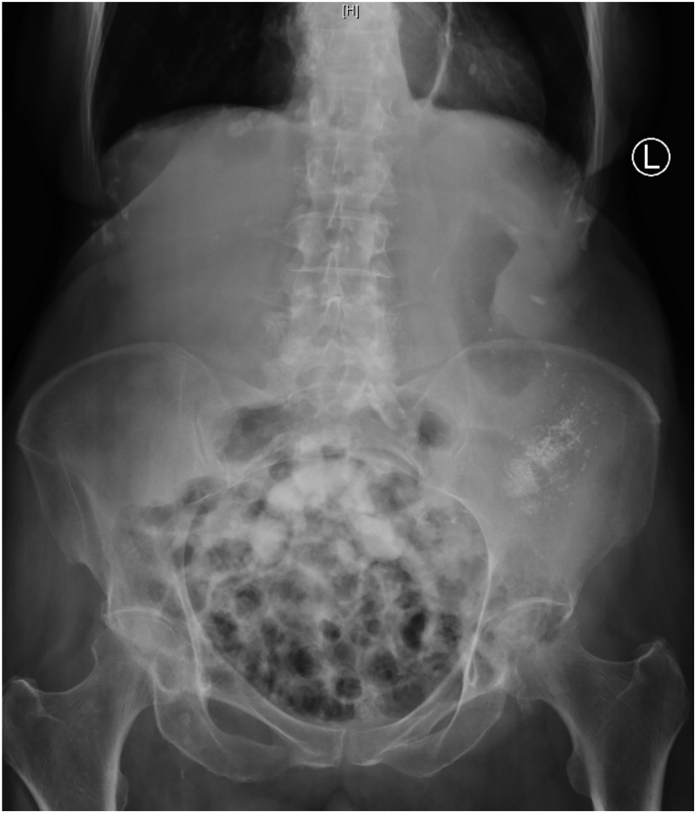
Small bowel obstruction continued to improve on conservative management and while the patient was still receiving Gastrografin, a decision to perform an abdominal x-ray was made. The x-ray demonstrated contrast medium was passing the point of obstruction (Figure 4). Because the obstruction was resolving, a decision to discharge the patient on the seventh day of admission was made. This decision was made with input from the patient, her family and the medical team looking after her.
Figure 4 .
Abdominal x-ray showing small bowel loops containing the contrast medium, Gastrografin
In cases in which conservative management fails to resolve the obstruction, surgical intervention is considered for resection of the non-viable duodenum or fixation of the viable segments.4
Outcome and follow-up
Conservative management successfully resolved patient’s symptoms. By the seventh day of admission, the patient had no complaint of abdominal pain, had good stoma output and was eating and drinking well. Considering the patient’s age and wishes, no surgical management was organised and therefore no general surgical follow-up was required. The patient and her family were informed and educated regarding seeking medical attention if the patient suffers from similar symptoms again.
Discussion
Very limited literature is available reporting cases of duodenal volvulus, reflecting its low incidence. From the limited cases seen, it is evident that duodenal volvulus can present in patients with or without a background of abdominal surgery; however, this case is the first to report duodenal volvulus in a patient with a background of bowel resection and laparotomy. Reported cases of duodenal volvulus, including this patient, were in elderly people supporting the aetiology of progressive laxity and downward migration of the duodenum with age.
Conclusion
The treatment option lies between conservative management or surgical management. In this patient and in the cases reported by Roux et al1 and Gavin et al,4 the patients were all managed conservatively due to the absence of complications. Surgical exploration is highly considered if patients fail to settle on conservative management. A third management option for patients with complex comorbidities is palliation.
References
- 1.Roux JW, Kahn AZ, Forshaw MJet al. Duodenal volvulus: report of a case. Surg Today 2007; 37: 434–436. [DOI] [PubMed] [Google Scholar]
- 2.Tsang CLN, Joseph CT, De Robles MSB, Putnis S. Primary small bowel volvulus: an unusual cause of small bowel obstruction. Cureus 2019; 11: e6465. [DOI] [PMC free article] [PubMed] [Google Scholar]
- 3.Large bowel Obstruction. BMJ Best Practice. https://bestpractice.bmj.com/topics/en-gb/3000125/aetiology (cited October 2021).
- 4.Gavin DJ, Wilkie BD, Ali N. A rare case of transient duodenal volvulus in an elderly patient. Journal of Medical Imaging and Radiation Oncology 2021; 65: 79–81. [DOI] [PubMed] [Google Scholar]
- 5.Peterson CM, Anderson JS, Hara AKet al. Volvulus of the gastrointestinal tract: appearances at multimodality imaging. RadioGraphics 2009; 29: 1281–1293. [DOI] [PubMed] [Google Scholar]
- 6.NELA Risk Calculator. https://data.nela.org.uk/riskcalculator/ (cited October 2021).
- 7.Haule C, Ongom PA, Kimuli T. Efficacy of Gastrografin® compared with standard conservative treatment in management of adhesive small bowel obstruction at Mulago National referral hospital. J Clin Trials 2013; 3: 1000144. [DOI] [PMC free article] [PubMed] [Google Scholar]


