Abstract
Gilthead seabream is among the most important farmed fish species in the Mediterranean Sea. Several approaches are currently applied to assure a lower impact of diseases and higher productivity, including the exploration of the fish microbiome and its manipulation as a sustainable alternative to improve aquaculture practices. Here, using 16S rRNA gene high-throughput sequencing, we explored the microbiome of farmed seabream to assess similarities and differences among microbial assemblages associated to different tissues and compare them with those in the surrounding environment. Seabream had distinct associated microbiomes according to the tissue and compared to the marine environment. The gut hosted the most diverse microbiome; different sets of dominant ASVs characterized the environmental and fish samples. The similarity between fish and environmental microbiomes was higher in seawater than sediment (up to 7.8 times), and the highest similarity (3.9%) was observed between gill and seawater, suggesting that gills are more closely interacting with the environment. We finally analyzed the potential connections occurring among microbiomes. These connections were relatively low among the host’s tissues and, in particular, between the gut and the others fish-related microbiomes; other tissues, including skin and gills, were found to be the most connected microbiomes. Our results suggest that, in mariculture, seabream microbiomes reflect only partially those in their surrounding environment and that the host is the primary driver shaping the seabream microbiome. These data provide a step forward to understand the role of the microbiome in farmed fish and farming environments, useful to enhance disease control, fish health, and environmental sustainability.
Keywords: Gilthead seabream, Fish microbiome, Aquaculture sustainability, Mediterranean Sea
Introduction
Aquaculture is the fastest-growing–food-production sector in the world and is predicted to contribute to more than half of the fish consumed globally by 2030 [1]. Gilthead seabream (Sparus [S.] aurata) is among the most important fish species farmed in the Mediterranean Sea [2, 3], with production increased by 77% over the last decade [4, 5] and with the EU representing the biggest producer worldwide for this species (https://ec.europa.eu/fisheries/sites/fisheries/files/docs/body/sea-bream_en.pdf). Aquaculture, including seabream farming, poses serious environmental concerns [6], represented mainly by organic wastes release into the surrounding environment, compromising the water quality and the benthic ecosystem nearby [7]. In addition, fish farms often experience diseases outbreaks due to microbial agents [8, 9], which cause further economic losses. Several solutions have been tested in order to assure a lower impact of farmed fish diseases while guaranteeing a higher productivity, most of them being based on the use of antimicrobials or vaccines [10, 11]. Solutions are likewise being used to reduce the organic enrichment of farm sediments by bioremediation actions [2]. An overall safer, more sustainable production of farmed fish is today demanded at the global level and developing under the principles of One Health to achieve beneficial health and well-being outcomes for people, non-human organisms, and their shared environment [12].
In this perspective, research efforts are being increasingly performed to improve the quality of farmed fish production by the exploitation and modulation of the fish microbiome [8, 9, 11, 13]. To do this, an improved and complete understanding of the teleost microbiome is required. However, while in the last years several works have been published on the fish microbiome, thanks to the application of high-throughput sequencing (HTS) [13], our knowledge is still in its infancy [13]. Most of the recent studies have been devoted to study fish gut and/or feces microbiome [14–16] with less works exploring at the same time the skin, gill, and other tissue’s microbiomes [3, 17, 18]. Also, it still remains unclear how microbiome diversity is partitioned among the different niches within teleosts, and how this partitioning might potentially be linked to the fish health.
Learning more about the microbiome of fish is essential given that, as for humans and other animals, the fish microbiome is crucial in protecting the health status of its host and in boosting the performance of the host immune system, well-being, food digestion, and synthesis of vital nutrients [8, 13, 18]. Mucosal surfaces such as skin and gills act as primary barriers to disease [3, 17–19] and play a key role in fish innate immunity [20]. At the same time, the gut microbiome has an active role in modulating the host’s physiology, assisting the intestinal development and physiology, as well as the overall development, growth, health, energy homeostasis, and immune response of the fish host [21]. Along with host-related benefits, the fish microbiome is increasingly being recognized as a means to obtain safe and high-quality food products for human consumption; understanding the prevalence and ecology of microorganisms, including those pathogenic and spoilage, associated to the fish tissues (e.g., skin, gill, gut) and products (e.g., fillet) along the production chain, has been also recently suggested to contribute to the development and application of new intervention strategies [9, 22] and a step forward to address the One Health concept also in aquaculture [12].
Among the drivers of the fish microbiome, diet is known to exert a major impact with particular reference to the gut [8], but also host-related factors, such as fish hosts’ genetic features, can play a role. Inter- and intra- host genetic diversity can drive the inclusion of beneficial or neutral microbes and the exclusion of potential pathogens [23]. However, the composition of the fish microbiome can also be affected by the environmental microbial communities, such as those associated with seawater and sediment [8]. Several studies have been performed on the inner (gut) and outer (gill and/or skin) fish microbiomes, with findings generally supporting the view that the environment is a source from which fishes acquire their microbiome [8, 16, 24, 25]. This aspect has not yet been clarified in Mediterranean sea bream, where information on the interactions between this species and the surrounding environment has been assessed only in early stages reared in tanks [26]. Moreover, it still remains under debate whether, and to what extent, the marine environment represents a source or a sink of the teleost microbiome in different compartments. To the best of our knowledge, no studies have been conducted in open mariculture settings to simultaneously assess the environmental microbiomes (sediment and seawater) and those associated with different seabream body tissues such as gut, skin, and gill; in addition, data on the presence, diversity, and abundance of microorganism in fish muscles (fillet) is still underexplored and up to debate.
In this study, by means of HTS 16S rRNA data, we aimed at exploring the microbiome of farmed Mediterranean gilthead seabream, the main farmed fish in the Mediterranean Sea, by (a) analyzing and identifying differences in the skin-, gill-, gut-, and fillet-associated microbial assemblages; (b) comparing the fish microbiome to environmental microbial communities (i.e., seawater and sediments); and (c) deciphering the potential connections occurring among microbiomes. Our study provides a step forward to the understanding of the diversity of the farmed sea bream microbiome in relation to the different fish tissues and the surrounding environment and aims at supporting future actions for enhancing disease control, fish health, and environmental sustainability in open-sea aquaculture settings.
Material and Methods
Study Area and Sampling Activities
Sampling activities were performed on 25th September 2019 at a marine fish farm located in the harbour of Licata (Sicily, Mediterranean Sea; coordinates 37.087713° N, 13.943773° E), within a semi-enclosed and sheltered area, characterized by a limited hydrodynamic circulation and a shallow depth (~ 10 m); further details on the sampling site are reported in [2] and [27].
Five adult individuals of S. aurata (avg. 273.2 g ± 32.3 g) fed a commercial diet were collected by a fishing net from a single sea cage. Fish specimens were immediately euthanized with a lethal dose (0.5 g/L) of Tricaine methanesulfonate (MS222), according to the ethical principles and national legislative context, and placed on ice until arrival at the laboratory around 1 h later; weight, total, and standard length were measured. Fish specimens were dissected within 4 h of capture.
For each fish specimen, we isolated: (1) fish skin (a 2 × 2 cm square from the left size), (2) fish gill (second gill arch on the left gill), (3) fish gut (fore-, mid-, and hindgut were separated; feces or digesta were collected by gentle squeezing, mucosae were collected by scraping of the internal side of the gut tube, and remaining gut tissue was also collected), (4) fish fillet (2 × 2 cm square at full depth from the left side). Except for fish feces, all the other tissue samples were rinsed with sterile phosphate buffer solution to remove possible loosely attached microorganisms. All described types of samples were obtained by dissecting fish specimens using sterile scalpels and scissors, immediately placed in sterile tubes, and stored at − 20 °C until further analyses.
Surface seawater and surface (0–1 cm) sediment samples were also collected at three sites, including the seabream sampling cage (37.086667°N, 13.943611°E) and at two additional sites (37.089732°N, 13.937469°E, and 37.091949°N, 13.933703°E), using a sterile plastic bottle and plexiglas cores, respectively. Further details on seawater and sediment sampling are reported in [27].
DNA Extraction and Sequencing
Seawater samples (one liter) were filtered onto 0.22 μm cellulose nitrate membrane filters (Sartorius, ø 47 mm) and stored at − 20 °C until processing; the entire membrane filters were used for DNA extraction. One g of the top 1 cm of sediment cores previously extruded and stored at − 20 °C was used for DNA extraction. DNA from sea bream skin, gill, fillet, feces and gut tissues, seawater, and sediment samples was extracted using the DNeasy PowerSoil Kit (Qiagen) as previously described in [27]. Extracted DNA samples were quantified with NanoDrop ND-1000 (NanoDrop Technologies, Wilmington, DE, USA) and stored at − 20 °C until processing. The PCR amplification of the V3-V4 hypervariable region of the 16S rRNA gene was carried out using the primer pair 341F-785R [28] and the PCR product purified as described in [27]. Nextera library indexing and preparation and Illumina MiSeq sequencing (2 × 300 bp paired-end protocol) were performed as described in [27]. The obtained raw sequences are submitted to the SRA—Sequence Read Archive (BioProject PRJNA692072).
Data Analysis
Primer and adapter sequences were removed from raw reads with Cutadapt [29]; paired-end reads were then imported and analyzed in RStudio (RStudio Team, 2020) using the DADA2 package [30]. Quality check and trimming of the reads were performed following the package instructions (at 280 and 230 bp for forward and reverse reads, respectively; max estimated error higher than 2 and 2 per 100 bp for forward and reverse reads, respectively). Paired-end reads were subsequently merged in amplicon sequence variants (ASVs, i.e., clusters sharing 100% sequence identity); chimeric sequences were identified and removed from the dataset. Finally, prokaryotic taxonomy was assigned using a native implementation of the naive Bayesian classifier method against the silva database (v132; https://www.arb-silva.de/documentation/release-132/). For alpha and beta diversity, all libraries obtained from gut samples (i.e., fore-, mid-, hindgut tissues, mucosae, and feces) were pooled per each fish sample. Chloroplast and eukaryotic sequences were removed from the ASV table obtained from DADA2; the table was normalized to the lowest number of reads present among the samples (n = 13,121) with the vegan package [31].
All statistical analyses were performed in Rstudio (RStudio Team, 2020). For the analysis of alpha diversity, ASV richness was calculated using the vegan package [31]. The occurrence of statistical differences among richness values in the different types of samples was assessed with a Kruskal–Wallis test (stats package) considering all possible comparisons. Non-metric multidimensional scaling (nMDS) was performed using a Bray–Curtis dissimilarity matrix and average linkage approach and plotted with the ggplot2 package. Significant differences in prokaryotic community composition between sample types (i.e., fish vs. environmental) as well as among matrices (i.e., skin, gill, gut, fillet, sediment, and seawater) and among fish tissues (i.e., skin, gill, gut, fillet) were calculated by using PERMANOVA through the adonis function (vegan package) in R, based on a Euclidean distance matrix calculated on the Hellinger transformed ASV table. A bubble plot including the most abundant ASVs within each sample was plotted using the ggplot2 package [32].
With the aim of exploring the connections occurring between microbiomes associated to the different fish tissues and the surrounding environment and in order to preliminarily hypothesize the ecological meaning of such connections, we performed network analysis using the network-based analysis performed by the make_otu_network.py script in QIIME v 1.9.0 [33]. This approach is used to analyze and display how ASVs were exclusive or shared among samples, with the aim to emphasize taxonomic similarities and differences among microbiomes. The network data were imported in Gephi (www.gephi.org) for visualization; in the obtained plot, the more similar are the samples, the closer they are positioned in the plot. The Venn diagrams were calculated using the R package Venn (https://github.com/dusadrian/venn), and the results from this analysis were reported in a table, as described in the “Results” section.
Results
Prokaryotic Diversity and Community Composition
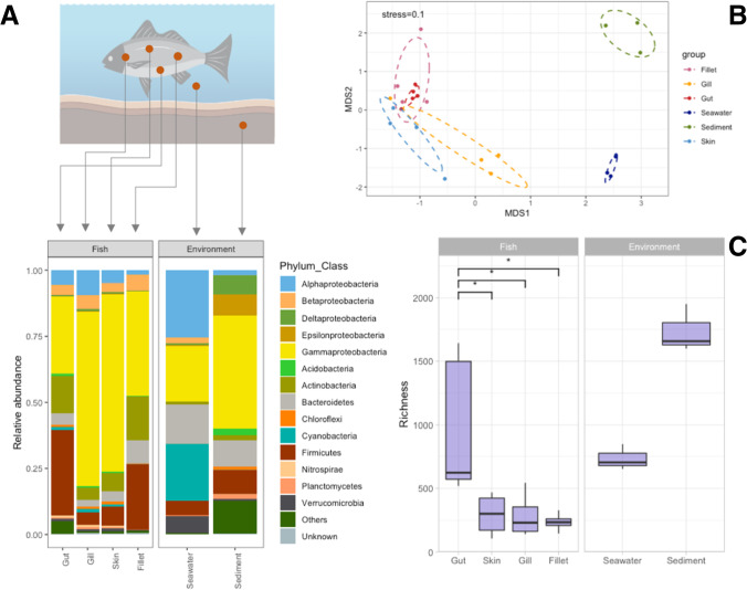
Taxonomic composition at the phylum level (Fig. 1A) highlighted the dominance of Proteobacteria in all samples; in particular, fish communities were particularly enriched in Gammaproteobacteria (avg. 49.24 ± 31.65%), followed by Alpha- (avg. 5.39 ± 6.67%) and Betaproteobacteria (avg. 4.61 ± 5.8%). Higher proportion of Alpha (25.03 ± 0.62% and 1.83 ± 0.27% in seawater and sediment, respectively), Delta- (0.71 ± 0.3% and 7.34 ± 1.53%), and Epsilonproteobacteria (0.22 ± 0.07% and 8.04 ± 10.48%) were overall observed in environmental samples. Fish samples also displayed high relative abundances of Actinobacteria (avg. 10.73 ± 10.56%) and Firmicutes (avg. 18.17 ± 14.19%), with particular regard to gut (14.27 ± 6.07% and 32.46 ± 11.07% for Actinobacteria and Firmicutes, respectively) and fillets (16.52 ± 18.44% and 24.73 ± 7.84% for Actinobacteria and Firmicutes, respectively) microbiomes (Fig. 1A). Cyanobacteria were observed mainly in seawater (avg. 21.63 ± 0.54%); higher proportions of Bacteroidetes finally characterized environmental samples (14.94 ± 0.92% and 9.76 ± 6.42% in seawater and sediment, respectively).
Fig. 1.
Panel A: barplot showing the prokaryotic community composition (as relative abundance) at the phylum and class level (for Proteobacteria only). Taxa with an average relative abundance across all samples < 1% were aggregated as “Others”. “Unknown” includes all those reads that did not match any known taxa. Panel B: nonmetric multidimensional scaling (NMDS) ordination of community composition of fish and environmental microbiomes based on Bray–Curtis dissimilarity matrix. Panel C: richness calculated for the different types of samples; asterisks indicate the occurrence of significant differences as calculated by the Kruskal–Wallis test (p < 0.05); non-significant comparisons are not reported in the plot. Richness in environmental samples also significantly differed from those observed in fish-associated microbiomes (Kruskal–Wallis p < 0.05). The fish figure has been created in BioRender (https://biorender.com/)
Non-metric multidimensional scaling (NMDS) (Fig. 1B) underlined the clear separation between fish and environmental samples, while showing a partial overlapping of communities belonging to the different fish tissue types. PERMANOVA analysis confirmed what previously observed for richness: microbiomes associated with fish tissues were significantly different from those in the surrounding environment (adonis, p < 0.001), while microbiomes associated with the different fish tissues, significantly differed among each other (adonis, p = 0.006).
Alpha diversity (i.e., ASV richness) (Fig. 1C) indicated an overall higher diversity in environmental microbiomes (i.e., seawater and sediments) than in those associated with fish, with sediment hosting the most diverse assemblages of the entire dataset. In more detail, statistical differences occurred between the number of ASVs harbored by fish- and environment-related samples (p = 0.004). Considering microbiomes from the fish specimens only, we observed a significantly higher diversity occurring in gut samples as compared to skin, gill, and fillet samples (p < 0.05) (Fig. 1C).
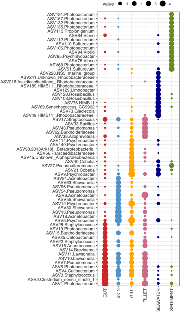
The analysis of the most abundant ASVs represented in the different types of samples (Fig. 2) highlighted that different sets of abundant ASVs characterized the environmental and fish-associated microbiomes. ASVs identified at the genus level as Photobacterium, Vibrio, and Sulfurovum were particularly abundant in sediment samples, whereas the seawater microbiome was enriched in ASVs identified as Rhodobacteraceae, Synechococcus, NS5 marine group, Limnobacter, Glaciecola, and Roseibacillus. Fish gut samples contained, at high abundances, ASVs belonging to the genus Photobacterium but different from those observed in sediment and seawater; ASVs identified as Streptococcus, Staphylococcus, Anaerococcus, Lawsonella, Pseudomonas, Cutibacterium, Clostridium, and Brevinema were also abundant in these type of samples. ASVs assigned to the genera Pseudomonas, Acinetobacter, Psychrobacter, and Shewanella were present as the most abundant in fish skin samples; moreover, some ASVs identified as Lawsonella, Staphylococcus, Pseudomonas, and Cutibacterium appeared to be shared also with gut, gill, and fillet microbiomes. Gill samples were enriched in ASVs belonging to the genera Psychrobacter, Cobetia, and Pseudoalteromonas; even in this case, several ASVs were shared with the other fish microbiomes’ most abundant ASVs. Finally, fillet microbiomes displayed a set of abundant ASVs and mainly belonging to the genera Streptococcus, Bacillus, Pseudomonas, Shewanella, Photobacterium, Staphylococcus, Lawsonella, and Cutibacterium.
Fig. 2.
Bubble chart reporting the proportion and taxonomic identification of the most abundant ASVs (avg. > 0.5% across the dataset) observed in each fish and environmental microbiome. The dimension of each bubble is proportional to the log of the relative abundance of the ASV
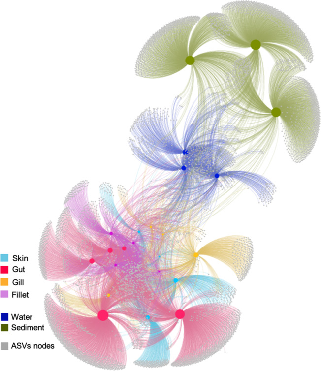
We used the make_otu_network.py script implemented in QIIME in order to visualize how ASVs were shared among different fish tissues and environmental samples (Fig. 3). The results of this analysis showed that unique ASVs occurred in all fish tissues and environmental samples, with the gut being the tissue hosting the largest number of unique ASVs for fish samples (n = 1945, 73.8% of total ASVs for gut samples) and the fillet being the one with the smallest (n = 181, 30.8% of total ASVs for fillet samples). Notably, sediment samples included the highest number of exclusive ASVs for the whole dataset (n = 2486, 93.8% of total ASVs for sediment samples). More in detail, only six ASVs were found to be present in all samples (ASV5 Psychrobacter; ASV3 Staphylococcus; ASV1 Photobacterium; ASV4 Cutibacterium; ASV9 Psychrobacter; ASV6 Photobacterium) and, in general, the fish microbiomes shared a lower percentage of ASVs with the environmental communities from sediment (0.6–0.8%) than seawater (Table 1). Interestingly, the highest number of shared ASVs between fish and environmental samples was observed when considering seawater and gill microbiomes (5.3%), with a value that doubled those observed when comparing ASVs shared between seawater and the other fish microbiome, and even exceeding the percentage of ASVs shared between seawater and sediment microbiomes (4.1%). Considering only fish microbiomes, the gut microbiome shared less than 13% of ASVs with each fish tissue, whereas skin and fillet shared 18.8% of their ASVs (Table 1).
Fig. 3.
Network-based analysis to display and analyze how ASVs are partitioned between samples. In the network diagram, there are two kinds of “nodes” represented, ASV-nodes (grey) and sample-nodes (colored according to the type of sample: red, gut; yellow, gill; cyan, skin; pink, fillet; dark blue, seawater, green, sediment). The network has been calculated excluding singletons. Sample nodes’ size is proportional to their degree, i.e., the number of connections with ASVs nodes. Edges (i.e., lines between nodes) connect ASVs to the samples where they are present; shorter distances between the sample and ASV nodes reflect larger numbers of sequences from that ASV found in the connected sample. Finally, ASVs occurring in the center of the network are more evenly distributed between all the samples, while those on the edges occur preferentially in certain samples
Table 1.
Summary of ASVs (excluding singletons) shared among different types of samples analyzed in this study, i.e., including both fish- and environment-related microbiomes. Reported are (i) the number of shared ASVs, (ii) the total ASV number, and (iii) the percentage of shared ASVs between pairs of sample types
| Type of sample | # of shared ASVs | Total ASVs | % of shared ASVs | |
|---|---|---|---|---|
| Fish gut | Fish fillet | 354 | 2886 | 12.4 |
| Fish gill | 316 | 2633 | 12.0 | |
| Fish skin | 410 | 3459 | 11.9 | |
| Sediment | 39 | 5243 | 0.7 | |
| Seawater | 74 | 3386 | 2.2 | |
| Fish fillet | Fish gill | 185 | 1194 | 15.5 |
| Fish skin | 224 | 1189 | 18.8 | |
| Sediment | 18 | 3218 | 0.6 | |
| Seawater | 36 | 1378 | 2.6 | |
| Fish gill | Fish skin | 218 | 1400 | 15.6 |
| Sediment | 28 | 3413 | 0.8 | |
| Seawater | 82 | 1537 | 5.3 | |
| Fish skin | Sediment | 27 | 3448 | 0.8 |
| Seawater | 40 | 1613 | 2.5 | |
| Seawater | Sediment | 138 | 3338 | 4.1 |
Discussion
The microbiome affects many properties of host phenotypes and metabolism [34]. Hence, an in-depth knowledge of fish microbiome is prompted aimed at understanding its role in the host organism [16]. This applies with particular reference to commercially relevant farmed fish species, given the possible implications of the microbiome to their production management, the safety, and quality of fish food produced as well as fish production’s environmental sustainability [9]. Here, for the first time to the best of our knowledge, we report on the different farmed Mediterranean gilthead seabream tissues’ microbiomes and their surrounding environment to test whether different tissues select for different microbial communities and to clarify the extent of the surrounding environment in contributing to the composition of fish-associated microbial communities. Furthermore, we explored the potential interplay among tissues’ microbiomes to shed light on the connections occurring between the different microbiomes and to hypothesize future directions in fish microbiome exploration.
Overall, our study showed clear distinctions between the fish and the surrounding environment microbiomes as well as among the different fish tissues. At the same time, we also observed the presence of connections among fish tissues and the environment. This finding supports the concept that such complex relationships should be studied more in detail and taken into account when designing and applying microbiome-based approaches to improve and manage farmed fish productions. In agreement with previous studies, our results indicate that fish have distinct associated microbiomes compared with the external environment [18, 35], confirming the notion that fish hosts select for specific assemblages, and that host species rather than the surrounding environment is the primary driver shaping microbial communities in fish [36]. Overall, the percentage of shared ASVs between fish microbiomes and the environment was higher with seawater than with sediments, suggesting that connections between fish and the environment mainly occur with the pelagic compartment, as expected for a pelagic species such as S. aurata. However, more experimental studies are needed to clarify this point.
Our results showed a higher diversity in environmental samples than in fish microbiomes, in contrast to previous observations [35]; however, it is noteworthy mentioning that different HTS approaches were used (454 vs. Illumina) and that such a discrepancy could have been caused by the different methodological approaches applied. As expected, planktonic communities were composed of typical marine classes such as the Alphaproteobacteria and Cyanobacteria; similarly, benthic assemblages were composed of common surface sediment-associated taxa, such as Gamma-, Delta-, and Epsilonproteobacteria [37]. Additionally, Proteobacteria, Firmicutes, and Actinobacteria, that are generally recognized as the dominant phyla in the fish microbiome with particular reference to the intestinal content and gut microbiome [16], were also similarly dominant in our study. Interestingly, we found lower Bacteroidetes in fish than in environmental samples, in contrast to several studies indicating this phylum as one of the most abundant in fish [16]; however, it is noteworthy mentioning that Bacteroidetes commonly dominate herbivores fish gut microbiomes [38], whereas S. aurata exerts a carnivore feeding style, which may explain the such discrepancy. At the ASV level, different sets of dominant ASVs characterized environmental and fish samples, suggesting the presence of discrete and potentially consistent signatures of microbial communities unique to the investigated tissues [39], as well as to the environmental samples. We speculate that such specific signatures may exert a role in the maintenance of the host health, an aspect that surely deserves further clarifications.
A further level of differentiation was observed when considering the environmental and fish microbiomes separately. Overall, as expected, we find higher richness values in sediment than in seawater, with several taxa, including Vibrio and Sulfurovum, as well as Rhodobacteraceae and Synechococcus, being differentially displayed in the two matrices and confirming established previous notions on the composition of benthic and pelagic bacterial communities [40]. Within microbiome fish tissues, higher richness values were found in the gut than in the other fish tissues, as generally reported for sea bream [41, 42]. Based on the evidence that increased microbial diversity is associated with improved physiology and homeostasis in humans [43]; we speculate that our finding supports a key role of gut microbes in fish health. Although gill microbiomes have been generally reported to display a lower richness compared to those of skin and water [17, 18], diversity in seabream skin and gills has been described as not significantly different [3], supporting the results of our study. Moreover, despite a partial overlapping of the overall community composition depicted by the nMDS analysis, the gut microbiome significantly differed from that of gill, skin, and fillet, suggesting that, although in communication with each other, each tissue favors the establishment of a specific community, likely in response to host-specific organizing factors.
Taxonomic characterization of seabream gut showed, in our dataset, the dominance of Photobacterium, Pseudomonas, and Staphylococcus genera, which have been already reported as important taxa in fish microbiomes. Photobacterium species are found widespread in marine habitat [44] as pathogens or decomposers of dead fish, as well as commensals in the gut of many marine organisms [45], although some Photobacterium species are also found on the skin and gills of marine fish [46, 47]. Notably, Photobacterium exerts contrasting behaviors in association with fish, with a member of this genus being associated with healthy individuals [18, 46] and others, such as Photobacterium damselae subsp. piscicida, representing the aetiological agent of pasteurellosis [48], suggesting an opportunistic lifestyle towards the fish host. The Pseudomonas genus was found in the core gut microbiome of 5 sympatrically farmed marine fish species [49] as well as in the core mid-gut microbiota of Mediterranean seabass and seabream [15] whereas Staphylococcus epidermidis represented one of the core OTUs in S. aurata in the Aegean Sea [15]. Pseudomonas members have been often reported in carnivorous teleosts and reported the most abundant shared bacterial species in the gut of S. aurata and D. labrax [50], and have been reported to contribute to teleosts’ digestion through the secretion of several digestive enzymes [50 and references therein]. Interestingly, Pseudomonas, together with Acinetobacter, Psychrobacter, and Shewanella, was also abundant in skin, although different ASVs were associated with the different host tissues.
Among the most represented taxa in skin samples, we found ASVs assigned to the genera Pseudomonas, Acinetobacter, Psychrobacter, and Shewanella. Some of these taxa, such as Acinetobacter, have been often associated with fish external microbiomes [19, 36]. Interestingly, despite being reported as fish spoilage agents [51], Pseudomonas and Psychrobacter strains have repeatedly shown their potential as probiotics in aquaculture [52, 53]. Similarly, a strain of Shewanella (e.g., SpPdp11), isolated from the skin of healthy specimens of S. aurata, is being widely tested as a fish health modulator; the capacity of this strain on wounded skin healing [54], as well as its regulatory role in skin inflammatory and epithelial barrier function [55] has been demonstrated in several fish species. Recently, a tight connection between fish skin-associated microbes and intestinal barrier functioning was suggested in a study investigating the use of this probiotic strain of Shewanella in skin-injured gilthead seabream [54].
Gill samples were enriched in ASVs belonging to the genera Psychrobacter, Cobetia, and Pseudoalteromonas. In a recent study by Rosado et al. [3], such taxa were not reported among those most abundantly found in Mediterranean Sea bream gills collected in Portugal. However, considering that our results indicate the gill microbiome as the one displaying the highest similarity with bacterioplankton communities compared to the other tissues, we hypothesize that such a discrepancy may be explained by the differences in the environmental microbial community composition at different sites. Although little is known about the presence of these taxa in gilthead seabream, all these genera were defined among those significantly abundant OTUs in mucosal surfaces (i.e., skin and gills) of healthy individuals in Yellowtail Kingfish [18]. Gills are vital for fish health, as fishes breathe and excrete waste through them, and they are also potential sites of pathogen invasion and colonization by other microbes [17] as well as a barrier preventing pathogen invasion. However, we know little about the gill microbiome and the factors shaping their diversity. Taken together, these results suggest that a set of potential beneficial microorganisms are enriched in the skin and gill microbiomes, with possible implications of the general health of seabream individuals that deserve further clarifications. Lastly, only a little information on fillet-associated microbiome diversity is reported in the literature for seabream [56, 57]. Interestingly, most of the fillet abundant taxa were shared with the other fish microbiomes (e.g., Photobacterium, Staphylococcus, Pseudomonas), and included some taxa previously described as fillet-associated in a study on other finfish species [58]. However, it must be pointed out that, in such studies, the word “fillet” refers to a (minimally) processed food product whose microbiome might have been impacted by contamination occurring during processing and storage. This substantially differs from our fillet samples, which were basically represented by a portion of fish muscle isolated from the entire fish avoiding, to the greatest possible extent, contamination from working surfaces or fish skin. Based on available knowledge, the muscles of healthy fishes are usually considered sterile, despite the debate on this aspect is still open [22]. Indeed, contamination of fish muscles is possible when immunological resistance is compromised [59], which may likely occur in aquaculture settings under stressful conditions. Although a potential, unavoidable cross-contamination between skin and fillet may have occurred during sampling, our results showing the sharing of bacterial ASVs between the fillet and other tissues, such as the gut or gills (which are unlikely to be caused by cross-contamination), highlight the need for more investigations to fully decipher the presence and role of bacteria in fish muscles.
Considering the level of shared ASVs as a proxy of the connections between different microbiomes, we found that, overall, connections were relatively low among the host’s microbiomes (i.e., skin, gut, gill, and fillet) and, in particular, between the gut and the others fish-related microbiomes. Despite further studies are needed to ascertain whether this is an established pattern in seabream, a recent study on fish skin and gut microbiomes reports that skin-associated communities were highly dependent upon environmental factors, conversely to gut-associated assemblages, suggesting that the stability of abiotic conditions within each tissue (e.g., intestinal pH) may have a role in promoting specialization of the microbiomes [39] and likely supports the lower level of ASVs sharing of the gut with the other fish microbiomes. Nevertheless, given the tight link between the fish microbiome and health [60, 61] and the knowledge acquired in the exploration of other animal microbiomes (e.g., human), we hypothesize that the connections between the fish gut and other fish tissues occur not only in terms of exchanges in microbial taxa but also through the production of metabolites by microorganisms associated to the different fish compartments [13]. A preliminary support to this hypothesis has been provided by [18], who showed the link between fish gut health status on the structuring of skin and gill bacterial assemblages. We also report here that skin, gills, and fillet were found to be the most connected microbiomes in seabream, suggesting the possibility of easier communications taking place among these compartments. If, from one side, close connections between skin and gills appear to be expectable, due to their continuous exchange with seawater and the resuspended sediments, more should be clarified about their connection with fillet. Only a small set of ASVs was shared among all the different seabream microbiomes, suggesting that such bacteria might represent the core members of seabream microbiomes and that these members are able to adapt to different conditions. Finally, a lower similarity was observed between the fish microbiomes and the environment. The higher number of ASVs shared between seawater and gills (around 4%) underlines the closer relationship occurring between this mucosal tissue and the surrounding environment and highlights the role of site features in structuring the outer microbiome of fish [39]. Overall, our results suggest that, although at different levels and depending on the tissue, the seabream microbiome is somehow connected with the environment and the surrounding environment acts as a source of microbes for the fish microbiome [8 and references therein].
Conclusions
The tight link between microbiome composition and fish health is being increasingly explored in teleosts, including the commercially relevant S. aurata species. Our understanding of the complex interactions occurring among fish and environmental microbiomes is still limited and, to better clarify all the aspects, dedicated studies including multi-omics able to trace and combine taxonomic and functional contributions are needed. The information here provided on fish microbiome composition according to the different types of tissue (skin, gill, gut, fillet) and its potential relationships with the surrounding environment (seawater, sediment) provides an additional step towards the exploration of the fish microbiome, useful also to implement future microbiome-based applications improving aquaculture practices. We show that the seabream microbiome, despite differentially sharing a number of microbial taxa with the surrounding seawater and sediments, which suggest the occurrence of relationships between the teleost and the surrounding marine microbiome, displays tissue-specific communities with a highly specific microbial signature. Future research is required aimed at better understanding the nature of the interactions occurring among a host’s microbiomes and how this affects fish health.
Acknowledgements
We thank Valeria Montalto, Francesca Ape, and Marco Martinez for their help in sampling activities and Massimo Cafarella for his support during laboratory analyses; we are also grateful to Prof. Alessio Bonaldo (University of Bologna) for his useful and kind advice and opinions on fish physiology. The research leading to these results has been conceived under the International PhD Program “Innovative Technologies and Sustainable Use of Mediterranean Sea Fishery and Biological Resources (www.FishMed-PhD.org). This study represents partial fulfillment of the requirements for the PhD thesis of M. Basili at the FishMed-PhD course.
Author Contribution
Conceptualization: GMQ, GML; formal analysis: GMQ, RP, MB; investigation: GMQ, GM, SM, EM; visualization: GMQ, RP, MB; writing—original draft: GMQ, RP, GML; writing—review and editing: all authors; resources: AMS; Funding acquisition; MC, GML.
Funding
This work was supported by the “Controlling Microbiomes Circulations for Better Food Systems” (CIRCLES) project, funded by the European Union’s Horizon 2020 research and innovation program under grant agreement no. 818290. This publication reflects only the author’s view and the Agency is not responsible for any use that may be made of the information it contains.
Data Availability
Raw sequence reads are available in the NCBI’s SRA—Short Read Archive under accession PRJNA692072.
Declarations
Competing Interests
The authors declare no competing interests.
Footnotes
Grazia Marina Quero and Roberta Piredda contributed equally to this work.
References
- 1.FAO . The state of world fisheries and aquaculture 2020. Rome: Sustainability in action; 2020. [Google Scholar]
- 2.Ape F, Manini E, Quero GM, Luna GM, Sara G, Vecchio P, Brignoli P, Ansferri S, Mirto S. Biostimulation of in situ microbial degradation processes in organically-enriched sediments mitigates the impact of aquaculture. Chemosphere. 2019;226:715–725. doi: 10.1016/j.chemosphere.2019.03.178. [DOI] [PubMed] [Google Scholar]
- 3.Rosado D, Perez-Losada M, Severino R, Cable J, Xavier R. Characterization of the skin and gill microbiomes of the farmed seabass (Dicentrarchus labrax) and seabream (Sparus aurata) Aquaculture. 2019;500:57–64. doi: 10.1016/j.aquaculture.2018.09.063. [DOI] [Google Scholar]
- 4.Neofitou N, Vafidis D, Klaoudatos S. Spatial and temporal effects of fish farming on benthic community structure in a semi-enclosed gulf of the Eastern Mediterranean. Aquac Environ Interact. 2010;1:95–105. doi: 10.3354/aei00010. [DOI] [Google Scholar]
- 5.Rosa R, Marques A, Nunes ML. Impact of climate change in Mediterranean aquaculture. Rev Aquac. 2012;4:163–177. doi: 10.1111/j.1753-5131.2012.01071.x. [DOI] [Google Scholar]
- 6.Quero GM, Ape F, Manini E, Mirto S, Luna GM. Temporal changes in microbial communities beneath fish farm sediments are related to organic enrichment and fish biomass over a production cycle. Front Mar Sci. 2020;7:524. doi: 10.3389/fmars.2020.00524. [DOI] [Google Scholar]
- 7.Mirto S, Gristina M, Sinopoli M, Maricchiolo G, Genovese L, Vizzini S, Mazzola A. Meiofauna as an indicator for assessing the impact of fish farming at an exposed marine site. Ecol Indic. 2012;18:468–476. doi: 10.1016/j.ecolind.2011.12.015. [DOI] [Google Scholar]
- 8.de Bruijn I, Liu Y, Wiegertjes GF, Raaijmakers JM. Exploring fish microbial communities to mitigate emerging diseases in aquaculture. FEMS Microbiol Ecol. 2018;94:fix161. doi: 10.1093/femsec/fix161. [DOI] [PubMed] [Google Scholar]
- 9.Luna GM, Quero GM, Kokou F, Kormas K. Time to integrate biotechnological approaches into fish gut microbiome research. Curr Opin Biotechnol. 2022;73:121–127. doi: 10.1016/j.copbio.2021.07.018. [DOI] [PubMed] [Google Scholar]
- 10.Bentzon-Tilia M, Sonnenschein EC, Gram L. Monitoring and managing microbes in aquaculture—towards a sustainable industry. Microb Biotechnol. 2016;9:576–584. doi: 10.1111/1751-7915.12392. [DOI] [PMC free article] [PubMed] [Google Scholar]
- 11.Perry WB, Lindsay E, Payne CJ, Brodie C, Kazlauskaite R. The role of the gut microbiome in sustainable teleost aquaculture. Proc R Soc B: Biol Sci. 2020;287:20200184. doi: 10.1098/rspb.2020.0184. [DOI] [PMC free article] [PubMed] [Google Scholar]
- 12.Stentiford GD, Bateman IJ, Hinchliffe SJ, Bass D, Hartnell R, Santos EM, Devlin MJ, Feist SW, Taylor NGH, Verner-Jeffreys DW, van Aerle R, Peeler EJ, Higman WA, Smith L, Baines R, Behringer DC, Katsiadaki I, Froehlich HE, Tyler CR. Sustainable aquaculture through the One Health lens. Nat Food. 2020;8:468–474. doi: 10.1038/s43016-020-0127-5. [DOI] [PubMed] [Google Scholar]
- 13.Legrand TP, Wynne JW, Weyrich LS, Oxley AP. A microbial sea of possibilities: current knowledge and prospects for an improved understanding of the fish microbiome. Rev Aquac. 2020;12:1101–1134. doi: 10.1111/raq.12375. [DOI] [Google Scholar]
- 14.Mente E, Nikouli E, Antonopoulou E, Martin SA, Kormas KA. Core versus diet-associated and postprandial bacterial communities of the rainbow trout (Oncorhynchus mykiss) midgut and faeces. Biol Open. 2018;7:bio034397. doi: 10.1242/bio.034397. [DOI] [PMC free article] [PubMed] [Google Scholar]
- 15.Nikouli E, Meziti A, Antonopoulou E, Mente E, Kormas KA. Gut bacterial communities in geographically distant populations of farmed sea bream (Sparus aurata) and sea bass (Dicentrarchus labrax) Microorganisms. 2018;6:92. doi: 10.3390/microorganisms6030092. [DOI] [PMC free article] [PubMed] [Google Scholar]
- 16.Ghanbari M, Kneifel W, Domig KJ. A new view of the fish gut microbiome: advances from next-generation sequencing. Aquaculture. 2015;448:464–475. doi: 10.1016/j.aquaculture.2015.06.033. [DOI] [Google Scholar]
- 17.Pratte ZA, Besson M, Hollman RD, Stewart FJ. The gills of reef fish support a distinct microbiome influenced by host-specific factors. Appl Environ Microbiol. 2018;84:e00063–e118. doi: 10.1128/AEM.00063-18. [DOI] [PMC free article] [PubMed] [Google Scholar]
- 18.Legrand TP, Catalano SR, Wos-Oxley ML, Stephens F, Landos M, Bansemer MS, Stone DA, Qin JG, Oxley A. The inner workings of the outer surface: skin and gill microbiota as indicators of changing gut health in yellowtail kingfish. Front Microbiol. 2018;15(8):2664. doi: 10.3389/fmicb.2017.02664. [DOI] [PMC free article] [PubMed] [Google Scholar]
- 19.Larsen AM, Bullard SA, Womble M, Arias CR. Community structure of skin microbiome of gulf killifish, Fundulus grandis, is driven by seasonality and not exposure to oiled sediments in a Louisiana salt marsh. Microb Ecol. 2015;70:534–544. doi: 10.1007/s00248-015-0578-7. [DOI] [PubMed] [Google Scholar]
- 20.Gourzioti E, Kolygas MN, Athanassopoulou F, Babili V. Tenacibaculosis in aquaculture farmed marine fish. J Hell Vet. 2016;67:21–32. [Google Scholar]
- 21.Butt RL, Volkoff H. Gut microbiota and energy homeostasis in fish. Front Endocrinol. 2019;10:9. doi: 10.3389/fendo.2019.00009. [DOI] [PMC free article] [PubMed] [Google Scholar]
- 22.Sheng L, Wang L. The microbial safety of fish and fish products: recent advances in understanding its significance, contamination sources, and control strategies. Compr Rev Food Sci Food Saf. 2021;20:738–786. doi: 10.1111/1541-4337.12671. [DOI] [PubMed] [Google Scholar]
- 23.Hooper LV, Littman DR, Macpherson AJ. Interactions between the microbiota and the immune system. Science. 2012;336:1268–1273. doi: 10.1126/science.1223490. [DOI] [PMC free article] [PubMed] [Google Scholar]
- 24.Huang Q, Sham RC, Deng Y, Mao Y, Wang C, Zhang T, Leung KM. Diversity of gut microbiomes in marine fishes is shaped by host-related factors. Molec Ecol. 2020;29:5019–5034. doi: 10.1111/mec.15699. [DOI] [PMC free article] [PubMed] [Google Scholar]
- 25.Wu S, Wang G, Angert ER, Wang W, Li W, Zou H. Composition, diversity, and origin of the bacterial community in grass carp intestine. PLoS ONE. 2012;7:e30440. doi: 10.1371/journal.pone.0030440. [DOI] [PMC free article] [PubMed] [Google Scholar]
- 26.Nikouli E, Meziti A, Antonopoulou E, Mente E, Kormas KA. Host-associated bacterial succession during the early embryonic stages and first feeding in farmed gilthead sea bream (Sparus aurata) Genes. 2019;10:483. doi: 10.3390/genes10070483. [DOI] [PMC free article] [PubMed] [Google Scholar]
- 27.Palladino G, Rampelli S, Scicchitano D, Musella M, Quero GM, Prada F, Mancuso A, Seyfarth AM, Turroni S, Candela M, Biagi E. Impact of marine aquaculture on the microbiome associated with nearby holobionts: the case of Patella caerulea living in proximity of Sea Bream aquaculture cages. Microorganisms. 2021;9:455. doi: 10.3390/microorganisms9020455. [DOI] [PMC free article] [PubMed] [Google Scholar]
- 28.Klindworth A, Pruesse E, Schweer T, Peplies J, Quast C, Horn M, Glöckner FO. Evaluation of general 16S ribosomal RNA gene PCR primers for classical and next-generation sequencing-based diversity studies. Nucleic Acids Res. 2013;41:e1. doi: 10.1093/nar/gks808. [DOI] [PMC free article] [PubMed] [Google Scholar]
- 29.Martin M. Cutadapt removes adapter sequences from high-throughput sequencing reads. EMBnet J. 2011;17:10–12. doi: 10.14806/ej.17.1.200. [DOI] [Google Scholar]
- 30.Callahan BJ, McMurdie PJ, Rosen MJ, Han AW, Johnson AJ, Holmes SP. DADA2: high-resolution sample inference from Illumina amplicon data. Nat Methods. 2016;13:581–583. doi: 10.1038/nmeth.3869. [DOI] [PMC free article] [PubMed] [Google Scholar]
- 31.Oksanen J, Blanchet FG, Kindt R, Legendre P, Minchin PR, O’hara RB, Simpson GL, Solymos P, Stevens MH, Wagner H (2013) Community ecology package. R package version 2(0)
- 32.Wickham H (2016) Programming with ggplot2. In ggplot2 2016 (pp. 241–253). Springer, Cham
- 33.Caporaso JG, Kuczynski J, Stombaugh J, Bittinger K, Bushman FD, Costello EK, Fierer N, Peña AG, Goodrich JK, Gordon JI, Huttley GA. QIIME allows analysis of high-throughput community sequencing data. Nat Methods. 2010;7:335–336. doi: 10.1038/nmeth.f.303. [DOI] [PMC free article] [PubMed] [Google Scholar]
- 34.Carnevali O, Maradonna F, Gioacchini G. Integrated control of fish metabolism, wellbeing and reproduction: the role of probiotic. Aquaculture. 2017;472:144–155. doi: 10.1016/j.aquaculture.2016.03.037. [DOI] [Google Scholar]
- 35.Chiarello M, Villeger S, Bouvier C, Bettarel Y, Bouvier T. High diversity of skin-associated bacterial communities of marine fishes is promoted by their high variability among body parts, individuals and species. FEMS Microbiol Ecol. 2015;91:fix061. doi: 10.1093/femsec/fiv061. [DOI] [PubMed] [Google Scholar]
- 36.Larsen A, Tao Z, Bullard SA, Arias CR. Diversity of the skin microbiota of fishes: evidence for host species specificity. FEMS Microbiol Ecol. 2013;85:483–494. doi: 10.1111/1574-6941.12136. [DOI] [PubMed] [Google Scholar]
- 37.Basili M, Quero GM, Giovannelli D, Manini E, Vignaroli C, Avio CG, De Marco R, Luna GM. Major role of surrounding environment in shaping biofilm community composition on marine plastic debris. Front Mar Sci. 2020;7:262. doi: 10.3389/fmars.2020.00262. [DOI] [Google Scholar]
- 38.Miyake S, Ngugi DK, Stingl U. Diet strongly influences the gut microbiota of surgeonfishes. Molec Ecol. 2015;24:656–672. doi: 10.1111/mec.13050. [DOI] [PubMed] [Google Scholar]
- 39.Sylvain FÉ, Holland A, Bouslama S, Audet-Gilbert É, Lavoie C, Val AL, Derome N. Fish skin and gut microbiomes show contrasting signatures of host species and habitat. Appl Environ Microbiol. 2020;86:e00789–e820. doi: 10.1128/AEM.00789-20. [DOI] [PMC free article] [PubMed] [Google Scholar]
- 40.Elifantz H, Horn G, Ayon M, Cohen Y, Minz D. Rhodobacteraceae are the key members of the microbial community of the initial biofilm formed in Eastern Mediterranean coastal seawater. FEMS Microbiol Ecol. 2013;85:348–357. doi: 10.1111/1574-6941.12122. [DOI] [PubMed] [Google Scholar]
- 41.Estruch G, Collado MC, Peñaranda DS, Tomás Vidal A, Jover Cerdá M, Pérez Martínez G, Martinez-Llorens S. Impact of fishmeal replacement in diets for gilthead sea bream (Sparus aurata) on the gastrointestinal microbiota determined by pyrosequencing the 16S rRNA gene. PLoS ONE. 2015;10:e0136389. doi: 10.1371/journal.pone.0136389. [DOI] [PMC free article] [PubMed] [Google Scholar]
- 42.Parma L, Candela M, Soverini M, Turroni S, Consolandi C, Brigidi P, Mandrioli L, Sirri R, Fontanillas R, Gatta PP, Bonaldo A. Next-generation sequencing characterization of the gut bacterial community of gilthead sea bream (Sparus aurata, L.) fed low fishmeal based diets with increasing soybean meal levels. Anim Feed Sci Technol. 2016;222:204–216. doi: 10.1016/j.anifeedsci.2016.10.022. [DOI] [Google Scholar]
- 43.Eckburg PB, Bik EM, Bernstein CN, Purdom E, Dethlefsen L, Sargent M, Gill SR, Nelson KE, Relman DA. Diversity of the human intestinal microbial flora. Science. 2005;308:1635–1638. doi: 10.1126/science.1110591. [DOI] [PMC free article] [PubMed] [Google Scholar]
- 44.Ruby EG, Hastings JW. Formation of hybrid luciferases from subunits of different species of Photobacterium. Biochemistry. 1982;19:4989–4993. doi: 10.1021/bi00563a009. [DOI] [PubMed] [Google Scholar]
- 45.Martini S, Al Ali B, Garel M, Nerini D, Grossi V, Pacton M, Casalot L, Cuny P, Tamburini C. Effects of hydrostatic pressure on growth and luminescence of a moderately-piezophilic luminous bacteria Photobacterium phosphoreum ANT-2200. PLoS ONE. 2013;8:e66580. doi: 10.1371/journal.pone.0066580. [DOI] [PMC free article] [PubMed] [Google Scholar]
- 46.Egerton S, Culloty S, Whooley J, Stanton C, Ross RP. The gut microbiota of marine fish. Front Microbiol. 2018;9:873. doi: 10.3389/fmicb.2018.00873. [DOI] [PMC free article] [PubMed] [Google Scholar]
- 47.Moi IM, Roslan NN, Leow AT, Ali MS, Rahman RN, Rahimpour A, Sabri S. The biology and the importance of Photobacterium species. Appl Microbiol Biotechnol. 2017;101:4371–4385. doi: 10.1007/s00253-017-8300-y. [DOI] [PubMed] [Google Scholar]
- 48.Romalde JL. Photobacterium damselae subsp. piscicida: an integrated view of a bacterial fish pathogen. Int Microbiol. 2002;5:3–9. doi: 10.1007/s10123-002-0051-6. [DOI] [PubMed] [Google Scholar]
- 49.Nikouli E, Meziti A, Smeti E, Antonopoulou E, Mente E, Kormas KA. Gut microbiota of five sympatrically farmed marine fish species in the Aegean Sea. Microb Ecol. 2021;81:460–470. doi: 10.1007/s00248-020-01580-z. [DOI] [PubMed] [Google Scholar]
- 50.Panteli N, Mastoraki M, Lazarina M, Chatzifotis S, Mente E, Kormas KA, Antonopoulou E. Configuration of gut microbiota structure and potential functionality in two teleosts under the influence of dietary insect meals. Microorganisms. 2021;9:699. doi: 10.3390/microorganisms9040699. [DOI] [PMC free article] [PubMed] [Google Scholar]
- 51.Boziaris IS, Parlapani FF (2017) Specific spoilage organisms (SSOs) in fish. In The microbiological quality of food (pp. 61–98). Woodhead Publishing
- 52.Yang HL, Sun YZ, Ma RL, Li JS, Huang KP. Probiotic Psychrobacter sp. improved the autochthonous microbial diversity along the gastrointestinal tract of grouper Epinephelus coioides. J Aquac Res Dev. 2011;1:10–4172. [Google Scholar]
- 53.Denner EB, Mark B, Busse HJ, Turkiewicz M, Lubitz W. Psychrobacter proteolyticus sp. nov., a psychrotrophic, halotolerant bacterium isolated from the Antarctic krill Euphausia superba Dana, excreting a cold-adapted metalloprotease. Syst Appl Microbiol. 2001;24:44–53. doi: 10.1078/0723-2020-00006. [DOI] [PubMed] [Google Scholar]
- 54.Chen Z, Ceballos-Francisco D, Guardiola FA, Esteban M. Dietary administration of the probiotic Shewanella putrefaciens to experimentally wounded gilthead seabream (Sparus aurata L.) facilitates the skin wound healing. Sci Rep. 2020;10:1–3. doi: 10.1038/s41598-020-68024-z. [DOI] [PMC free article] [PubMed] [Google Scholar]
- 55.Cordero H, Guardiola FA, Tapia-Paniagua ST, Cuesta A, Meseguer J, Balebona MC, Moriñigo MÁ, Esteban MÁ. Modulation of immunity and gut microbiota after dietary administration of alginate encapsulated Shewanella putrefaciens Pdp11 to gilthead seabream (Sparus aurata L.) Fish Shell Immunol. 2015;45:608–618. doi: 10.1016/j.fsi.2015.05.010. [DOI] [PubMed] [Google Scholar]
- 56.Parlapani FF, Kormas KA, Boziaris IS. Microbiological changes, shelf life and identification of initial and spoilage microbiota of sea bream fillets stored under various conditions using 16S rRNA gene analysis. J Sci Food Agricult. 2015;95:2386–2394. doi: 10.1002/jsfa.6957. [DOI] [PubMed] [Google Scholar]
- 57.Parlapani FF, Meziti A, Kormas KA, Boziaris IS. Indigenous and spoilage microbiota of farmed sea bream stored in ice identified by phenotypic and 16S rRNA gene analysis. Food Microbiol. 2013;33:85–89. doi: 10.1016/j.fm.2012.09.001. [DOI] [PubMed] [Google Scholar]
- 58.Shehata HR, Mitterboeck TF, Hanner R. Characterization of the microbiota of commercially traded finfish fillets. Food Res Int. 2020;137:109373. doi: 10.1016/j.foodres.2020.109373. [DOI] [PubMed] [Google Scholar]
- 59.Guzmán MC, de los Angeles Bistoni M, Tamagnini LM, González RD. Recovery of Escherichia coli in fresh water fish, Jenynsia multidentata and Bryconamericus iheringi. Wat Res. 2004;38:2368–2374. doi: 10.1016/j.watres.2004.02.016. [DOI] [PubMed] [Google Scholar]
- 60.Gomez D, Sunyer JO, Salinas I. The mucosal immune system of fish: the evolution of tolerating commensals while fighting pathogens. Fish Shell Immunol. 2013;35:1729–1739. doi: 10.1016/j.fsi.2013.09.032. [DOI] [PMC free article] [PubMed] [Google Scholar]
- 61.Kelly C, Salinas I. Under pressure: interactions between commensal microbiota and the teleost immune system. Front Immunol. 2017;8:559. doi: 10.3389/fimmu.2017.00559. [DOI] [PMC free article] [PubMed] [Google Scholar]
Associated Data
This section collects any data citations, data availability statements, or supplementary materials included in this article.
Data Availability Statement
Raw sequence reads are available in the NCBI’s SRA—Short Read Archive under accession PRJNA692072.


