Abstract
Background: The goal of surgery, when treating a patient with a traumatized hand, is to restore function. The importance of the aesthetics on a patient’s psychological well-being should also be considered. The biomechanical ideals for creating a useful hand after digit amputation have been defined; however, ideal aesthetic levels for finger amputation have not been elucidated. The purpose of this study was to determine the general population’s visual preferences for different levels of digit amputation in the hand. Methods: In all, 310 participants were surveyed to identify preferences of different levels of single digit amputations in dorsal and volar views. A normal hand was digitally manipulated to simulate various levels of digit amputation. The aesthetics of amputation at the distal interphalangeal (DIP) joint, proximal interphalangeal (PIP) joint, metacarpophalangeal (MCP) joint, and ray amputation were compared to one another via rank order. Average rank for each level of amputation for a digit was determined. Results: Amputation at the DIP was favored over all other levels; however, ray amputation was the second most aesthetic, particularly in the middle and ring fingers even when compared to amputation at the PIP level. Conclusion: When presented a choice at which level to perform a completion amputation or a primary amputation of a digit, and functionality at multiple levels of amputation is equivocal, aesthetic outcomes should be considered. Amputation at the DIP joint is preferable, but ray amputation is aesthetically more pleasing than amputation at the PIP or MCP joints in the index, middle, ring, and small fingers.
Keywords: aesthetics, hand, anatomy, amputation, trauma, diagnosis, digit, cosmetic
Introduction
The hand is one of the most visible and interactive appendages of the body. Deformities have a negative impact to the psychological well-being of the patient.1,2 Williams et al 3 identified high levels of psychological distress in patients who sustained upper extremity trauma in the acute setting, defined as at least 1 month after injury. Additionally, Richards et al 4 found similar symptoms in patients at an average of 16 months from injury to the upper extremity. In both studies, hand disability was strongly related to symptoms of posttraumatic stress disorder (PTSD) and depression for nearly 1/3 of the patients. Pain and aesthetics are 2 factors following hand injury that have been demonstrated to statistically predict symptoms of psychological distress. 5 Finger length, proportionality, and visual subunits of the hand have been identified as key aesthetic components6-8; however, there are currently no outcome assessment tools that allow one to effectively determine the cosmesis of the hand following surgical intervention. 9
Ray amputation of traumatized fingers may be more visually balanced than leaving a traumatized finger, so long as it does not sacrifice function. While ray amputation to improve cosmesis seems counterintuitive, a partially amputated finger disrupts the smooth arc of the hand and may not be functionally beneficial to patients.10-12 Additionally, even minimal disruption of this arc leads to a mutilated appearance and less patient satisfaction. 13 In this study, we aimed to compare aesthetic acceptability of ray amputation and amputation at the metacarpophalangeal (MCP), proximal interphalangeal (PIP), or distal interphalangeal (DIP) joints of the hand, suspecting ray amputation may be more aesthetic than other levels of single digit amputation.
Materials and Methods
Participants
After review by the institutional review board, participants were recruited via word of mouth, primarily through email chains and listservs at the authors’ respective institutions and convenience sampling in public spaces. A total of 388 surveys were started, with 310 subjects fully completing the study. Two hundred six (66.45%) were female and 104 (33.55%) were male (Table 1). Additional demographics that were surveyed included age and race in an effort to identify patient specific demographics that would help identify groups in which preference for one position of amputation is preferred over another. Participants were presented images of a normal hands that were digitally manipulated to simulate various single digit amputations.
Table 1.
Survey Demographics.
| Demographics | Number (Percent) |
| Age | N (%) |
| 29 years or younger | 167 (55.3) |
| 30-39 | 22 (7.28) |
| 40-49 | 29 (9.60) |
| 50-59 | 63 (20.86) |
| 60 years or older | 21 (6.95) |
| Gender | N (%) |
| Male | 104 (33.55) |
| Female | 206 (66.45) |
| Race | N (%) |
| Asian | 27 (8.71) |
| Black | 8 (2.58) |
| Native American | 1 (0.32) |
| Other | 10 (3.23) |
| White | 264 (85.16) |
Technique
Digital manipulation was performed in Adobe Photoshop CC using puppet warp, distort, and blur using similar methods employed by Kościński 7 for digital manipulation of the hand. The hand was manipulated in standard anatomical views, both dorsal and volar, to fully demonstrate maximum differences in length between varying levels of amputation that may not be fully appreciated in other natural hand positions that occur at rest. The lasso tool was used to remove the “amputated” portion of the digit, then anchor points were placed for each image in order to puppet warp the digit to simulate the desired level of amputation, as well as to maintain the surrounding anatomical landmarks and their relationship to one another. The blur tool was then used to smooth out and refine any remaining areas so as to make the hand appear more natural.
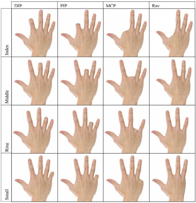
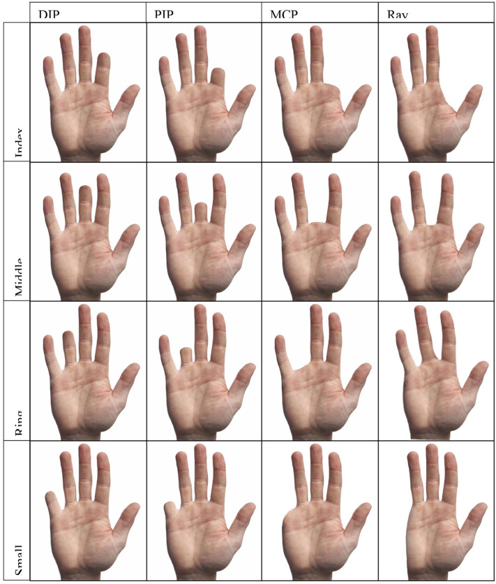
Each question contained 4 levels of amputation in either dorsal or volar view for a given digit: amputations at the distal interphalangeal (DIP) joint, proximal interphalangeal (PIP) joint, metacarpophalangeal (MCP) joint, and ray amputation (Figures 1 and 2). Participants ranked these images in order of most aesthetic (1) to least aesthetic (4) for each digit.
Figure 1.
The above figure comprises all of the digitally manipulated images in the dorsal view utilized in the survey.
Note. DIP = distal interphalangeal; PIP = proximal interphalangeal; MCP = metacarpophalangeal.
Figure 2.
The above figure comprises all of the digitally manipulated images in the volar view utilized in the survey.
Note. DIP = distal interphalangeal; PIP = proximal interphalangeal; MCP = metacarpophalangeal.
Statistics
Descriptive statistics were used to summarize the ranking data. We calculated the summary statistics based on the definitions from Lee and Yu. 14 The mean rank for each level of amputation for a given digit was determined to measure the preference of each level of amputation (Table 2). Additionally, the pairwise frequency was determined to allow for comparison of how frequently 1 level of amputation was selected more favorably over one other level of amputation, regardless of rank (Table 3). The marginal distribution for each question shows how many times a given level of amputation was selected for each rank (Table 4). Furthermore, to test whether the ranking of 4 levels of amputation was uniform, chi-squared tests were used. P value < .05 was considered statistically significant, implying all possible rankings for the 4 levels of amputation are not equally likely to appear (some are more preferable than the others). The whole analysis is performed in R (R 3.6.2) using R package pmr.
Table 2.
Mean Ranks of Digit Amputation.
| Level | Volar index | Volar middle | Volar ring | Volar small | Dorsal index | Dorsal middle | Dorsal ring | Dorsal small |
|---|---|---|---|---|---|---|---|---|
| DIP | 1.371 | 1.600 | 1.461 | 1.322 | 1.316 | 1.298 | 1.444 | 1.449 |
| PIP | 2.555 | 2.997 | 2.806 | 2.534 | 2.556 | 2.800 | 2.760 | 2.712 |
| MCP | 2.942 | 3.132 | 3.348 | 2.847 | 3.227 | 3.342 | 3.218 | 2.873 |
| Ray | 3.132 | 2.271 | 2.384 | 3.297 | 2.902 | 2.560 | 2.578 | 2.966 |
| P value | 1.70e-75 | 7.55e-61 | 7.77e-77 | 5.35e-33 | 2.44e-61 | 8.59e-66 | 7.19e-50 | 4.19e-44 |
Note. DIP = distal interphalangeal; PIP = proximal interphalangeal; MCP = metacarpophalangeal.
The table above presents mean ranks of digit amputation where a lower number indicates higher level of preference amongst study participants. P-values < .05 indicate preference amongst choices exists and choices were not uniformly distributed.
Table 3.
Pairwise Frequency.
| View | Level | DIP | PIP | MCP | Ray |
|---|---|---|---|---|---|
| Volar Index | DIP | 0 | 281 | 274 | 260 |
| PIP | 29 | 0 | 211 | 208 | |
| MCP | 36 | 99 | 0 | 193 | |
| Ray | 50 | 102 | 117 | 0 | |
| Volar middle | DIP | 0 | 284 | 253 | 207 |
| PIP | 26 | 0 | 173 | 112 | |
| MCP | 57 | 137 | 0 | 75 | |
| Ray | 103 | 198 | 235 | 0 | |
| Volar ring | DIP | 0 | 283 | 285 | 219 |
| PIP | 27 | 0 | 215 | 128 | |
| MCP | 25 | 95 | 0 | 82 | |
| Ray | 91 | 182 | 228 | 0 | |
| Volar small | DIP | 0 | 109 | 104 | 103 |
| PIP | 9 | 0 | 78 | 86 | |
| MCP | 14 | 40 | 0 | 82 | |
| Ray | 15 | 32 | 36 | 0 | |
| Dorsal index | DIP | 0 | 206 | 210 | 188 |
| PIP | 19 | 0 | 173 | 133 | |
| MCP | 15 | 52 | 0 | 107 | |
| Ray | 37 | 92 | 118 | 0 | |
| Dorsal middle | DIP | 0 | 213 | 209 | 186 |
| PIP | 12 | 0 | 164 | 94 | |
| MCP | 16 | 61 | 0 | 71 | |
| Ray | 39 | 131 | 154 | 0 | |
| Dorsal ring | DIP | 0 | 209 | 199 | 167 |
| PIP | 16 | 0 | 155 | 108 | |
| MCP | 26 | 70 | 0 | 80 | |
| Ray | 58 | 117 | 145 | 0 | |
| Dorsal small | DIP | 0 | 107 | 97 | 97 |
| PIP | 11 | 0 | 71 | 70 | |
| MCP | 21 | 47 | 0 | 65 | |
| Ray | 21 | 48 | 53 | 0 |
Note. DIP = distal interphalangeal; PIP = proximal interphalangeal; MCP = metacarpophalangeal.
The above table shows how frequently one level of amputation was selected more favorably over another level of amputation, regardless of rank. Shaded boxes highlight differences between the selections between PIP and Ray level of amputation in particular.
Table 4.
Marginal Distribution.
| View | Level | 1 | 2 | 3 | 4 |
|---|---|---|---|---|---|
| Volar index | DIP | 237 | 43 | 18 | 12 |
| PIP | 11 | 163 | 89 | 47 | |
| MCP | 16 | 59 | 162 | 73 | |
| Ray | 46 | 45 | 41 | 178 | |
| Volar middle | DIP | 196 | 61 | 34 | 19 |
| PIP | 7 | 95 | 100 | 108 | |
| MCP | 13 | 58 | 114 | 125 | |
| Ray | 94 | 96 | 62 | 58 | |
| Volar ring | DIP | 208 | 70 | 23 | 9 |
| PIP | 9 | 116 | 111 | 74 | |
| MCP | 10 | 29 | 114 | 157 | |
| Ray | 83 | 95 | 62 | 70 | |
| Volar small | DIP | 97 | 11 | 3 | 7 |
| PIP | 4 | 67 | 27 | 20 | |
| MCP | 4 | 28 | 68 | 18 | |
| Ray | 13 | 12 | 20 | 73 | |
| Dorsal index | DIP | 182 | 25 | 8 | 10 |
| PIP | 5 | 126 | 58 | 36 | |
| MCP | 7 | 18 | 117 | 83 | |
| Ray | 31 | 56 | 42 | 96 | |
| Dorsal middle | DIP | 184 | 23 | 10 | 8 |
| PIP | 2 | 87 | 90 | 46 | |
| MCP | 7 | 24 | 79 | 115 | |
| Ray | 32 | 91 | 46 | 56 | |
| Dorsal ring | DIP | 159 | 42 | 14 | 10 |
| PIP | 6 | 96 | 69 | 54 | |
| MCP | 7 | 30 | 95 | 93 | |
| Ray | 53 | 57 | 47 | 68 | |
| Dorsal small | DIP | 91 | 8 | 12 | 7 |
| PIP | 3 | 58 | 27 | 30 | |
| MCP | 7 | 28 | 56 | 27 | |
| Ray | 17 | 24 | 23 | 54 |
Note. DIP = distal interphalangeal; PIP = proximal interphalangeal; MCP = metacarpophalangeal.
The marginal distribution presented in Table 4 reflects how frequently a given level of amputation was selected for a particular rank.
Results
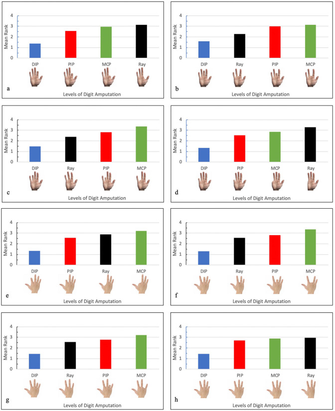
In order to best determine preference of 1 amputation position over another for a given digit, we must look to Table 2 and Figure 3. Looking at the output for the mean rank, smaller mean rank value indicates that the amputation position is more preferable (aesthetic) than the others on average. Across all positions of digit amputation, in both dorsal and volar views, amputation at the DIP was determined to be the most aesthetic as evidenced by DIP having the lowest mean rank value for all digits (Table 2, Figure 3). In the index volar view, PIP was second most aesthetic, followed by MCP and Ray. In the index dorsal view, however, the Ray amputation was determined to be more aesthetic than amputation at the MCP. The dorsal and volar views of the middle and ring fingers indicated the aesthetics of digit amputation from most to least aesthetic was DIP, Ray, PIP, then MCP. In the small finger, the preference was DIP, PIP, MCP, and then Ray amputation as least aesthetic in both volar and dorsal views.
Figure 3.
(a) Volar index mean rank, (b) volar middle mean rank, (c) volar ring mean rank, (d) volar small mean rank, (e) dorsal index mean rank, (f) dorsal middle mean rank, (g) dorsal ring mean rank, and (h) dorsal small mean rank.
Note. DIP = distal interphalangeal; PIP = proximal interphalangeal; MCP = metacarpophalangeal.
A chi-squared test was subsequently performed using the mean rank data, with the null hypothesis being that uniformity exists between the mean ranks and each position of amputation has the same numerical mean rank. A P-value less than .05 was significant, indicating the ranking data is not uniformly distributed. For each digit and view, P-values presented in Table 2 were very small, much less than .05, allowing rejection of the null hypothesis. This evidence shows there is a certain preference among the 4 levels of amputation for survey participants, as mean rank was not the same between positions of amputation as demonstrated in Table 2.
The pairwise frequency and marginal distribution further reflect the preferences demonstrated by mean rank in the index finger, long finger, ring finger, and small finger (Tables 3 and 4). For example, when looking at the volar index data in Table 3, DIP was selected more favorably over PIP for 281 times, while PIP was only selected more favorably over DIP for 29 times, which represents a preference for DIP. In the marginal distribution table (Table 4), DIP usually has the highest frequency in rank “1.” However, the marginal distribution also indicates ray amputation was selected with the second highest frequency as “1,” for most aesthetic, across all digits. In looking at the pairwise frequency, DIP was selected most frequently over all other levels of amputation as more aesthetic. Additionally, participants ranked ray amputation more aesthetic with a higher frequency in comparison to PIP amputation in the long and ring fingers, both dorsal and volar.
Discussion
Results of this study indicate efforts should be made to preserve the middle phalanx following traumatic injury to a digit; however, when this is not possible, in the long and ring fingers, ray resection results in the most cosmetically appealing hand. A number of studies have demonstrated differences in hand functionality that occur with ray amputation of certain digits. Ray amputation of the border digits, the index, and little fingers, has been shown to decrease grip strength and 3-point pinch, as well as palm width.10,15,16 Following index ray resection, however, the long finger takes over much of the function of the index finger.10,15,16 Similar changes to function were noted following amputation of the long and ring fingers with a reduction of at least 20% to 30% for grip, key pinch, and 3-point pinch strength. 11 In spite of the acquired functional deficits following ray resection, patients reported satisfaction with the remaining function and cosmesis.10,16,17 Consistent with the wider literature, this study indicates that ray resection should be used preferentially to amputation at the PIP and MCP following traumatic injury to the long and ring fingers for the most cosmetically pleasing result. This option should be considered unless the patient requires strong grip strength and pinch strength for their occupation or lifestyle.
The present study has several limitations. The images presented in the study are not of actual amputated hands and thus do not contain the scars associated with digit amputation. It is possible that disruption of the functional aesthetic subunits of the hand described by Rehim et al 18 would influence preference. Additionally, the images are presented in anatomical views, exposing the maximal visual length discrepancy between positions of amputation, instead of how one might see another’s hand in day to day activities like shaking hands, holding an instrument to write, holding utensils when eating or in repose. In these alternative, naturally occurring positions, the length discrepancy between the varying levels of amputation may not be as obvious. Furthermore, preference of varying levels of amputation may change in the ring finger with the addition of a ring in the digitally manipulated images.
The current study offers preliminary data regarding preference in the cosmesis of the various digit amputations. Of participants surveyed, the majority reported demographic information placing them in a younger age group below 30, of the female sex, and identifying with white race. While the significance of this data is incomplete without further study, it does suggest aesthetic preference for preservation of digit length and natural arc of the hand for young, female patients who experience digital amputation. The data generated from this study are insufficient for comparing preference based off demographics as gender choice was limited to just male/female, participant response numbers differed significantly based on age, and 85% of participants selected white when answering the question of race. Another area of consideration for future studies would be to include digital manipulations of hands more representative of the varying demographics. For example, to include hands belonging to both males and females, younger and older persons, and hands of varying skin color. The results of this study should be replicated in an independent, balanced, diverse sample and consider more demographic factors that may influence preference.
While no significant difference was found in preference based on gender in this study, cosmetic preference of the hand may differ across cultural or ethnic background, race, or age. Nishizuka et al 19 compared attitudes toward digital replantation in the United States and Japan, and found survey participants in Japan rated appearance of the hand as more important for replantation than did American participants. Other studies evaluating aesthetic preference from laypersons and surgeons for the lips, face, breast, and nose further support that aesthetic preference differs by age, sex, gender, culture, and ethnicity.20-23
The study demonstrates that every effort should be made to limit amputations of mangled digits to the distal joint if a functional finger can be expected. However, in more severe injuries, or in cancer operations, when a more proximal amputation is unavoidable, cosmetic results can be achieved with a ray amputation rather than attempting to preserve the metacarpal phalangeal joint. Specifically, ray amputation at the middle and ring fingers offers a more cosmetically acceptable hand than amputation at the MCP or PIP joints. With respect to the index and small fingers, ray amputation is the least aesthetically pleasing and should be considered for patients in whom the functional benefits outweigh the aesthetic drawbacks.
Footnotes
Ethical Approval: This study was approved by our institutional review board.
Statement of Human and Animal Rights: All procedures followed were in accordance with the ethical standards of the responsible committee on human experimentation (institutional and national) and with the Helsinki Declaration of 1975, as revised in 2008 (5). Informed consent was obtained from all patients for being included in the study.
Statement of Informed Consent: Informed consent was obtained from all individual participants included in this study.
The author(s) declared no potential conflicts of interest with respect to the research, authorship, and/or publication of this article.
Funding: The author(s) received no financial support for the research, authorship, and/or publication of this article.
ORCID iD: John Collar III  https://orcid.org/0000-0001-9679-2601
References
- 1.Andersson GB, Gillberg C, Fernell E, et al. Children with surgically corrected hand deformities and upper limb deficiencies: self-concept and psychological well-being. J Hand Surg Eur Vol. 2011;36(9):795-801. [DOI] [PubMed] [Google Scholar]
- 2.Gustafsson M, Amilon A, Ahlström G.Trauma-related distress and mood disorders in the early stage of an acute traumatic hand injury. J Hand Surg Br. 2003;28(4):332-338. [DOI] [PubMed] [Google Scholar]
- 3.Williams AE, Newman JT, Ozer K, et al. Posttraumatic stress disorder and depression negatively impact general health status after hand injury. J Hand Surg Am. 2009;34(3):515-522. [DOI] [PubMed] [Google Scholar]
- 4.Richards T, Garvert DW, McDade E, et al. Chronic psychological and functional sequelae after emergent hand surgery. J Hand Surg Am. 2011;36(10):1663-1668. [DOI] [PubMed] [Google Scholar]
- 5.Opsteegh L, Reinders-Messelink HA, Groothoff JW, et al. Symptoms of acute posttraumatic stress disorder in patients with acute hand injuries. J Hand Surg Am. 2010;35(6):961-967. [DOI] [PubMed] [Google Scholar]
- 6.Jakubietz RG, Jakubietz MG, Kloss D, et al. Defining the basic aesthetics of the hand. Aesthetic Plast Surg. 2005;29(6):546-551. [DOI] [PubMed] [Google Scholar]
- 7.Kościński K.Determinants of hand attractiveness—a study involving digitally manipulated stimuli. Perception. 2011;40(6):682-694. [DOI] [PubMed] [Google Scholar]
- 8.Higgins JP, Seruya M.Visual subunits of the hand: proposed guidelines for revision surgery after flap reconstruction of the traumatized hand. J Reconstr Microsurg. 2011;27(9):551-557. [DOI] [PubMed] [Google Scholar]
- 9.Johnson SP, Sebastin SJ, Rehim SA, et al. The importance of hand appearance as a patient-reported outcome in hand surgery. Plast Reconstr Surg Glob Open. 2015;3(11):e552. [DOI] [PMC free article] [PubMed] [Google Scholar]
- 10.Peimer CA, Wheeler DR, Barrett A, et al. Hand function following single ray amputation. J Hand Surg Am. 1999; 24(6):1245-1248. [DOI] [PubMed] [Google Scholar]
- 11.Blazar PE, Garon MT.Ray resections of the fingers: indications, techniques, and outcomes. J Am Acad Orthop Surg. 2015; 23(8):476-484. [DOI] [PubMed] [Google Scholar]
- 12.Monreal R.Reconstructive surgery of the amputated ring finger. Int Orthop. 2017;41(8):1617-1622. [DOI] [PubMed] [Google Scholar]
- 13.del Piñal F. The indications for toe transfer after “minor” finger injuries. J Hand Surg Br. 2004;29(2):120-129. [DOI] [PubMed] [Google Scholar]
- 14.Lee PH, Yu PLH. An R package for analyzing and modeling ranking data. BMC Med Res Methodol. 2013;13(1):65. [DOI] [PMC free article] [PubMed] [Google Scholar]
- 15.Murray JF, Carman W, MacKenzie JK.Transmetacarpal amputation of the index finger: a clinical assessment of hand strength and complications. J Hand Surg Am. 1977;2(6):471-481. [DOI] [PubMed] [Google Scholar]
- 16.Melikyan EY, Beg MS, Woodbridge S, et al. The functional results of ray amputation. Hand Surg. 2003;8(1):47-51. [DOI] [PubMed] [Google Scholar]
- 17.Nuzumlali E, Orhun E, Oztürk K, et al. Results of ray resection and amputation for ring avulsion injuries at the proximal interphalangeal joint. J Hand Surg Br. 2003;28(6):578-581. [DOI] [PubMed] [Google Scholar]
- 18.Rehim SA, Kowalski E, Chung KC.Enhancing aesthetic outcomes of soft-tissue coverage of the hand. Plast Reconstr Surg. 2015;135(2):413e-428e. [DOI] [PMC free article] [PubMed] [Google Scholar]
- 19.Nishizuka T, Shauver MJ, Zhong L, et al. A comparative study of attitudes regarding digit replantation in the United States and Japan. J Hand Surg Am. 2015;40(8):1646-1656, 1656.e1. [DOI] [PMC free article] [PubMed] [Google Scholar]
- 20.Heidekrueger PI, Szpalski C, Weichman K, et al. Lip attractiveness: a cross-cultural analysis. Aesthet Surg J. 2017;37(7): 828-836. [DOI] [PubMed] [Google Scholar]
- 21.Broer PN, Juran S, Liu YJ, et al. The impact of geographic, ethnic, and demographic dynamics on the perception of beauty. J Craniofac Surg. 2014;25(2):e157-e161. [DOI] [PubMed] [Google Scholar]
- 22.Broer PN, Buonocore S, Morillas A, et al. Nasal aesthetics: a cross-cultural analysis. Plast Reconstr Surg. 2012;130(6): 843e-850e. [DOI] [PubMed] [Google Scholar]
- 23.Broer PN, Juran S, Walker ME, et al. Aesthetic breast shape preferences among plastic surgeons. Ann Plast Surg. 2015;74(6):639-644. [DOI] [PubMed] [Google Scholar]


