Abstract
Background
Skin dullness has long been a major concern of Japanese women. It is usually evaluated and judged visually by experts. Although several factors are recognized to play a role, it is unclear to what extent such physiological characteristics contribute to skin dullness. The purpose of this study is to establish an objective method for evaluation, which will assist in developing cosmetics products targeting skin dullness.
Methods
We conducted a skin measurement study on 50 Japanese women in their 30–50s, where skin dullness was visually assessed by a group of experts to obtain an average dullness score, and several skin parameters were obtained. We then developed a regression model that explains the visual assessment score using these physiological parameters.
Results
The results of partial least squares analysis of the dullness perception and physiological characteristics showed that skin dullness can be defined by colorimetric, optical, and skin surface microtopography parameters. Additionally, the contribution of each parameter to the model was determined. Our results suggest that dullness perception is highly affected by the melanin content and yellowness of the skin, followed by skin reddishness, roughness, and translucency score, whereas glossiness has less effect. Strikingly, the contribution ratio of each parameter varied among age groups. Furthermore, we confirmed that the predicted value of skin dullness increases with age.
Conclusion
Our results will help the design of cosmetics targeting factors specific to age groups in developing effective solutions for skin dullness.
Keywords: glossiness, melanin, optical properties, partial least squares analysis, skin dullness, skin parameters, skin roughness, translucency, visual assessment
1. INTRODUCTION
Skin dullness is a constant skin concern of women of almost all ethnic backgrounds. Various expressions are used to describe skin dullness in different languages (e.g., the term “kusumi” is used in Japanese, the term “fūsè àn chén, fūsè àn huáng” in Chinese and the term “rookhaapan” in Hindi), and these terms are mostly interpreted as “paleness”, “lack of energy”, “gloominess” or “loss of smoothness” and referring to unhealthy discoloration and a lack of radiance or an uneven texture. However, these are vague terms based on the visual perception of the individual or a third person. 1 In the cosmetics industry, it is important to target such vague concerns of consumers, and research has been conducted to clarify the causes of skin dullness and to establish a method of evaluating skin dullness. Previous studies have suggested that inherent factors, including skin color intensity due to melanin accumulation and quality of blood flow associated with hemoglobin level, as well as factors of light reflectance and skin roughness correlate with skin dullness. 2 , 3 The Japan Cosmetic Industry Association proposed that skin dullness increases with a decrease in the reddishness of skin tone due to poor blood circulation, yellowing of the skin due to aging, diffused melanin deposition, a decrease in translucency (light transmission) due to thickening of the stratum corneum, reduced glossiness due to skin surface reflection and shadowing by an uneven surface due to the loss of skin elasticity. 4 It is unclear whether these factors play a role alone or in combination, or how much they contribute to skin dullness. Other reports have revealed the contributions of skin parameters such as the melanin content, hemoglobin content, water content, and fine texture in determining skin translucency; although “translucency” is often considered the opposite of “dullness” in linguistic surveys, these parameters are not opposites of one another. 5 Additionally, a yellowish tone of skin has been related to the deterioration of translucency. 6 , 7 However, the similarity of the perception of a lack of translucency and the perception of dullness remains largely unknown, and we thus aim to develop an evaluation method to determine the effects of physiological parameters involved in assessing skin dullness.
To calculate the weights of skin parameters, we adopted partial least squares (PLS) analysis as a method of multiple regression analysis. 8 PLS analysis is a combination of principle component (PC) analysis and multiple linear regression, and it is particularly useful when predicting a set of dependent variables (e.g., dullness) from a large set of explanatory variables (e.g., skin parameters) that can be highly correlated with each other, which is referred to as collinearity. Collinearity is diminished by projecting the explanatory variables to a completely new set of unrelated orthogonal factors, known as latent variables. A subset of such latent variables that best describes the dependent variable is then selected through regression model fitting. Several methods such as the bootstrap and jackknife (leave‐one‐out) techniques are commonly used for such cross‐validation (CV). 9 In the case of the bootstrap method, a set of randomly selected training data is used to build the regression model, and the remaining data are used to test the model. In the case of the jackknife technique, one set of data is removed from the data set at a time, and the remaining points are used to construct a model for the prediction of the excluded point. The optimal number of latent variables is selected when the model has least regression error and highest prediction power, as assessed using the explanation rate (R 2). R 2 > 0.5 represents a high level of confidence in the prediction whereas 0.3 < R 2 < 0.5 indicates acceptable prediction accuracy. PLS analysis is adopted in a wide range of research fields, including skin sensory evaluation and drug efficacy evaluation. 10 , 11 , 12 , 13 , 14 , 15 In the present study, we applied PLS analysis to explain the sensory evaluation of skin dullness using skin parameters and proposed that the perception of dullness is age dependent among Japanese women. Such methodology and this paper's insights into physiological changes can be applied to define the perception of dullness on a global scale in the future.
2. MATERIALS AND METHODS
2.1. Study participants
Fifty Japanese women in their 30s−60s were recruited according to three criteria. (1) The participants usually take care of their face with a maximum of three simple products, such as lotions, essences and creams, after washing. (2) The participants do not suffer chronic skin conditions, such as atopic dermatitis, and are not pregnant or lactating. (3) The participants have no previous experience of cosmetic treatment, hormone replacement therapy or special facial care regimes. The purpose of the study was clearly explained to all participants, who provided informed consent.
2.2. Test conditions
The study complied with ethical principles established by the Declaration of Helsinki and was approved by the Shiseido Ethics Committee (approval number: B01729). All 50 participants visited a local facility in Yokohama city during March. To reduce intervening factors that could have affected the precision of the results, all participants performed facial cleansing and waited for 20 min to allow their skin to return to normal conditions before their skin conditions were evaluated. The examination room was maintained at a constant temperature of 23 ± 1°C and relative humidity of 45% ± 5%.
2.3. Visual assessment of skin dullness
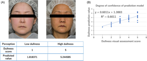
The skin dullness score is a grading of dullness, as defined by the Japan Cosmetic Industry Association, 4 on a five‐point scale (1: slightly dull, 2: partially dull, 3: moderately dull, 4: quite dull, 5: extremely dull). Representative images are shown in Figure 1. Full‐face images of the participants were captured using a digital camera (D5300 with a 18−140‐mm lens; Nikon Corporation, Tokyo, Japan) under good lighting immediately after skin acclimatization. The color was balanced using ColorChecker Passport Photo 2 (MSCCPP‐B, X‐Rite, Tokyo, Japan). Three trained experts observed and graded the captured images, and the average score was calculated for each image.
FIGURE 1.

Establishment of a mathematical model to estimate the perception of dullness. (A) Examples of participants with low and high perceived dullness scores and the dullness predicted using the mathematical model. (B) Degree of confidence in the results of the prediction model obtained in Bland–Altman analysis. R 2 > 0.5 indicates high prediction accuracy.
2.4. Skin color measurement
A contact‐type spectrophotometer (CM‐700d; Konica Minolta Inc., Tokyo, Japan) was used to obtain the spectral reflectance of the cheek, specifically at the intersection of a vertical line drawn downward from the outer corner of the eye and a horizontal line drawn outward from the outside corner of the nose. The measurement was made three times and the average was taken. L* (lightness), b* (yellowness) and a* (redness) in the CIE Lab color space (1976 CIELAB space) under the conditions of illuminant D65 and a 2° visual field and the melanin index, hemoglobin index and Hb SO2 index (i.e., the hemoglobin oxygen saturation calculated as the ratio of oxyhemoglobin to total hemoglobin) were calculated from the spectral reflectance data by a proprietary algorithm 16 , 17 in skin analysis software (CM‐SA; Konica Minolta).
2.5. Measurement of optical properties of the skin
2.5.1. Measurement of the angular polarized reflectance
The glossiness of the skin was determined from the specular reflection component (S) using a polarized goniophotometer in which two polarized filters were set in front of the light‐emitting device and light‐receiving device. 5 In this system, the angle of the incident light on the skin is fixed at 45° against the skin plane, and the angle of the light reflected from the skin can be measured simultaneously in the range from 0 to 68°. At each of the reflection angles in the range from 0 to 68°, the diffuse reflection component (D) is acquired by arranging the polarizing filters at right angles to both the light‐emitting device and light‐receiving device. The specular reflection component (S) is then measured independently by subtracting the amount of light of the diffuse reflection component from the total amount of light measured when the polarizing filters are arranged in parallel. The components are
where I is the measured light intensity, and the subscripts s and p respectively indicate the direction of the polarization filters on the incident and receiving sides of the light.
2.5.2. Measurement of subsurface scattering light from the skin
Light entering the interior of the skin is first reflected diffusely by the microstructure of the stratum corneum and then reaches the deeper skin layers, including the epidermis, dermis and subcutaneous tissue. Finally, part of the light is emitted from the skin and returns to the initial medium (air) through subsurface scattering. We measured the subsurface light scattering from the skin using the system proposed by Kuwahara et al. 18 The system transmits light from a light source through a narrow slit on the skin. The slit blocks any reflection on the skin surface, and only the light that passes through the inner layers of the skin is detected by the charge‐coupled device camera. The distribution of the subsurface scattering light is expressed as an exponential function of distance:
where R slit is the distribution of brightness, r is the distance from the slit, and C1 and C2 are variables. The skin translucency index (T) is then calculated from the parameters that describe the rate of the attenuation of brightness with distance (C2):
2.6. Skin surface microtopography
Skin surface microtopography (SSMT) was conducted by taking replicas from the cheek using Silflo (Amique Group Co., Ltd, Tokyo, Japan). A replica was taken at the intersection of a vertical line drawn downward from the outer corner of the eye and a horizontal line drawn outward from the outside corner of the nose for surface texture‐related parameters, including the parameters of ridges and furrows, and a replica was taken at the center of the cheek for skin pore‐related parameters. The three‐dimensional (3D) surface morphology was captured from the replica using a confocal laser scanning microscope (HD100D; Lasertek Corporation, Yokohama, Japan) and adjacently tiled to obtain an integrated scan area of 6.76 × 6.76 mm. 3D data, including the 3D roughness (Sa), 2D roughness (Ra), distance between furrow and ridges (Sm), furrow depth and ridge count, were processed for the skin texture. As parameters relating to skin pores, the pore area, pore volume, pore depth, pore count, and pore aspect ratio were calculated using a previously published protocol. 19
2.7. Statistical analysis
2.7.1. Single‐correlation analysis
Single‐correlation analysis of the perceived dullness and facial parameters was conducted by performing Kendall's tau correlation test to consider nonparametric tied values in the dullness score. Parameters with an absolute coefficient value τ > 0.3 and p‐value < 0.05 were considered highly correlated with the dullness score. Table S1 summarizes the results.
2.7.2. Calculation of weights of parameters
To find which factors contribute to the perception of skin dullness, we performed a PLS analysis, whereby a regression model was established to find the relation between multiple parameters and to exclude the effects of multicollinearity among colorimetric (L* and melanin index) or surface (Ra and Sa) parameters. Parameters were initially transformed into a set of PCs, and CV was conducted using the jackknife methods with 50 leave‐one‐out segments to determine the optimal number of PCs that generate a model with minimum regression error (CV) and maximum rate of explanation (R 2). Table S2 gives the CV and R 2 values for all sets of PCs. Furthermore, a Bland–Altman comparison was made to determine the prediction accuracy of the model using the R 2 index. The coefficients of the parameters were then calculated using the regression model (Figure 2). All statistical analyses were conducted using R software (version 3.6.1) (https://cran.r‐project.org/bin/windows/base/old/3.6.1/).
FIGURE 2.

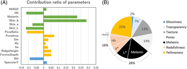
Contributions of skin parameters to the perception of skin dullness (combined model). (A) Coefficient of variables calculated from the regression model of the combined age groups (30−50s). (B) Definition of skin dullness based on the contribution ratio of colorimetric, optical, and microtopographic parameters.
3. RESULTS
3.1. Establishment of a mathematical model to estimate the perception of skin dullness based on skin parameters
The purpose of the present study was to establish a model that can be used to estimate the perception of skin dullness. To develop a standard for perception, we chose three experts to assess the dullness of facial skin objectively. Figure 1A shows examples of participants assessed to have low dullness (left) and high dullness (right). Next, we performed single‐correlation analysis of the dullness score of each participant and corresponding colorimetric, optical, and SSMT parameters of the skin. A total of 31 skin parameters were analyzed, and 10 parameters, including the melanin index, b*, intensity of diffuse reflection, pore area, and furrow depth, were found to be strongly correlated (|τ| > 0.3) with the dullness score. The results are given in Table S1. To establish the model for estimating skin dullness, we selected parameters according to not just their high correlation but also their importance in describing skin tone. This process excluded parameters with high collinearity, such as the intensity of diffuse reflection, but included parameters such as L*, a*, the intensity of specular reflection, and the subsurface scattering component. We performed PLS regression analysis, whereby parameters were converted to multiple PCs. Estimates of the root‐mean‐square error of prediction (RMSEP) and rate of explanation (R 2) for each PC are given in Table S2. The optimal number of PCs for the combined model was two, having the minimum RMSEP value and maximum R 2 value. In determining the prediction accuracy of the designed model, Bland–Altman analysis was performed to match the predicted value of dullness with the visual judgment (Figure 1B). The model showed high predictive power (R 2 = 0.6), suggesting that the model is suitable for the estimation of skin dullness.
3.2. Weights of individual skin parameters in dullness perception and distinct contributions among age groups
Using the regression model, we calculated the coefficients of variables to deduce the weights of individual parameters. The results are shown in Figure 2A. Among pigment‐related parameters, a higher melanin index and higher b* (skin yellowness) contributed to the perception of dullness, whereas lower L* (skin lightness), lower a* (skin redness), and lower Hb (hemoglobin index) were related with dullness. The highest contribution among the colorimetric parameters was that of skin yellowness, followed by that of the melanin content.
SSMT data obtained for skin replica can be classified into skin‐pore‐related and skin‐texture‐related data. A larger pore area and more pore sagging (lower pore aspect ratio) led to a higher perception of dullness. A higher ridge height and furrow depth were texture‐related properties associated with dullness. Light reflectance properties such as the subsurface scattering light component, which refers to skin translucency, negatively affected the dullness perception. Meanwhile, specular reflection, which corresponds to glossiness, made little contribution. Figure 2B summarizes the contribution levels of the skin parameters.
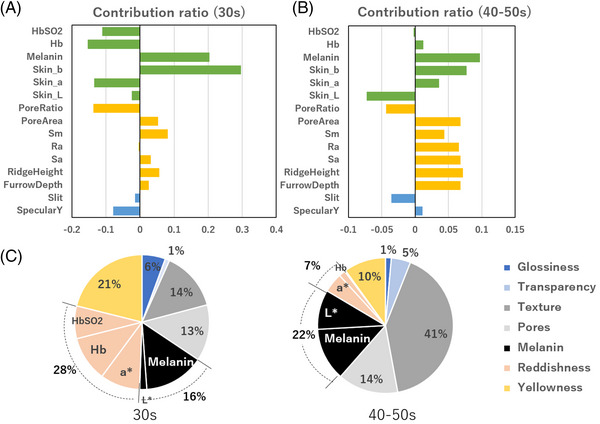
Furthermore, when we redesigned the model by dividing the participants by age group (30 and 40−50s age groups), we found a difference in the contribution ratios of several parameters (Figure 3A,B). Skin yellowness was the predominant cause of dullness for women in their 30s, whereas the melanin index had the highest weight in women in their 40 and 50s. The contribution of reddishness was lower in the older age group. Interestingly, even though the combined model showed little effect of the intensity of specular reflection (glossiness) on dullness perception, the intensity made an appreciable contribution in the 30s age group. In contrast, the subsurface scattering component (translucency) had a lower effect for the women in their 30s than for the women in their 40s and 50s. In the 40s−50s age group, there was almost no contribution of the intensity of specular reflection but the weight of the subsurface scattering component was moderate. Additionally, the SSMT parameters had strikingly different effects between the age groups, with the contributions of the pore area and 3D unevenness being greater for the 40−50s group. The contributions for each age group are summarized in Figure 3C.
FIGURE 3.

Contributions of skin parameters by age group. Coefficients of variables calculated with the regression model of (A) the 30s age group and (B) the 40−50s age group. (C) Definition of skin dullness based on the contribution ratio of colorimetric, optical, and microtopographic parameters in the 30 and 40−50s age groups.
3.3. Change in skin dullness with increasing age
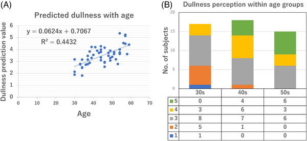
To determine whether the perception of dullness is affected by age, we calculated prediction values of dullness and plotted them against the age of the participant (Figure 4A). The graph shows a high correlation of dullness increasing with age. Similar results were obtained when the visual judgment score was plotted against the age group, with a younger age group having a lower score and an older age group having more participants with higher dullness scores (Figure 4B).
FIGURE 4.

Change in dullness with age. (A) Increase in the dullness prediction with age. (B) Number of participants for each dullness score 1 to 5 in each age group (30s, 40s, 50s).
4. DISCUSSION
In this study, we aimed to define skin dullness from an objective point of view by analyzing its relationship with skin‐related parameters. To date, skin dullness has been considered a vague but consistent skin concern among Japanese women in almost all age groups. We selected age groups ranging from 30 to 50s, in which primary changes in facial appearance, such as pigmentation and wrinkling, start to appear. Although one's subjective evaluation of oneself or perception by nonexperts is essential to interpreting the needs of consumers and the effectiveness of cosmetic products, we showed that an evaluation by experts and its correspondence with the quantitative data of skin parameters are equally important in clarifying the key modulators of skin dullness. Although single‐correlation analysis did not detect the association of skin reddishness or skin translucency with dullness, through the careful selection of variables in PLS analysis, we excluded the possibility of multicollinearity and deduced the importance of these parameters in assessing dullness. The prediction accuracy was adequate for all models, with the 30s age group having the highest correspondence (R 2 > 0.8) and the 40−50s age group having slightly weaker correspondence (R 2 > 0.3) (data not shown). We speculate that this result was due to a lower number of participants having a low dullness score in the higher age groups, in terms of providing sufficient data for constructing the regression model (Figure 4B).
The contributions of colorimetric parameters agreed well with the results of previous studies. An increase in the melanin content and yellowing of the skin due to aging have long been considered root causes of dullness. 1 , 2 , 7 Additionally, a lack of reddishness due to poor blood circulation has been linked with dullness in skin tone. 3 , 4 Furthermore, skin lightness is a highly contributing factor and is itself affected by the major skin chromophores, melanin and hemoglobin. 20 One interesting result was the difference in the contribution balance of the melanin index and yellowness among age groups. In the younger age groups, the contribution of yellowness was higher, whereas the melanin index had a higher weight in the older group. An increase in the melanin content of skin occurs because of the excessive synthesis or insufficient disposal of melanin in the epidermis, whereas skin yellowness involves the carbonylation of dermal proteins. 7 , 21 , 22 , 23 Both these phenotypes are induced by photoaging; however, the drastic signs of pigmentary changes can substantially affect dullness perception with the advancement of age. Additionally, we note a higher recognition of skin reddishness in the younger age groups. It would be intriguing to identify what causes such a difference in perception.
Skin roughness has been implicated as a critical factor in defining skin dullness. 1 , 3 In our study, roughness referred to a greater skin pore area, pore aspect ratio (pore sagging) and depth and anisotropy of furrows. Conspicuous pores seemed to have higher contribution in the younger age groups, whereas the depth and irregularity of the skin fine texture affected perception in the higher age groups. This difference might be due to the worsening of microtopographic parameters with increasing age, 19 upon development of facial micro‐wrinkles in the cheek zone, affecting the overall impression of the skin.
Previous studies found skin dullness recognition to be linguistically associated with the terms “lack of translucency” and “loss of radiance” of the skin. 24 , 25 Indeed, our results confirm the role of translucency in describing dullness. Although we could not decipher the importance of glossiness in the combined model, it seemed to have a moderate effect in younger age groups.
Other reports have presented the effects of skin color and roughness in determining the optical properties of skin, such as glossiness and translucency. 5 , 24 Despite the categorization of parameters as pigment‐related, optical, and surface‐related parameters in this study, it is necessary to understand that such parameters are also closely associated, and we thus mainly compared their contributions among age groups or within each category. We also acknowledge that it is not possible to directly compare the coefficient values of mathematical models constructed for different objective variables, however we consider it is acceptable to use the relative contribution of the parameters within each model for interpretation of data across models.
Recently, several other factors have been suggested to induce dullness, including surface friction, skin dryness, the deterioration of skin turnover, the glycosylation of proteins, a lack of exposure to sunlight, sleep deprivation, poor nutritional health, diurnal variations, exposure to ultraviolet light, poor blood circulation, and the clogging of skin pores. Mechanical stimuli induce melanin production, 26 skin water content has been shown to be linked with translucency, 5 and skin turnover is required for the excretion of melanin. Meanwhile, the glycosylation of proteins (advanced glycation end products), clogging of skin pores, and accumulation of bilirubin in the epidermis have been associated with yellowish discoloration of the skin. 27 , 28 , 29 The accumulation of brownish pigments, such as like lipofuscin, in blood vessels has been proposed to accelerate with aging. 30 , 31 Such external stimuli may lead to chronic changes in the physiological parameters described in this study and trigger the onset of skin dullness. Further molecular studies addressing how stimulating factors affect dullness‐related skin parameters are required. Although the term “dullness” and its perception may vary largely with culture and trends, the physiological changes associated with aging are consistent. The careful analysis and targeting of age‐specific factors will assist in improving the efficacy of cosmetic products for dullness.
5. CONCLUSION
In this study, we developed a mathematical model that defines skin dullness according to colorimetric, optical, and surface micro‐topographical parameters in Japanese women. Using the model, we clarified the age‐dependent perception of skin dullness and its major contributing factors. We believe that our results will open avenues for conducting future mechanistic research to help develop personalized cosmetic solutions aimed toward specific age groups.
CONFLICT OF INTEREST STATEMENT
The authors declare no conflict of interest.
Supporting information
Supporting Infomation
ACKNOWLEDGMENTS
The authors thank Motoki Oguri, Tomohiro Kuwahara, and Tomoyuki Katsuyama for technical assistance with the SSMT analysis, slit light sensor and polarized goniophotometer respectively. The authors further thank Edanz (https://jp.edanz.com/ac) for editing a draft of this manuscript. This research was funded by Shiseido Co. Ltd.
Nurani AM, Kikuchi K, Iino M, et al. Development of a method for evaluating skin dullness: A mathematical model explaining dullness by the color, optical properties, and microtopography of the skin. Skin Res Technol. 2023;29:e13407. 10.1111/srt.13407
DATA AVAILABILITY STATEMENT
Author elects to not share data in accordance with the terms of the privacy‐preserving agreement with the subject.
REFERENCES
- 1. Kaneda Y, Muramatsu Y, Takahasi K, et al. Study on darkness of the face skin in Japanese women. J Soc Cosmet Chem Jpn. 1993;26(4):280‐288. [Google Scholar]
- 2. Kaneko O, Tsukada H, Ishikawa Y, Kawaguchi Y. Measuring apparent darkening of the skin (first report): factors inherent in light reflected from the skin. J Soc Cosmet Chem Jpn. 1997;31(1):44‐51. [Google Scholar]
- 3. Kaneko O, Kawaguchi Y, Ishikawa Y, Inagaki K. Measuring apparent darkening of the skin (second report): relationship between age‐associated changes in physical properties of the skin and apparent darkening. J Soc Cosmet Chem Jpn. 1997;31(4):429‐438. [Google Scholar]
- 4. Naganuma M. History of the effectiveness of cosmetics. J Cos Sci Soc. 2015;39(4):275‐285. [Google Scholar]
- 5. Masuda Y, Kunizawa N, Takahashi M, et al. Methodology for evaluation of skin transparency and the efficacy of an essence that can improve skin transparency. J Soc Cosmet Chem Jpn. 2005;39(3):201‐208. [Google Scholar]
- 6. Iwai I, Kuwahara T, Hirao T, et al. Decrease in the skin transparency induced by protein carbonylation in the stratum corneum. J Soc Cosmet Chem Jpn. 2008;42(1):16‐21. [Google Scholar]
- 7. Ogura Y, Kuwahara T, Akiyama M, et al. Dermal carbonyl modification is related to the yellowish color change of photo‐aged Japanese facial skin. J Dermatol Sci. 2011;64(1):45‐52. [DOI] [PubMed] [Google Scholar]
- 8. Abdi H. Partial least squares regression and projection on latent structure regression (PLS Regression). Wiley Interdiscipl Rev Comput Stat. 2010;2:97‐106. [Google Scholar]
- 9. Efron B, Tibshirani RJ. An Introduction to the Bootstrap. Chapman & Hall; 1993. [Google Scholar]
- 10. Flament F, Ye C, Mercurio DG, et al. Evaluating the respective weights of some facial signs on the perceived radiance/glow in differently aged women of six countries. Skin Res Technol. 2020;27:1116‐1127. [DOI] [PubMed] [Google Scholar]
- 11. Silva FALS, Brites G, Ferreira I, et al. Evaluating skin sensitization via soft and hard multivariate modeling. Int J Toxicol. 2020;39(6):547‐559. [DOI] [PubMed] [Google Scholar]
- 12. Wu YW, Ta GH, Lung YC, Weng CF, Leong MK. In silico prediction of skin permeability using a two‐QSAR approach. Pharmaceutics. 2022;14(5):961. [DOI] [PMC free article] [PubMed] [Google Scholar]
- 13. Guinot C, Latreille J, Tenenhaus M, Malvy DJ. Global classification of human facial healthy skin using PLS discriminant analysis and clustering analysis. Int J Cosmet Sci. 2001;23(2):67‐73. [DOI] [PubMed] [Google Scholar]
- 14. Suh E, Woo Y, Kim H, et al. Determination of water content in skin by using a FT near infrared spectrometer. Arch Pharm Res. 2005;28(4):458‐462. [DOI] [PubMed] [Google Scholar]
- 15. Liu YQ, Xu CY, Liang FY, et al. Selecting and characterizing tyrosinase inhibitors from Atractylodis macrocephalae rhizoma based on spectrum‐activity relationship and molecular docking. J Anal Methods Chem. 2021;2021:5596463. [DOI] [PMC free article] [PubMed] [Google Scholar]
- 16. Masuda Y, Yamashita T, Hirao T, Takahashi M. An innovative method to measure skin pigmentation. Skin Res Technol. 2009;15(2):224‐229. [DOI] [PubMed] [Google Scholar]
- 17. Kikuchi K, Masuda Y, Hirao T, et al. Imaging of hemoglobin oxygen saturation ratio in the face by spectral camera and its application to evaluate dark circles. Skin Res Technol. 2013;19(4):499‐507. [DOI] [PubMed] [Google Scholar]
- 18. Kuwahara T. Measurement of skin translucency. Photonics. 2010;39(11):524‐528. [Google Scholar]
- 19. Masuda Y, Oguri M, Morinaga T, Hirao T. Three‐dimensional morphological characterization of the skin surface micro‐topography using a skin replica and changes with age. Skin Res Technol. 2014;20(3):299‐306. [DOI] [PubMed] [Google Scholar]
- 20. Igarashi T. Resent research on color and chromophores of the skin: a short survey. J Color Sci Assoc Jpn. 2018;42(2):65‐73. [Google Scholar]
- 21. Lambert MW, Maddukuri S, Karanfilian KM, Elias ML, Lambert WC. The physiology of melanin deposition in health and disease. Clin Dermatol. 2019;37(5):402‐417. [DOI] [PubMed] [Google Scholar]
- 22. Taylor SC. Photoaging and pigmentary changes of the skin. In: Burgess CM, eds. Cosmetic Dermatology.Berlin, Heidelberg, Germany: Springer; 2005. doi: 10.1007/3-540-27333-6_3 [DOI] [Google Scholar]
- 23. Zucchi H, Pageon H, Asselineau D, Ghibaudo M, Sequeira I, Girardeau‐Hubert S. Assessing the role of carbonyl adducts, particularly malondialdehyde adducts, in the development of dermis yellowing occurring during skin photoaging. Life (Basel). 2022;12(3):403. [DOI] [PMC free article] [PubMed] [Google Scholar]
- 24. Masuda Y, Yagi E, Oguri M, Kuwahara T. Development of a quantitative method for evaluation of skin radiance and its relationship with skin surface topography. J Soc Cosmet Chem Jpn. 2017;51(3):211‐218. [Google Scholar]
- 25. Yasumori H, Saegusa C, Okiyama N, Kurotani N. Difference in effects of skin gloss on facial impression perception depending on facial features. J Color Sci Assoc Jpn. 2018;42(6):56‐57. [Google Scholar]
- 26. Maki Y, Morita M, Asai S, Morita S. The involvement of nitric oxide in skin dullness induced by mechanical stimulation. J Soc Cosmet Chem Jpn. 2016;51(1):12‐17. [Google Scholar]
- 27. Ohshima H, Oyobikawa M, Tada A, et al. Melanin and facial skin fluorescence as markers of yellowish discoloration with aging. Skin Res Technol. 2009;15(4):496‐502. [DOI] [PubMed] [Google Scholar]
- 28. Kikuchi M, Ozawa T, Kirino A, et al. Improvement of dullness of the skin by cleaning keratotic plugs. Paper presented at: 86th SCCJ Research Symposium, Tokyo, JAPAN; July 15th, 2021. [Google Scholar]
- 29. Fang B, Card PD, Chen J, et al. A potential role of keratinocyte‐derived bilirubin in human skin yellowness and its amelioration by sucrose laurate/dilaurate. Int J Mol Sci. 2022;23(11):5884. [DOI] [PMC free article] [PubMed] [Google Scholar]
- 30. Kakimoto Y, Okada C, Kawabe N, et al. Myocardial lipofuscin accumulation in ageing and sudden cardiac death. Sci Rep. 2019;9(1):3304. [DOI] [PMC free article] [PubMed] [Google Scholar]
- 31. Sakamoto K, Fujimoto R, Nakagawa S, et al. Juniper berry extract containing Anthricin and Yatein suppresses lipofuscin accumulation in human epidermal keratinocytes through proteasome activation, increases brightness, and decreases spots in human skin. Int J Cosmet Sci. 2023. 10.1111/ics.12876 [DOI] [PubMed] [Google Scholar]
Associated Data
This section collects any data citations, data availability statements, or supplementary materials included in this article.
Supplementary Materials
Supporting Infomation
Data Availability Statement
Author elects to not share data in accordance with the terms of the privacy‐preserving agreement with the subject.


