Abstract
Background
Human babesiosis is a worldwide disease caused by intraerythrocytic protozoa of the genus Babesia. It is transmitted by bites from ixodid ticks, and mechanically transmitted by blood transfusion. It is primarily treated with quinine and/or atovaquone, which are not readily available in China. In this study, we developed a novel treatment regimen involving doxycycline monotherapy in a patient with severe Babesia venatorum infection as an alternative therapeutic medication. The aim of our study is to provide a guidance for clinical practice treatment of human babesiosis.
Case presentation
A 73-year-old man who had undergone splenectomy and blood transfusion 8 years prior, presented with an unexplained fever, headache, and thrombocytopenia, and was admitted to the Fifth Medical Center of the PLA General Hospital. He was diagnosed with B. venatorum infection by morphological review of thin peripheral blood smears, which was confirmed by multi-gene polymerase chain reaction (PCR), and sequencing of the entire 18s rRNA and partial β-tubulin encoding genes, as well as isolation by animal inoculation. The doxycycline monotherapy regimen (peros, 0.1 g bisindie) was administered following pharmacological guidance and an effective outcome was observed. The patient recovered rapidly following the doxycycline monotherapy. The protozoan load in peripheral blood samples decreased by 88% in hematocrit counts after 8 days, and negative PCR results were obtained after 90 days of follow-up at the hospital. The treatment lasted for 3 months without any side effects or sequelae. The nine-month follow-up survey of the patient did not reveal any signs of recrudescence or anti-babesial tolerance.
Conclusions
We have reported a clinical case of successful doxycycline monotherapy for human babesiosis caused by B. venatorum, which provides an optional medical intervention for human babesiosis.
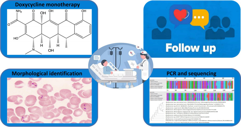
Graphical Abstract
Supplementary Information
The online version contains supplementary material available at 10.1186/s40249-023-01111-1.
Keywords: Human babesiosis, Babesia venatorum, Doxycycline monotherapy, China
Background
Human babesiosis is a zoonotic disease caused by an intraerythrocytic protozoan of the genus Babesia. Babesia is a parasite seen in the red blood cells of mammals, and is transmitted mainly by tick bites and blood transfusions [1]. Currently, more than 100 species of Babesia are recognized as pathogenic to various wild and domestic animal hosts, including humans [2]. Since the first case of human babesiosis was reported in 1957 in a Croatian farmer (formerly Yugoslavia), who underwent splenectomy, the number of cases has increased dramatically, particularly in the Northern Hemisphere [3]. In the United States alone, the annual cumulative number of cases of human babesiosis was estimated to range from 20,000 to 24,000 between 2006 and 2018, and it has been designated as a notifiable infectious disease [4]. In China, several Babesia species have been recorded at least 317 human patients [5], including Babesia divergens [6], B. venatorum [7], B. microti [8], B. crassa-like pathogens [9], and other unassigned species [5].
B. venatorum, formerly known as Babesia sp. EU1, was named after the Austrian and Italian Babesia-infected splenectomized hunters [10]. Since then, protozoan species have been documented to naturally infect the host roe deer Capreolus capreolus, and the vector species Ixodes ricinus and I. persulcatus [11, 12]. In epidemiological surveys of Chinese patients, few human cases of infection with B. venatorum have been reported in the endemic regions of China [7, 13]. The overall disease burden of B. venatorum in China is typically underestimated, because of its mild manifestations and asymptomatic appearance. Consequently, human infections with B. venatorum tend to be ignored. An accurate diagnosis and effective treatment regimen for infection with B. venatorum is therefore urgently needed for clinical development and practice.
In this study, a patient with unexplained febrile symptoms sought medical services at the Fifth Medical Center of the PLA General Hospital. A morphological microscopic review of thin peripheral blood smear examination and multi-gene polymerase chain reaction (PCR) amplification testing, confirmed infection with B. venatorum, and appropriate treatment with sensitive doxycycline was administered.
Three generations of anti-Babesia drugs have been approved for clinical use. Quinine, a traditional drug used against Babesia and Plasmodium species, inhibits hemozoin biocrystallisation via the heme detoxification pathway, which facilitates the aggregation of cytotoxic heme and results in parasite death [2, 14, 15]. As a unique naphthoquinone with broad-spectrum anti-protozoal activity, atovaquone acts against susceptible parasites by inhibiting the mitochondrial electron transport chain at the site of the cytochrome bc1 complex (complex III), ultimately blocking the synthesis of nucleic acids and adenosine triphosphatase [16–18]. According to the clinical practice guidelines for diagnosis and management of human babesiosis, it is commonly recommended to treat human babesiosis with either quinine or atovaquone, in combination with clindamycin or azithromycin [2], since the two antibiotics are protein synthesis inhibitors of apicoplast, a plastid organelle in apicomplexan parasites [19–21]. Doxycycline, a broad-spectrum tetracycline-class antibiotic, is frequently used to treat Plasmodium spp. by killing erythrocytic-stage parasites that target the apicoplast [22], in combination with quinine. Several attempts have been made to demonstrate its distinctive anti-babesial effects in experimental animals [23–25]. However, to date, no trials with doxycycline have been conducted in human patients with Babesia infections.
Case presentation
Clinical course of the case
A 73-year-old male underwent splenectomy after a traffic accident in 2014, and blood transfusion was performed during surgery. He had no history suggestive of any chronic diseases, or drug or alcohol abuse. The patient, with no recorded family history, had quit smoking 10 years ago and lived with his family in an urban area of Hulunbuir grassland (E115°31’–126°04’, N 47°05’–53°20’) Inner Mongolia Autonomous Region, China.
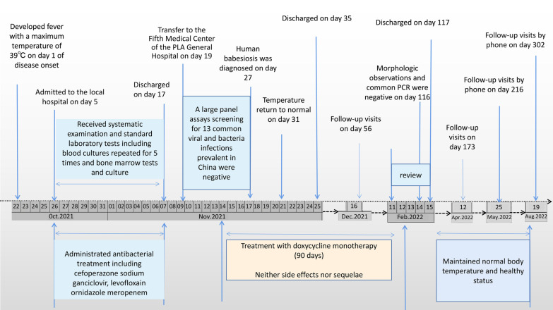
The patient developed fever, with a maximum body temperature of 39 °C on October 22, 2021, defined as the first day of the onset of the disease (Fig. 1). Headache, nausea, and vomiting persisted for several days. On October 26, the patient was admitted to the local hospital, where he underwent physical examination, standard laboratory tests, including blood cultures (five repeats) and bone marrow check and culture. He was administered antibacterial treatment, including cefoperazone sodium, ornidazole, and meropenem, over the following time (Additional file 1: Fig. S1). No relevant signs were found, except for a few abnormal biochemical tests, including thrombocytopenia (counts of platelets = 34 × 109/L), increased C-reactive protein (CRP, 88.1 mg/L), and weakly positive tuberculosis-interferon gamma release assay. However, the patient’s condition did not improve.
Fig. 1.
Timeline of doxycycline monotherapy for the patient infected with Babesia venatorum (Babesiidae, Sporozoa)
On November 9, 2021, the patient was transferred to the Fifth Medical Center of the PLA General Hospital in Beijing due to uncontrolled body temperature, with complain of no remission of fever, and dependence on indomethacin suppository twice daily. After admission, a detailed physical examination revealed no positive signs, except for abdominal scars owing to the previous splenectomy surgery. Assays for 11 pathogenic bacteria and two viruses (Additional file 1: Table S1) were performed at the hospital laboratory and no positive results were indicated. Therefore, we ruled out common bacterial and viral infections in this patient. Also, the erythrocyte sedimentation rate, pure protein derivative test, and computed tomography images of the lung did not indicate infection with tuberculosis. Notably, thrombocytopenia (counts of platelets = 31 × 109/L) and extremely high ferritin levels (> 2000 ng/ml) were observed, and a tentative suspicion of hematological disorder or malignant tumor bone metastases was considered. To validate our suspicion, a thin smear of the peripheral blood sample was reviewed, on November 11, 2021. However, the clinical laboratory in the hospital did not report definitive positive results. We empirically used doxycycline monotherapy [peros (po), 0.1 g bisindie (Bid)] to treat some undetectable pathogens, as the patient’s symptoms deteriorated, and conventional the anti-infective therapy was insufficient. During the first two days of doxycycline administration, the patient’s body temperature gradually decreased, and the symptoms resolved. Hence, we submitted the blood sample drawn on November 11 to the Beijing Institute of Microbiology and Epidemiology for further pathogen detection. On November 17, a laboratory specialist reported that Babesia protozoan was found in the blood smears submitted on November 11 (Fig. 2). To confirm the result, we collected fresh blood samples on November 17 for smear observation, and submitted them for PCR testing and sequencing. Babesia was also found as expected in blood smears, and B. venatorum was identified with sequences annotation. Additionally, positron emission tomography-computed tomography on November 13, did not indicate any high metabolic lesions, and two aspirations for bone marrow on November 11 and 23, did not reveal any specific abnormalities. The decreased hemoglobin level was observed at day of 19, 20, 21, 32 and 35 (Additional file 1: Table S1), which suggest the anemia status of the patient. Meanwhile, the elevated lactate dehydrogenase demonstrated at day of 19, 20, 23 also indicated the hemolysis occurred in the acute phase, which consistent with the increased CRP level until the day 32 (Additional file 1: Table S2). Therefore, the patient was diagnosed with human babesiosis.
Fig. 2.
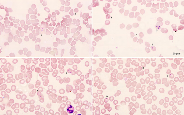
Photomicrographs of red cells infected with Babesia venatorum on a thin peripheral blood smear of the patient. A Tetrad; B Ring form trophozoites; C Extraerythrocytic Babesia parasites; D Schizont form
Babesia species identification
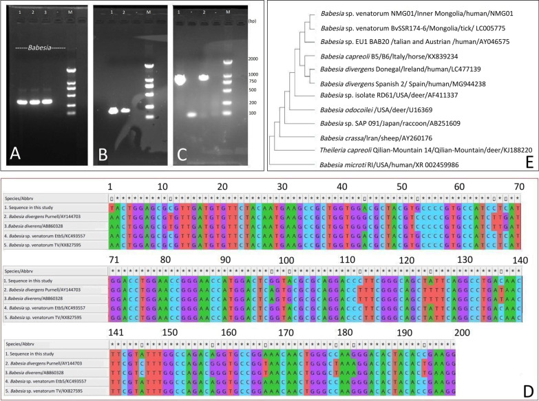
Morphological examination, PCR testing, sequencing, parasitemia surveillance and isolation of inoculated experimental mice were performed to identify the babesia infection for the patient (Additional file 1). On microscopically reviewing the thin peripheral blood smears using an oil-immersion lens, typical intraerythrocytic ring-form trophozoites, tetrads, and paired pyriform (Fig. 2A–D) were discovered without any pigments and lesser quantities of merozoites, and the parasitemia level was calculated to be as high as 12,000 parasites/µl in a blood smear on November 11, 2021. The PCR results were positive only for Babesia spp. (Fig. 3A), but negative for other known tick-borne agents. The 373 bp sequence obtained indicated that the patient was infected with B. venatorum. A 199 bp β-tubulin gene amplicon also confirmed infection with Babeisa spp. (Fig. 3B), and its sequence (GenBank accession No. OP 522105) was identical to that of B. venatorum isolated from ticks (Ixodes persulcatus) (KX827595) (Fig. 3D). Two expected amplicons for the entire 18s rRNA gene (Fig. 3C) were also obtained, and its nearly full-length 18 S rRNA gene (1665 bp, OP559478) further confirmed the infection of B. venatorum, with 99.82% similarity to B. venatorum isolated from ticks (LC005775) in Mongolia and human patients in Europe (AY046575) within the same clade after phylogenetic analysis, the statistical software MEGA11 (Tamura, Stecher, and Kumar 2021,USA). (Fig. 3E). Red blood cells infected with Babesia were also seen in the blood smears of the inoculated severe combined immune deficiency (SCID) mice on days 3, 6, and 12. Finally, human babesiosis was diagnosed according to the recommended laboratory criteria specified by the Centers for Disease Control and Prevention (CDC) [2].
Fig. 3.
Molecular identification of the parasite in the patient from Inner Mongolia, China. A Electrophoresis of the partial18S rRNA target gene of Babesia spp. from human whole blood (Lanes 1−3); B Electrophoresis of the amplicon of a β-tubulin gene of Babesia venatorum from human whole blood (Lanes 1−2); C Electrophoresis of two amplicons of the entire 18S rRNA gene from Babesia venatorum; D Comparison analysis of the 199 base-pair nucleotide sequence alignment of babesia β-tubulin gene. E Phylogenetic analysis of the sequence from patient strain NMG01 and other members of the order Piroplasmide based on nearly entire 18S rRNA gene sequences, inferred using the use of the maximum likelihood method and bootstrap analysis of 1000 replicates to assess the reliability of the reconstructed phylogenies
Treatment, outcomes and follow up
After the patient was diagnosed with human babesiosis, following the expert opinion on anti-babesial theory on European human babesiosis specified in 2020 [19], we were advised to treat the patient with atovaquone or quinine, accompanied by either clindamycin or azithromycin, since B. venatorum is the closest relatives of B. divergens in Babesia sensu stricto lineage. However, an emergency prescription for either quinine or atovaquone is inaccessible in Beijing. As an alternative chemical, doxycycline has been shown to have sensitive antiparasitic activities by inhibiting the synthesis of apicoplast protein in Plasmodium species, and successfully treating B. canis and B. gibsoni infection in dogs [23, 24]. It is reported that doxycycline was also aimed at other possible pathogens [26]. Notably, the patient’s clinical symptoms gradually improved following the administration of doxycycline. Therefore, we continued our monotherapy of doxycycline (po, 0.1 g Bid), followed by strict supervision and regular monitoring of parasite loads. As expected, the patient recovered rapidly, and the Babesia protozoan burden decreased dramatically from the pretreated 12,000/µl to 3840/µl [3 days post days post treatment (dpt)], 2400/µl (5 dpt), and 1440/µl (8 dpt) (Fig. 4). Broken or fragmented merozoites were frequently recorded during the regimen. Body temperature decreased after the two-day therapy, returned to normal after seven-day therapy, and then maintained normally thereafter (Fig. 4). The clinical manifestations subsided (Additional file 1: Table S3).
Fig. 4.
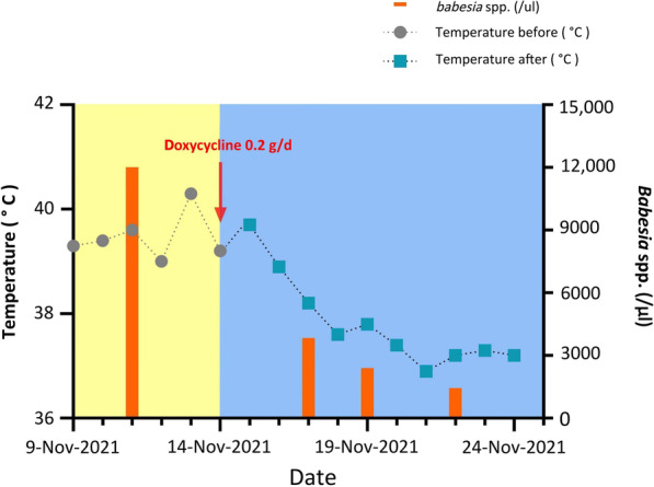
Dynamic change in the patient’s temperature and Babesia spp. infected erythrocyte count following the administration of doxycycline monotherapy
The patient was discharged on the 35th day after the onset of the disease. Since then, a consecutive follow-up survey of the patient with a continuing course of doxycycline treatment was performed for 90 days. The patient was readmitted to the hospital for follow-up on February 11, 2022. We performed a detailed physical examination and found no abnormalities. The same laboratory protocol was used to monitor the Babesia burden in patients. Morphological examination and PCR tests were negative for whole blood samples collected on February 15, 2022, 117 days after the onset time (Fig. 1). No doxycycline-related side effects or sequelae, including erythema or hepatic insufficiency, were observed. The patient maintained a normal body temperature and health status from the time the chemical therapy was stopped to the final follow-up visit on August 19, 2022 (Fig. 1 and Additional file 1: Table S3).
Discussion and conclusion
We confirm a case of human babesiosis in Inner Mongolia, China according to the guidelines on diagnosis and management of babesiosis [2]. The first obtained entire 18s rRNA and β-tubulin protein genes were from a patient infected with B. venatorum in China. To our knowledge, this is the first successful clinical application of doxycycline monotherapy for human babesiosis, providing an alternative therapeutic strategy with clinical trials and useful anti-babesial chemicals.
Cycline reportedly has satisfactory prophylactic effects against experimental infections with highly pathogenic strains of B. canis and B. bovis [23–25]. However, it is not recommended as a first-line treatment for Babesia infection, even when the parasite is doxycycline-sensitive, because the anti-parasite effect of doxycycline is delayed [27]. In the present case of human babesiosis, the patient maintained a normal body temperature and health status from the time that the therapy was stopped, until the final follow-up visit. This suggests that doxycycline is suitable for the treatment of babesiosis in humans.
An epidemiological survey of possible infection routes did not produce definitive results, similar to the previous cases of human babesiosis. Babesia spp. were most frequently transmitted by Ixodes spp. ticks such as B. microti detected in Dermacentor nuttallii, and Hyalomma asiaticum from the Ceke, Mandala, and Manzhouli areas of Inner Mongolia, China [28, 29]. Babesia infections may also occur as a result of blood transfusions [30]. Although no definitive tick bites were recorded in our patient, humans are often imperceptible to tick bites, and hence these cannot be excluded. Another possible mode of infection, caused by a previous blood transfusion surgery, should not be discounted. Cases of human babesiosis relevant to blood transfusion have been documented for B. microti since 1980 [31], and for B. duncani since 1994 [32]. The incubation period of these Babesia species ranges from 23 to 384 days, when transmitted by blood transfusion [33]. As for B. venatorum, no human babesiosis cases were recorded to be caused by blood transfusions or other relevant operations. One splenectomy patient from Germany without obvious exposure history for tick bite and blood product developed chronic B. venatorum (formerly named Babesia sp. EU1) parasitemia after treatment with quinine and clindamycin due to a relapse of Hodgkin’s disease, and long-term maintenance therapy with atovaquone was administered to clear the parasites [34]. Our case of human babesiosis remained undiagnosed for eight years after splenectomy, and blood transfusions had to be given. Consequently, B. venatorum exceeded the limit of incubation period in this patient, and the clinical manifestations might have been caused by a possible recrudescence, or an unrecorded tick bite, especially in the asymptomatic period.
Notably, monotherapy for human babesiosis has been successful. It is reasonable to attribute the inhibitory activity of doxycycline on protein synthesis in the apicoplast of the parasites. As both Babesia and Plasmodium parasites replicate inside the erythrocytes of the mammalian host, the apicoplast, a special organelle required to invade host erythrocytes, may serve as a candidate target for anti-parasite chemicals [30]. To inhibit protein synthesis in apicoplasts, future trials should address potential anti-babesial chemicals, such as tetracyclines (tetracycline, doxycycline, and minocycline), macrolides (spiramycin and azithromycin), and lincosamides (lincomycin, clindamycin, pirlimycin, and chloramphenicol) [35]. In 1996, the prophylactic treatment of experimental canine babesiosis (B. canis) with doxycycline achieved success, with satisfactory efficacy [23]. Another option is an alternative combination therapy regimen, which works well with metronidazole, clindamycin, and doxycycline for B. gibsoni (Asian genotype) infection in dogs in Hong Kong [24]. In China, clindamycin is used to treat patients with B. microti infections. Azithromycin and atovaquone was used for treatment of one pediatric patient infected with B. venatorum in 2014 [13], and three cases of B. microti [36–38]. Doxycycline, a tetracycline antibiotic, is widely used to treat various pathogens [26, 39, 40]. Currently, there is no standard course of treatment owing to the lack of medical records on doxycycline monotherapy for human Babesia infections. In the present study, we used doxycycline monotherapy for 90 days to prevent recrudescence, and found no adverse drug reactions, as revealed by the patient’s health status and laboratory evidence. This implies that doxycycline monotherapy for human infection with B. venatorum is suitable for up to 90 days.
In China, the actual number of infected individuals, including blood product donors may be significantly higher than expected, as many of them may be asymptomatic. However, most doctors and laboratory personnel in China lack awareness about human babesiosis, leading to underdiagnosis and an improper treatment. Similar to this case, laboratory personnel are also prone to miss the protozoan, as they are accustomed to fetching results directly from the hematology analyzer. Furthermore, due to insufficient awareness of the disease, first-line drugs for human babesiosis are still scarce in China. Therefore, doxycycline monotherapy should be considered in the future.
In summary, our successful experience treatment of human babesiosis caused by B. venatorum strongly suggests that doxycycline could be used as an alternative chemical to treat natural Babesia protozoal infections. However, it is necessary to expand the number of cases and perform more standard case-control studies to further study the efficacy and duration of doxycycline monotherapy.
Supplementary Information
Additional file 1. Figure S1. The administration time and the dosage daily of the drugs before the patient was transferred to the Sentinel Hospital of the FifthMedical Center of the PLA General Hospital. Table S1. The panel assaysscreening for 13 common viruses and bacteria. Table S2. Laboratory values observed during hospitalisation (Reference values are given in parentheses). Table S3. Symptoms and signs reported during thefirst hospital stay. Additional materials.
Acknowledgements
The authors would like to acknowledge all staffs of the Senior Department of Infectious Diseases, The Fifth Medical Center of Chinese PLA General Hospital.
Author contributions
J-FJ, F-SW, W-CC and LH designed the study. LH, D-DH, J-F J, YS and MX did the main statistical analysis, and wrote the paper. D-DH, NY, WH, W-MN, YL, H-CM, XZ and Y-GL, recruited the patients and gathered the data. MX, L-YX, NY, WH, LH, M-ZZ, D-YZ and YL did the laboratory tests. D-DH, R-HL, X-AH and J-YW did the statistical analysis. All authors contributed to the review and revision of the paper. All authors read and approved the final manuscript.
Funding
This study was funded by the State Key Research Development Program of China (2019YFC1200501), National Natural Science Foundation of China (81772185), and National Key R&D Program of China (2021YFC2301801, 2022YFC2304405).
Availability of data and materials
All data generated or analyzed relating to this study are presented within this published article.
Declarations
Ethics approval and consent to participate
This research was carried out according to the principles of the Declaration of Helsinki and was approved by the Ethics Committees of the Fifth Medical Center of Chinese PLA General Hospital (No.KY-2022-6-44-2). Written informed consent was obtained from the patient.
Consent for publication
Written informed consent for publication of the clinical details was obtained from the patient.
Competing interests
The authors declare that they have no competing interests.
Footnotes
The original version of this article was revised: “The affiliations assigned to Ming-Zhu Zhang and Jing-Yuan Wang have been updated due to a typing mistake.”
Lei Huang, Yi Sun, Dan-Dan Huo and Ming Xu contributed equally to this work.
Change history
7/27/2023
A Correction to this paper has been published: 10.1186/s40249-023-01123-x
Contributor Information
Wu-Chun Cao, Email: caowc@bmi.ac.cn.
Fu-Sheng Wang, Email: fswang302@163.com.
Jia-Fu Jiang, Email: jiangjf2008@139.com.
References
- 1.Vannier E, Krause PJ. Human babesiosis. N Engl J Med. 2012;366(25):2397–407. doi: 10.1056/NEJMra1202018. [DOI] [PubMed] [Google Scholar]
- 2.Krause PJ, Auwaerter PG, Bannuru RR, Branda JA, Falck-Ytter YT, Lantos PM, et al. Clinical practice guidelines by the infectious Diseases Society of America (IDSA): 2020 Guideline on diagnosis and management of Babesiosis. Clin Infect Dis. 2021;72(2):e49–e64. doi: 10.1093/cid/ciaa1216. [DOI] [PubMed] [Google Scholar]
- 3.Gray JS, Estrada-Pena A, Zintl A. Vectors of babesiosis. Annu Rev Entomol. 2019;64:149–65. doi: 10.1146/annurev-ento-011118-111932. [DOI] [PubMed] [Google Scholar]
- 4.Gray EB, Herwaldt BL. Babesiosis surveillance—United States, 2011–2015. MMWR Surveill Summ. 2019;68(6):1–11. doi: 10.15585/mmwr.ss6806a1. [DOI] [PubMed] [Google Scholar]
- 5.Xin CM, Bo XJ, Lin A, Peng S, Peng S, Xu CJ. Epidemic status and research progress of babesiosis in China. J Trop Dis Parasitol. 2022;20(3):149–57. [Google Scholar]
- 6.Wang J, Zhang S, Yang J, Liu J, Zhang D, Li Y, et al. Babesia divergens in human in Gansu province, China. Emerg Microbes Infect. 2019;8(1):959–61. doi: 10.1080/22221751.2019.1635431. [DOI] [PMC free article] [PubMed] [Google Scholar]
- 7.Jiang JF, Zheng YC, Jiang RR, Li H, Huo QB, Jiang BG, et al. Epidemiological, clinical, and laboratory characteristics of 48 cases of “Babesia venatorum” infection in China: a descriptive study. Lancet Infect Dis. 2015;15(2):196–203. doi: 10.1016/S1473-3099(14)71046-1. [DOI] [PubMed] [Google Scholar]
- 8.Wang HS, Hen P, Min ZH, Shao X. Investigation of Babesia spp. infections in blood donors in Guangxi, China. Acad J Second Mil Med Univ. 2016;283–7 (in Chinese).
- 9.Jia N, Zheng YC, Jiang JF, Jiang RR, Jiang BG, Wei R, et al. Human babesiosis caused by a Babesia crassa-like pathogen: a case series. Clin Infect Dis. 2018;67(7):1110–9. doi: 10.1093/cid/ciy212. [DOI] [PubMed] [Google Scholar]
- 10.Herwaldt BL, Caccio S, Gherlinzoni F, Aspock H, Slemenda SB, Piccaluga P, et al. Molecular characterization of a non-Babesia divergens organism causing zoonotic babesiosis in Europe. Emerg Infect Dis. 2003;9(8):942–8. doi: 10.3201/eid0908.020748. [DOI] [PMC free article] [PubMed] [Google Scholar]
- 11.Bajer A, Dwuznik-Szarek D. The specificity of babesia-tick vector interactions: recent advances and pitfalls in molecular and field studies. Parasit Vectors. 2021;4(1):507. doi: 10.1186/s13071-021-05019-3. [DOI] [PMC free article] [PubMed] [Google Scholar]
- 12.Kumar A, O’Bryan J, Krause PJ. The global emergence of human babesiosis. Pathogens. 2021;10(11):1447. doi: 10.3390/pathogens10111447. [DOI] [PMC free article] [PubMed] [Google Scholar]
- 13.Sun Y, Li SG, Jiang JF, Wang X, Zhang Y, Wang H, et al. Babesia venatorum infection in child, China. Emerg Infect Dis. 2014;20(5):896–7. doi: 10.3201/eid2005.121034. [DOI] [PMC free article] [PubMed] [Google Scholar]
- 14.Woodland JG, Hunter R, Smith PJ, Egan TJ. Shining new light on ancient drugs: preparation and subcellular localisation of novel fluorescent analogues of Cinchona alkaloids in intraerythrocytic Plasmodium falciparum. Org Biomol Chem. 2017;15(3):589–97. doi: 10.1039/C6OB02110G. [DOI] [PubMed] [Google Scholar]
- 15.Woodland JG, Hunter R, Smith PJ, Egan TJ. Chemical proteomics and super-resolution imaging reveal that chloroquine interacts with Plasmodium falciparum multidrug resistance-associated protein and lipids. ACS Chem Biol. 2018;13(10):2939–48. doi: 10.1021/acschembio.8b00583. [DOI] [PubMed] [Google Scholar]
- 16.Lawres LA, Garg A, Kumar V, Bruzual I, Forquer IP, Renard I, et al. Radical cure of experimental babesiosis in immunodeficient mice using a combination of an endochin-like quinolone and atovaquone. J Exp Med. 2016;213(7):1307–18. doi: 10.1084/jem.20151519. [DOI] [PMC free article] [PubMed] [Google Scholar]
- 17.Montazeri M, Mehrzadi S, Sharif M, Sarvi S, Shahdin S, Daryani A. Activities of anti-Toxoplasma drugs and compounds against tissue cysts in the last three decades (1987 to 2017), a systematic review. Parasitol Res. 2018;117(10):3045–57. doi: 10.1007/s00436-018-6027-z. [DOI] [PubMed] [Google Scholar]
- 18.Vaidya AB, Mather MW. Atovaquone resistance in malaria parasites. Drug Resist Updat. 2000;3(5):283–7. doi: 10.1054/drup.2000.0157. [DOI] [PubMed] [Google Scholar]
- 19.Smith RP, Hunfeld KP, Krause PJ. Management strategies for human babesiosis. Expert Rev Anti Infect Ther. 2020;18(7):625–36. doi: 10.1080/14787210.2020.1752193. [DOI] [PubMed] [Google Scholar]
- 20.Hildebrandt A, Zintl A, Montero E, Hunfeld KP, Gray J. Human babesiosis in Europe. Pathogens. 2021;10(9):1165. doi: 10.3390/pathogens10091165. [DOI] [PMC free article] [PubMed] [Google Scholar]
- 21.Hildebrandt A, Gray J, Montero E. Characteristics of human babesiosis in Europe. Pathogens. 2023;12(2):323. doi: 10.3390/pathogens12020323. [DOI] [PMC free article] [PubMed] [Google Scholar]
- 22.Pradel G, Schlitzer M. Antibiotics in malaria therapy and their effect on the parasite apicoplast. Curr Mol Med. 2010;10(3):335–49. doi: 10.2174/156652410791065273. [DOI] [PubMed] [Google Scholar]
- 23.Vercammen F, De Deken R, Maes L. Prophylactic treatment of experimental canine babesiosis (Babesia canis) with doxycycline. Vet Parasitol. 1996;66(3–4):251–5. doi: 10.1016/S0304-4017(96)01016-3. [DOI] [PubMed] [Google Scholar]
- 24.Almendros A, Burchell R, Wierenga J. An alternative combination therapy with metronidazole, clindamycin and doxycycline for Babesia gibsoni (asian genotype) in dogs in Hong Kong. J Vet Med Sci. 2020;82(9):1334–40. doi: 10.1292/jvms.20-0209. [DOI] [PMC free article] [PubMed] [Google Scholar]
- 25.Taylor SM, Elliott CT, Kenny J. Inhibition of Babesia divergens in cattle by oxytetracycline. Vet Rec. 1986;118(4):98–102. doi: 10.1136/vr.118.4.98. [DOI] [PubMed] [Google Scholar]
- 26.Paul A, Anandabaskar N, Mathaiyan J, Raj GM. Adrenergic agonists. In: Paul A, Anandabaskar N, Mathaiyan J, Raj GM eds. Introduction to basics of pharmacology and toxicology. Singapore: Springer; 2021. p. 41–53.
- 27.Dahl EL, Shock JL, Shenai BR, Gut J, DeRisi JL, Rosenthal PJ. Tetracyclines specifically target the apicoplast of the malaria parasite Plasmodium falciparum. Antimicrob Agents Chemother. 2006;50(9):3124–31. doi: 10.1128/AAC.00394-06. [DOI] [PMC free article] [PubMed] [Google Scholar]
- 28.Zhou X, Xia S, Huang JL, Tambo E, Zhuge HX, Zhou XN. Human babesiosis, an emerging tick-borne disease in the People’s Republic of China. Parasit Vectors. 2014;7:509. doi: 10.1186/s13071-014-0509-3. [DOI] [PMC free article] [PubMed] [Google Scholar]
- 29.Hao GF, Li H, Sun Y, Ge RP, Qiao GQ, Li B, et al. Detection of tick and tick-borne pathogen in some ports of Inner Mongolia. Chin J Epidemiol. 2009;30(4):365–7. [PubMed] [Google Scholar]
- 30.Herwaldt BL, Linden JV, Bosserman E, Young C, Olkowska D, Wilson M. Transfusion-associated babesiosis in the United States: a description of cases. Ann Intern Med. 2011;155(8):509–19. doi: 10.7326/0003-4819-155-8-201110180-00362. [DOI] [PubMed] [Google Scholar]
- 31.Jacoby GA, Hunt JV, Kosinski KS, Demirjian ZN, Huggins C, Etkind P, et al. Treatment of transfusion-transmitted babesiosis by exchange transfusion. N Engl J Med. 1980;303(19):1098–100. doi: 10.1056/NEJM198011063031906. [DOI] [PubMed] [Google Scholar]
- 32.Gerber MA, Shapiro ED, Krause PJ, Cable RG, Badon SJ, Ryan RW. The risk of acquiring Lyme disease or babesiosis from a blood transfusion. J Infect Dis. 1994;170(1):231–4. doi: 10.1093/infdis/170.1.231. [DOI] [PubMed] [Google Scholar]
- 33.Tonnetti L, Eder AF, Dy B, Kennedy J, Pisciotto P, Benjamin RJ, et al. Transfusion-transmitted Babesia microti identified through hemovigilance. Transfusion. 2009;49(12):2557–63. doi: 10.1111/j.1537-2995.2009.02317.x. [DOI] [PubMed] [Google Scholar]
- 34.Haselbarth K, Tenter AM, Brade V, Krieger G, Hunfeld KP. First case of human babesiosis in Germany—clinical presentation and molecular characterisation of the pathogen. Int J Med Microbiol. 2007;297(3):197–204. doi: 10.1016/j.ijmm.2007.01.002. [DOI] [PubMed] [Google Scholar]
- 35.Wiesner J, Seeber F. The plastid-derived organelle of protozoan human parasites as a target of established and emerging drugs. Expert Opin Ther Targets. 2005;9(1):23–44. doi: 10.1517/14728222.9.1.23. [DOI] [PubMed] [Google Scholar]
- 36.Yao J, Liu G, Zou Y, Jiang J, Li S, Wang H, et al. Babesia microti causing intravascular hemolysis in immunocompetent child, China. Emerg Infect Dis. 2023;29(3):667–9. doi: 10.3201/eid2903.220888. [DOI] [PMC free article] [PubMed] [Google Scholar]
- 37.Man SQ, Qiao K, Cui J, Feng M, Fu YF, Cheng XJ. A case of human infection with a novel Babesia species in China. Infect Dis Poverty. 2016;5:28. doi: 10.1186/s40249-016-0121-1. [DOI] [PMC free article] [PubMed] [Google Scholar]
- 38.He Wang WQ. A case report of human babesia infection in Beijing. Chin J Exp Clin Infect Dis. 2019;13(6):519–23. [Google Scholar]
- 39.Sanchez E, Vannier E, Wormser GP, Hu LT. Diagnosis, treatment, and prevention of Lyme disease, human granulocytic anaplasmosis, and babesiosis: a review. JAMA. 2016;315(16):1767–77. doi: 10.1001/jama.2016.2884. [DOI] [PMC free article] [PubMed] [Google Scholar]
- 40.Al-Tawfiq JA, Memish ZA. Antibiotic susceptibility and treatment of brucellosis. Recent Pat Antiinfect Drug Discov. 2013;8(1):51–4. doi: 10.2174/1574891X11308010010. [DOI] [PubMed] [Google Scholar]
Associated Data
This section collects any data citations, data availability statements, or supplementary materials included in this article.
Supplementary Materials
Additional file 1. Figure S1. The administration time and the dosage daily of the drugs before the patient was transferred to the Sentinel Hospital of the FifthMedical Center of the PLA General Hospital. Table S1. The panel assaysscreening for 13 common viruses and bacteria. Table S2. Laboratory values observed during hospitalisation (Reference values are given in parentheses). Table S3. Symptoms and signs reported during thefirst hospital stay. Additional materials.
Data Availability Statement
All data generated or analyzed relating to this study are presented within this published article.


