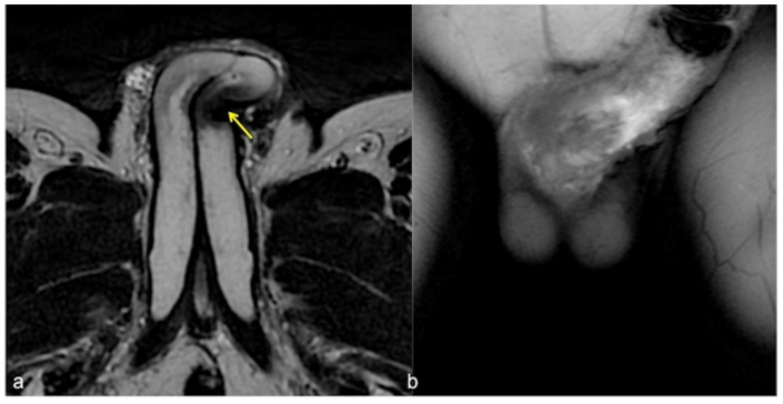
Figure 6.
Cav-MRI. SSFSE T2W axial acquisition (a) showing a thickened plaque of reduced signal intensity in the left cavernous body ((a), arrow); GRE T1W 3D FS coronal acquisition obtained after contrast agent administration in the cavernous bodies (b) documents a defect in the contrast agent distribution due to fibrotic distortion of the cavernous bodies.

