Abstract
Background
Breast cancer (BC) with low human epidermal growth factor receptor 2 (HER2) expression is attracting much attention due to the breakthrough progress of novel anti-HER2 antibody-drug conjugates. HER2 expression is examined in patients with HER2-low BC and their distant metastases in this study, so as to further clarify the dynamic characteristics of HER2 low status in the process of disease progression.
Methods
Patients diagnosed with HER2 low breast cancer (defined as IHC1+ or IHC2+/ISH-) between 2012 and 2021 were included in this study. We evaluated HER2 expression of primary sites and metastatic sites, compared the impact of different clinicopathological parameters on HER2 status of metastases and compared the overall survival and disease-free survival of patients with different HER2 status in metastases.
Results
Ninety-eight patients were included. All HER2 IHC scores were confirmed and the consistent rate with the original pathological report was 81.1%. 27.6% of the patients showed different HER2 status in metastases. The HER2 discordance rate differed among different metastatic sites (p = 0.040). The higher the T stage of the primary BC, the higher the rate of HER2 discordance was observed (p = 0.042). For the specimen type of metastasis, HER2 discordant rate was higher in surgical specimen than biopsy (p = 0.050). No difference of HER2 discordance rate was found between HER2-1+ and HER2-2+ patients. But comparing HER2 IHC score, HER2-2+ patients were less likely to have consistent metastatic HER2 levels than HER2-1+ patients (p = 0.006). No difference in survival outcomes was observed between patients with different HER2 status in metastases.
Conclusions
There is a possibility of HER2 expression alteration in the metastases of HER2-low breast cancer. And the rate of altered HER2 low expression was different among different metastatic sites, different T stages of primary BC and specimen type of metastasis. No prognostic significance was observed.
Supplementary Information
The online version contains supplementary material available at 10.1186/s12885-023-11134-4.
Keywords: HER2-low expression, HER2 discordance, Metastasis, Anti-HER2 antibody-drug conjugates, Breast cancer
Background
As one of the most frequently diagnosed cancer in the world [1], Breast cancer (BC) is a highly heterogenous disease, with significant biological diversity and different clinicopathologic features, prognosis and sensitivity to treatments. Despite complex biological diversity, in clinical practice breast cancer can be divided into several subgroups according to estrogen receptor (ER), progesterone receptor (PR) and human epidermal growth factor receptor 2 (HER2) status. For anti-HER2 targeted agents, the treatment-decision is usually made according to dichotomization. In the context of HER2-positive BC (which is defined as HER2 3+ on IHC(immunohistochemistry) score or HER2 gene amplification on ISH(in situ hybridization) assay [2]), which accounts for about 15% of all BCs [3], anti-HER2 agents has shown great clinical benefits [4, 5]. While HER2-negative BCs, the definition of which is IHC 0/1+ or IHC 2+ with no HER2 gene amplification [2], have been proved with no benefit from traditional HER2-targeted agents by several studies [6–8].
Recently, however, results from several clinical trials focusing on new anti-HER2 antibody-drug conjugates (ADCs) in breast cancer patients with HER2-low expression (defined as IHC1+ or IHC2+ without HER2 gene amplification by ISH) are changing the situation [9–11]. For instance, the results of a phase Ib study showed that Trastuzumab Deruxtecan (DS8201a) achieved a remarkable 10.4 months of median response duration and 37% of overall response rate in patients with HER2-low advanced breast cancer [9]. Likewise, in HER2-low breast cancer patients with hormone receptor positive (HR+) or HR-, SYD985 (Trastuzumab Duocarmazine) had an objective response rate (ORR) of 28% and 40% separately in phase Ib trials [10]. And the results of Destiny-Breast04, a phase III trial testing DS8201a in pretreated advanced HER2-low breast cancer patients showed that the median progression free survival (PFS) was 4.8 months longer in the DS8201a group compared with 5.1 months in the physician’s choice group (p < 0.001), and the overall survival (OS) was 6.6 months longer (p = 0.001) [11].
Based on all these results, HER2-low breast cancer is becoming a new important entity different from the old HER2-negative group. And several reports have investigated that the proportion of HER2-low breast cancer in the population is about 45–55% [12–14], which is not negligible. It has been reported that there exists HER2 status difference between primary and recurrent breast cancer [15–17]. And for HER2-low breast cancer, this phenomenon also has been reported [18, 19]. Nonetheless, most of the reported studies are about patients with HER2-neagtive breast cancer and the changes of HER2 expression in recurrent or advanced stage [18, 19], and few evidence has yet been reported concerning the evolution of HER2 status focusing on the HER2-low primary breast cancer and the matched distant metastasis.
In this study, our goal is to characterize the HER2 status of distant metastasis in patients with HER2-low primary breast cancer, evaluate the association of the HER2 status discordance with the clinicopathologic features and tumor-related factors, as well as exploring the possible prognostic value.
Methods
Sample selection
Breast cancer patients who had received biopsy or surgical resection of both primary lesion and distant metastasis at Fudan University Shanghai Cancer Center from July 4, 2012 to October 13, 2021 with available HER2 IHC score and, if necessary, HER2 FISH (fluorescence in situ hybridization) results of both primary and metastatic tumor were retrieved from a pathology database. HER2 IHC slides of all primary and metastatic tumor samples were retrieved from the Pathology Departments of our center. Patients clinicopathologic features including HER2 status, HR status, age, histological subtype and grade, site and timing of distant metastases, and survival status were recorded.
Evaluation of HER2 expression
All staining was performed by the auto-staining machine, which is Ventana BenchMark autostainer or Ventana BenchMark Ultra autostainer (Ventana Medical System Inc, Roche, Tucson, Arizona) with the BenchMark ULTRA advanced staining system operator guide. ULTRA Cell Conditioning Solution (ULTRA CC1, pH = 8.5) was used to perform antigen retrieval at 90 °C to 100 °C. HER2 expression was all tested by prediluted Ventana 4B5 antibody, and was reassessed by a pathologist according to the latest standard of HER2 diagnosis by ASCO/CAP Guidelines Update [2], and cases that were difficult to diagnose were reassessed by another pathologist. Tumors were considered HER2-low when the IHC score was 1+ or 2+ with negative ISH assay. And according to the latest ASCO/CAP Guidelines Update [2], IHC 0 is defined by no staining observed or membrane staining that is incomplete and is faint or barely perceptible and within ≤ 10% of the invasive tumor cells, and if the proportion is over 10%, tumor is defined as IHC 1+. The definition of IHC 2+ is invasive breast cancer with weak to moderate complete membrane staining observed in > 10% of tumor cells, and when over 10% of circumferential membrane staining is complete and intense, tumor is defined as IHC 3+.
Statistical analysis
SPSS 20.0 statistical software (IBM Corp, Armonk, NY, USA) was used to perform statistical analysis., For continuous variables, data was presented as median and interquartile range, and for categorical variables, data was presented as relative frequencies (percentage). Chi-square and Fisher’s exact tests were used to investigate differences by HER2-low status for categorical variables and Wilcoxon rank test for continuous variables. Kaplan-Meier curves were performed using GraphPad Prism 9.0 for survival analysis. P < 0.05 was considered statistically significant.
Results
Patients and their clinicopathologic features
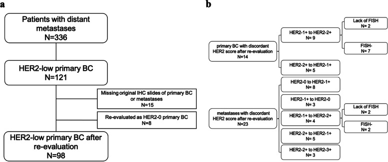
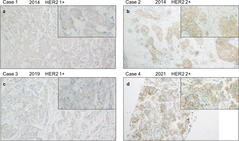
Three hundred thirty-six patients with available HER2 IHC score and, if necessary, FISH results on both primary BC and distant metastases were included during 2012 and 2021. Among these cases, 121 patients with HER2-low primary BC were selected. 15 cases lacking original HER2 IHC slides were ruled out. And 8 cases re-evaluated as HER2-0 of primary BC were ruled out (Fig. 1a). In total, 98 patients with HER2-low BC who had distant metastases were included in the analysis, including 55 (56.1%) patients with HER2-1+ and 43 (43.9%) patients with HER2-2+. And among the overall population, 78.6% of the patients were HR+, and 21.4% were triple negative breast cancer (TNBC). Table 1 summarized the clinical, pathological and immunohistochemical characteristics. All of the HER2 IHC-stained slides were rescored, and the concordance rate of HER2 IHC score with the original pathological report was 81.1% (Fig. 1b). For primary BC with HER2-low expression, the concordance rate between HER-1+ and HER2-2+ was 85.7%. And for metastatic sites, the concordant rate among different HER2 IHC scores was 76.5%. And 4 cases with HER2-1+ were re-evaluated as HER2-2+ which lacked FISH results. Considering their relatively weak HER2 staining, they were still classified as HER2-low for subsequent statistical analysis. For the possible fading of DAB staining, we also compared IHC slides of different years, and we believed that the fading didn’t affect the HER2 diagnosis (Fig. 2).
Fig. 1.
Case selection and grouping. a Flow diagram of the study. b Re-evaluation results of the HER2 IHC score. N number, BC breast cancer, IHC immunohistochemistry, FISH fluorescence in situ hybridization
Table 1.
Clinical, pathological and immunohistochemical characteristics of HER2 low breast cancers
| Variable | Overall (98) | HER2-1+ (55) | HER2-2+ (43) | P-value |
|---|---|---|---|---|
| N(%) or mean | N(%) or mean | N(%) or mean | ||
| Age at primary diagnosis/y | 50.5 ± 11.6 | 50.3 ± 11.3 | 51.7 ± 11.6 | 0.552 |
| Tumor size | 0.089 | |||
| - ≤ 2 cm | 22 (22.4) | 16 (29.1) | 6 (14.0) | |
| - > 2 cm | 47 (48.0) | 24 (43.6) | 23 (53.4) | |
| -Unavailable | 29 (29.6) | 15 (27.3) | 14 (32.6) | |
| Histological subtype | 1.000 | |||
| -Invasive breast carcinoma NOS | 93 (94.9) | 52 (94.5) | 41 (95.3) | |
| -Others | 5 (5.1) | 3 (5.5) | 2 (4.7) | |
| Histological grade | 0.459 | |||
| -II | 52 (51.0) | 31 (56.4) | 21 (48.8) | |
| -III | 46 (45.9) | 24 (43.6) | 22 (51.2) | |
| Estrogen receptor expression | 0.697 | |||
| -Positive | 77 (78.6) | 44 (80.0) | 33 (76.7) | |
| -Negative | 21 (21.4) | 11 (20.0) | 10 (23.3) | |
| Progesterone receptor expression | 0.579 | |||
| -Positive | 60 (61.2) | 35 (63.6) | 25 (58.1) | |
| -Negative | 38 (38.8) | 20 (36.4) | 18 (41.9) | |
| Ki67 | 0.371 | |||
| - < 20% | 15 (15.3) | 10 (18.2) | 5 (11.6) | |
| - ≥ 20% | 83 (84.7) | 45 (81.8) | 38 (88.4) | |
| Age at metastasis diagnosis | 53.0 ± 11.7 | 52.2 ± 11.3 | 53.9 ± 11.8 | 0.471 |
| Site of metastasis | 0.929 | |||
| -Bone | 13 (13.3) | 8 (14.5) | 5 (11.6) | |
| -Lung/Pleura | 28 (28.6) | 15 (27.3) | 13 (30.2) | |
| -Liver | 41 (41.8) | 24 (43.6) | 17 (39.5) | |
| -Skin and soft tissues | 11 (11.2) | 6 (10.9) | 5 (11.6) | |
| -Others | 5 (5.1) | 2 (3.6) | 3 (7.0) |
Fig. 2.
HER2 staining situation of different years. Case 1 (a) showing HER2 1+ staining situation of 2014. Case 2 (b) showing HER2 2+ staining situation of 2014. Case 3 (c) showing HER2 1+ staining situation of 2019. Case 4 (d) showing HER2 2+ staining situation of 2021. All of the insets are high-power field highlighting area of HER2 immunoreactivity
Discordance between primary breast cancer and distant metastases regarding HER2 status
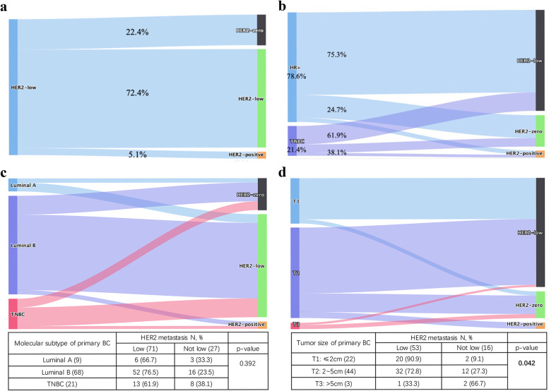
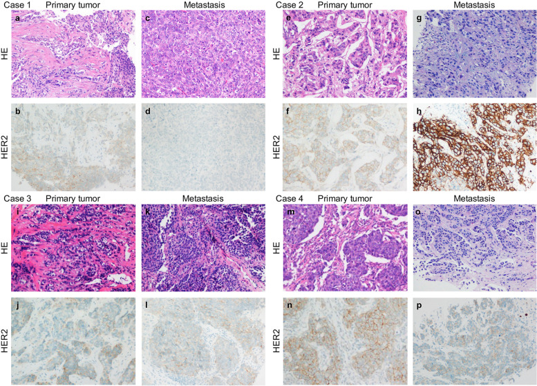
Figure 3 summarizes the discordance of HER2 status between primary breast cancer and distant metastases, while Fig. 4 shows examples of typical immunohistochemical staining of HER2. The overall HER2 discordant rate was 27.6% (n = 27). Among all these changed patients, 81.5% (n = 22/27) turned into HER2-0, while 19.5% (n = 5/27) became HER2-3+, of which 2 cases had positive FISH results due to the original score of HER2 2+ and others were lack of FISH test (Fig. 3a). And 4 cases with HER2-3+ in metastases were HER2-2+ in primary sites except 1 case being HER2-1+. The percentage of HER2 discordant cases did not differ among primary tumor phenotypes. In detail, as shown in Fig. 3b, cases with HER2 discordance in HR-positive and triple-negative breast cancers were 24.7% and 38.1% (p = 0.222). There was no significant difference in HER2 discordance rate when divided primary BC into Luminal A, Luminal B and TNBC (p = 0.392) (Fig. 3c). And the rate of HER2 discordance differed among different tumor sizes of the primary BC (p = 0.042), with lower rates observed for T1 tumors (≤ 2 cm) (9.1%), followed by T2 tumors (2.0 cm–5.0 cm) (27.3%) and T3 tumors (> 5.0 cm) (66.7%) (Fig. 3d).
Fig. 3.
HER2 evolution from primary breast cancer to distant metastases. a The overall rate of HER2 discordance. b HER2 discordant cases in HR-positive and triple-negative breast cancers respectively. c Evolution of the HER2 status between primary tumors and metastases stratified by molecular subtypes of primary BC (Chi-square test). d Evolution of the HER2 status between primary tumors and metastases stratified by T stages of primary BC (Fisher’s exact test). BC breast cancer, N number
Fig. 4.
Four examples of HER2 status evolution from primary breast cancer to distant metastases. High-power views of four primary breast carcinomas and their metastases, with corresponding HER2 IHC images. Case 1: a HER2-low (1+) primary tumor with a HER2-0 metastasis (a, c haematoxylin & eosin (HE) staining; b, d HER2 IHC). Case 2: a HER2-low (2+) primary tumor with a HER2-positive metastasis (e, g HE staining; f, h HER2 IHC). Case 3: a HER2-low (1+) primary tumor with a HER2-low (1+) metastasis (i, k HE staining; j, l HER2 IHC). Case 4: a HER2-low (2+) primary tumor with a HER2-low (1+) metastasis (m, o HE staining; n, p HER2 IHC)
Effect of metastatic tumor sample on HER2 status evolution
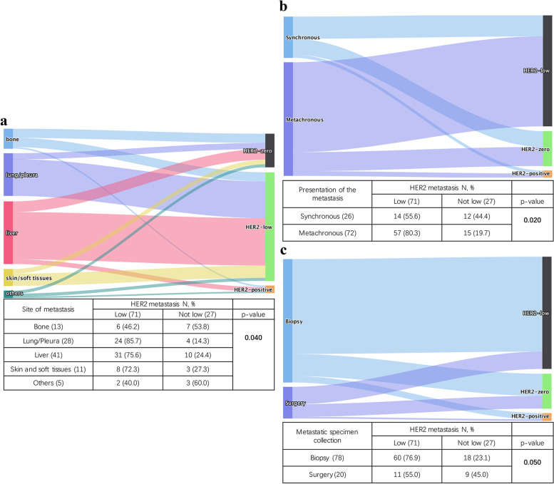
In our cohort, distant metastatic sites included lung/pleura (28.6%, n = 28), liver (41.8%, n = 41), bone (13.3%, n = 13), skin and soft tissue (11.2%, n = 11), and others (5.1%, n = 5). As shown in Fig. 5a, the HER2 discordant rate notably differed among sites of metastases (p = 0.040). In detail, higher rates were observed in other sites (60.0%) and bone (53.8%), and lower rates were observed in skin/soft tissue (27.3%), liver (24.4%) and lung/pleura (14.3%). For the 5 cases of other sites of metastases, two metastasized to rectum and posterior peritoneum separately, and remained HER2-low in metastases. Two cases metastasized to ovary and uterine appendages, and became HER2-0 in metastases. The last one metastasized to parietal lobe and became HER2-3+ in metastases. And there are 5 cases in our cohorts which had double metastatic sites, as shown in Table 2. For metastases with discordant HER2 status, the data of the first metastatic site was selected for analysis.
Fig. 5.
Effect of metastatic tumor sample on HER2 status evolution. The diagram shows the evolution of the HER2 status between primary tumors and metastases stratified by site of metastasis (a) (Fisher’s exact test), presentation of metastases (b) (Chi-square test) and metastatic specimen type (c) (Chi-square test). N, number
Table 2.
Cases with double metastatic sites
| Case number | Interval time of metastases | Metastatic sites | HER2 status of metastases |
|---|---|---|---|
| 1 | 0 months | Soft tissue | 0 |
| Nasopharynx | |||
| 2 | 24 months | Lung | 0 |
| Bone | 1+ | ||
| 3 | 36 months | Lung | 1+ |
| Liver | |||
| 4 | 20 months | Bone | 1+ |
| Liver | 3+ | ||
| 5 | 24 months | Soft tissue | 2+/FISH- |
| Liver |
HER2 discordance rate was also influenced by the presentation of the metastasis as synchronous (up to 6 months following the primary diagnosis) or metachronous (p = 0.020). Synchronous metastases had higher HER2 discordance rate (Fig. 5b). Moreover, HER2 discordance rate also differed according to type of metastatic BC sample (Fig. 5c). In detail, biopsy included core needle biopsy, endoscopic biopsy and incisional biopsy, and the discordance rate was 23.1% when metastatic BC was assessed on biopsies with being 45.0% on surgical samples, instead (p = 0.050).
Evolution of HER2 IHC score from primary sites to metastases
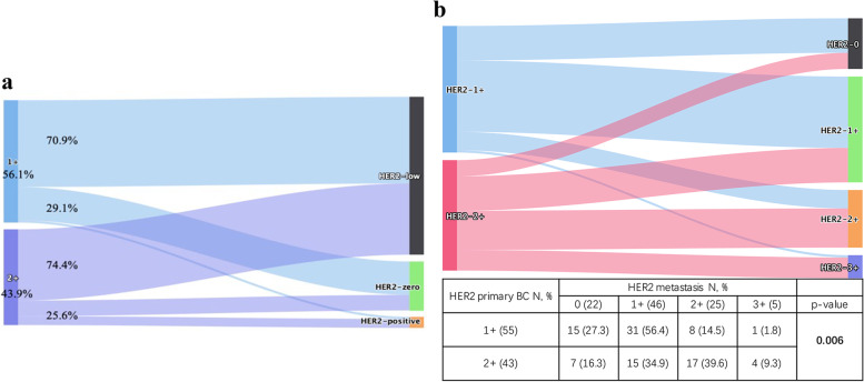
Between HER2-low primary BC with different HER2 IHC score, the discordance rate of metastases had no significant difference. In particular, discordant rate of HER2 status was 29.1% (n = 16/55) and 25.6% (n = 11/43) when primary BC was HER2-1+ and HER2-2+, respectively (p = 0.700) (Fig. 6a). When only comparing IHC score of HER2 in metastases, however, the rate of HER2 discordance was significantly lower in HER2-1+ primary BC cohorts (p = 0.006) (Fig. 6b). Among HER2-1+ cases, 56.4% (n = 31/55) stayed consistent in metastases, while 27.3% (n = 15/55) converted to HER2-0 with 14.5% (n = 8/55) and 1.8% (n = 1/55) switching to HER2-2+ and HER2-3+, respectively. Among HER2-2+ cases, only 39.6% (n = 17/43) had concordant HER2 status in metastases, while 16.3% (n = 7/43) and 34.9% (n = 15/43) converted to HER2-0 and HER2-1+ separately, and the percentage of patients switching to HER2-3+ was 9.3% (Fig. 6b).
Fig. 6.
The evolution of the HER2 IHC score from primary breast cancer to metastases. a Evolution of HER2 expression according to primary tumor HER2 IHC score. b Evolution of the HER2 IHC score from primary breast cancer to metastases (Fisher’s exact test)
Survival analysis
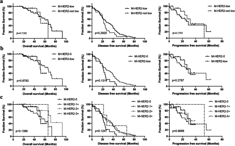
We compared OS, DFS and PFS separately according to HER2 status in metastatic sites. We didn’t observe significant difference among metastases with different HER2 status (Fig. 7a). Median OS was 67 months for HER2-low metastatic tumors compared with 52 months for HER2-not-low metastatic tumors (p = 0.718), median DFS was 28 months for HER2-low metastatic tumors compared with 22 months for HER2-not-low metastatic tumors (p = 0.292), and median PFS was 29 months for HER2-low metastatic tumors compared with 32 months for HER2-not-low metastatic tumors (p = 0.1765). The results didn’t change much when excluding cases that metastases of which changed to HER2-3+ (Fig. 7b). We also compared OS,DFS and PFS between metastases with different HER2 IHC score, and no significant difference was found, either (Fig. 7c).
Fig. 7.
Kaplan–Meier survival curve for overall survival, disease free survival and progression free survival. a Disease outcomes according to HER2 status in metastases. b Disease outcomes were compared between HER2-0 and HER2-low metastatic samples. c Disease outcomes were compared among different HER2 IHC scores in metastases
Discussion
Due to the new ADCs, HER2-low breast cancer is getting much more attention and becoming a new entity, which elevated the clinical complexity of BC. HER2 status changes between primary and metastatic breast cancer have been reported in some studies with inconsistent results (see Additional file 1). Different from previous studies about HER2-negative BCs [18, 19], our analysis investigated the changes of HER2 expression in patients with HER2-low BC and their distant metastases.
In our cohort, we included HER2-low BC patients with distant metastases, and the proportion of patients with HER2-1+ primary BC was higher than that of HER2-2+, which was consistent with reported evidence [20, 21]. Also, we found a significantly higher percentage of HR+ BC (78.6%) than that of TNBC, which was similar with previous studies which reported that proportion of HR+ cases was higher than TNBC among HER2-low breast tumors [21–23].
Mainly, we wanted to examine how HER2-low status evolved from primary sites to distant metastases, and the overall HER2 discordant rate was 27.6%. In particular, 22.4% of HER2-low primary tumor switched to HER2-0 in metastases, which was consistent with a previous study that 22% of HER2-low primary tumor switched to HER2-0 in the advanced stage [19]. Interestingly, we found an association between T stages of primary tumors and altered HER2 expression in metastases. In particular, the higher the T stage of the primary BC, the higher the rate of HER2 discordance. And the HER2 discordant rate was higher in surgical specimen than biopsy for the specimen type of metastasis. These may be explained – at least in part – by the intratumor heterogeneity of HER2. Previous studies have already proved that both HER2 expression and HER2 gene amplification have intratumor heterogeneity [24, 25], and in HER2-low BCs, the degree of intratumor heterogeneity is obviously higher [26]. For tumor with larger size, there may be higher frequency of coexistence of multiple tumor-cell subpopulations with distinct HER2 status, and this may influence the discordance between primary tumor and its metastases. And although, currently core-needle-biopsy is believed to be acceptable to get enough samples for HER2 expression assessment [27], our findings suggest that it may be necessary for surgical resection or at least multi-point biopsy of metastases in clinical practice to obtain accurate HER2 information. Similarly, higher intratumor heterogeneity in primary tumor, which has been reported being associated with breast cancer progression and worse prognosis [26, 28], may also lead to shorter metastatic intervals and resulted in higher discordance rate in synchronous metastases.
It’s worth to be noted that our cohort showed the rate of HER2 evolution varied between different metastatic sites, and the highest discordance rate was shown in bone metastases while lung metastases shown the lowest HER2 discordant rate. Although our analysis focused on HER2-low primary tumor and the matched metastases, these results were similar with previous study that included HER2-0 and HER2-low primary tumor and the matched recurrent tumor [18]. However, controversial results were also reported [19]. And the possible role of the decalcification process on the results may be doubted. Firstly, we checked the records of all bone metastases, and all samples were not decalcified. And there also has been reported that there is a significant correlation between ERBB2 mRNA and HER2 protein levels in bone metastases [29]. So, we tend to believe that the data on bone metastases is reliable. And one possible explanation for the higher discordance rate in bone metastases is that the biopsies of bone metastases were more likely to yield insufficient tissue for examination than other metastatic sites [30]. And relatively higher discordance rate in bone metastases were also reported in similar studies. A study about HER2-negative BC [18] and a review including HER2-negative and HER2-positive BC [31] both reported bone metastases had higher discordance rate.
Another worth-noting observation is that although it was reported that there were molecular biological differences between HR+ and HR- in people with low HER2 expression [21], the HER2 discordance rate didn’t differ between HR-positive and triple-negative breast cancers in our cohorts, which was inconsistent with the previous study [18]. And we didn’t find any difference of HER2 discordance between HER2-1+ and HER2-2+ patients, but we observed that HER2-2+ patients were less likely to have consistent metastatic HER2 levels than HER2-1+ patients when comparing HER2 IHC scores. This may indicate that more attention is needed for HER2 status in metastases when primary tumor being HER2-2+. And to investigate possible survival differences, we also performed survival analysis. Either OS, DFS or PFS was observed no difference among metastases with different HER2 status, which suggested that there was no prognostic value for HER2 discordance in metastases of HER2-low BC.
Our work has several strengths. Firstly, we rescored all of the HER2 IHC slides according to the latest standard of HER2 diagnosis by ASCO/CAP Guidelines Update [27]. On the one hand, the cut-offs for HER2-zero were downgraded since October 2013 according to the ASCO/CAP HER2 testing guideline [32], while we have cases before 2013 in our cohort. On the other hand, before the concept of HER2-low was proposed, the emphasis was on the distinction between HER2-negative and positive for clinical practice, and there could be errors in the diagnosis of HER2-0 and HER2-1+. So, we think it is necessary to re-evaluate the HER2 IHC score of all cases. Due to limited conditions, we didn’t choose old archival paraffin blocks or stored unstained paraffin slides for re-staining, which may also have the problem of antigenicity loss [33–35]. And considering the fact that positive and negative controls were set in all cases and HER2 IHC scores decreased in only 8 cases after reevaluation, we believe that the fading problem of DAB staining does not affect the reliability of rescored results. In addition, HER2 expression of all the cases was tested by Ventana 4B5 antibody on Ventana BenchMark autostainer using the same automatic staining protocol, with barcode generated by the autostainer on every slide (see Additional files 2 and 3), which ruled out the possible influence of different antibodies on HER2 discordance [36]. However, some limitations need to be emphasized as well. Firstly, patients included in the analysis received heterogenous systemic treatments, which might influence the HER2 status [37] and impair definitive conclusions on survival outcomes. Moreover, we only included patients with a historical primary score of HER2-low expression. This resulted in limited number of our cases, which may lead to some bias in our comparisons. And more HER2-low BCs may be found if we rescored patients with a historical primary score of HER2-0, considering the poor accuracy of HER2 IHC score in 0 and 1+ [38], which might expand our study cohort.
Conclusions
For primary BCs with HER2-low status, there is a possibility of HER2 status alteration in the metastases. The rate of altered HER2-low expression was different among different metastatic sites, and the discordant rate of bone metastasis was the highest. The discordant rates of HER2 were also different among different T stages of primary BC and different specimen type of metastasis. No difference of HER2 discordance rate was found between HER2-1+ and HER2-2+ patients. No prognostic significance was observed. These data further support development of best practices for identifying patients with HER2-low expression who could benefit from anti-HER2 ADCs.
Supplementary Information
Additional file 1. Literature on HER2 (all scores) changes between primary tumors and metastases [39–47].
Additional file 2. Laboratory accreditation certificate by China National Accreditation Service for Conformity Assessment (CNAS).
Additional file 3. Assessment of HER2 IHC by Nordic Immunohistochemical Quality Control (NordiQC).
Acknowledgements
Not applicable.
Abbreviations
- BC
Breast cancer
- HER2
Human epidermal growth factor receptor 2
- IHC
Immunohistochemistry
- ISH
In situ hybridization
- ER
Estrogen receptor
- PR
Progesterone receptor
- ADCs
Antibody-drug conjugates
- ORR
Objective response rate
- HR
Hormone receptor
- TNBC
Triple negative breast cancer
- PFS
Progression free survival
- OS
Overall survival
- FISH
Fluorescence in situ hybridization
- HE
Haematoxylin & eosin
Authors’ contributions
MC collected the samples, analyzed the data and prepared the draft of this manuscript. HL and ML helped to collect the samples. SZ, HL and WY independently helped to rescore the HER2 IHC slides. ML, RS and XX supervised the research. WY conceived the idea, supervised the research and revised this manuscript. All authors have read and approved the final version of this manuscript.
Funding
The authors have not declared a specific grant for this research from any funding agency in the public, commercial or not-for-profit sectors.
Availability of data and materials
All data generated or analysed during this study are included in this published article.
Declarations
Ethics approval and consent to participate
This study was approved by the institutional review board of Fudan University Shanghai Cancer Center. Written informed consent was obtained from all participants at the time of admission. All methods were carried out in accordance with relevant guidelines and regulations or Declaration of Helsinki.
Consent for publication
Not applicable.
Competing interests
The authors declare no competing interests.
Footnotes
Publisher’s Note
Springer Nature remains neutral with regard to jurisdictional claims in published maps and institutional affiliations.
References
- 1.Sung H, Ferlay J, Siegel RL, Laversanne M, Soerjomataram I, Jemal A, et al. Global cancer statistics 2020: GLOBOCAN estimates of incidence and mortality worldwide for 36 cancers in 185 countries. CA Cancer J Clin. 2021;71(3):209–249. doi: 10.3322/caac.21660. [DOI] [PubMed] [Google Scholar]
- 2.Wolff AC, Hammond MEH, Allison KH, Harvey BE, Mangu PB, Bartlett JMS, et al. Human epidermal growth factor receptor 2 testing in breast cancer: American Society of Clinical Oncology/College of American Pathologists clinical practice guideline focused update. J Clin Oncol. 2018;36(20):2105–2122. doi: 10.1200/JCO.2018.77.8738. [DOI] [PubMed] [Google Scholar]
- 3.Tarantino P, Hamilton E, Tolaney SM, Cortes J, Morganti S, Ferraro E, et al. HER2-low breast cancer: pathological and clinical landscape. J Clin Oncol. 2020;38(17):1951–1962. doi: 10.1200/JCO.19.02488. [DOI] [PubMed] [Google Scholar]
- 4.Piccart M, Procter M, Fumagalli D, de Azambuja E, Clark E, Ewer MS, et al. Adjuvant pertuzumab and trastuzumab in early HER2-positive breast cancer in the APHINITY trial: 6 years’ follow-up. J Clin Oncol. 2021;39(13):1448–1457. doi: 10.1200/JCO.20.01204. [DOI] [PubMed] [Google Scholar]
- 5.Swain SM, Miles D, Kim SB, Im YH, Im SA, Semiglazov V, et al. Pertuzumab, trastuzumab, and docetaxel for HER2-positive metastatic breast cancer (CLEOPATRA): end-of-study results from a double-blind, randomised, placebo-controlled, phase 3 study. Lancet Oncol. 2020;21(4):519–530. doi: 10.1016/S1470-2045(19)30863-0. [DOI] [PubMed] [Google Scholar]
- 6.Gianni L, Lladó A, Bianchi G, Cortes J, Kellokumpu-Lehtinen PL, Cameron DA, et al. Open-label, phase II, multicenter, randomized study of the efficacy and safety of two dose levels of Pertuzumab, a human epidermal growth factor receptor 2 dimerization inhibitor, in patients with human epidermal growth factor receptor 2-negative metastatic breast cancer. J Clin Oncol. 2010;28(7):1131–1137. doi: 10.1200/JCO.2009.24.1661. [DOI] [PMC free article] [PubMed] [Google Scholar]
- 7.Fehrenbacher L, Cecchini RS, Geyer CE, Jr, Rastogi P, Costantino JP, Atkins JN, et al. NSABP B-47/NRG oncology phase III randomized trial comparing adjuvant chemotherapy with or without trastuzumab in high-risk invasive breast cancer negative for HER2 by FISH and with IHC 1+ or 2. J Clin Oncol. 2020;38(5):444–453. doi: 10.1200/JCO.19.01455. [DOI] [PMC free article] [PubMed] [Google Scholar]
- 8.Burris HA, 3rd, Rugo HS, Vukelja SJ, Vogel CL, Borson RA, Limentani S, et al. Phase II study of the antibody drug conjugate trastuzumab-DM1 for the treatment of human epidermal growth factor receptor 2 (HER2)-positive breast cancer after prior HER2-directed therapy. J Clin Oncol. 2011;29(4):398–405. doi: 10.1200/JCO.2010.29.5865. [DOI] [PubMed] [Google Scholar]
- 9.Modi S, Park H, Murthy RK, Iwata H, Tamura K, Tsurutani J, et al. Antitumor activity and safety of trastuzumab deruxtecan in patients with HER2-low-expressing advanced breast cancer: results from a phase Ib study. J Clin Oncol. 2020;38(17):1887–1896. doi: 10.1200/JCO.19.02318. [DOI] [PMC free article] [PubMed] [Google Scholar]
- 10.Banerji U, van Herpen CML, Saura C, Thistlethwaite F, Lord S, Moreno V, et al. Trastuzumab duocarmazine in locally advanced and metastatic solid tumours and HER2-expressing breast cancer: a phase 1 dose-escalation and dose-expansion study. Lancet Oncol. 2019;20(8):1124–1135. doi: 10.1016/S1470-2045(19)30328-6. [DOI] [PubMed] [Google Scholar]
- 11.Modi S, Jacot W, Yamashita T, Sohn J, Vidal M, Tokunaga E, et al. Trastuzumab deruxtecan in previously treated HER2-low advanced breast cancer. N Engl J Med. 2022;387(1):9–20. doi: 10.1056/NEJMoa2203690. [DOI] [PMC free article] [PubMed] [Google Scholar]
- 12.Lal P, Salazar PA, Hudis CA, Ladanyi M, Chen B. HER-2 testing in breast cancer using immunohistochemical analysis and fluorescence in situ hybridization: a single-institution experience of 2,279 cases and comparison of dual-color and single-color scoring. Am J Clin Pathol. 2004;121(5):631–636. doi: 10.1309/VE7862V2646BR6EX. [DOI] [PubMed] [Google Scholar]
- 13.Schalper KA, Kumar S, Hui P, Rimm DL, Gershkovich P. A retrospective population-based comparison of HER2 immunohistochemistry and fluorescence in situ hybridization in breast carcinomas: impact of 2007 American Society of Clinical Oncology/College of American Pathologists criteria. Arch Pathol Lab Med. 2014;138(2):213–219. doi: 10.5858/arpa.2012-0617-OA. [DOI] [PubMed] [Google Scholar]
- 14.Shui R, Liang X, Li X, Liu Y, Li H, Xu E, et al. Hormone receptor and human epidermal growth factor receptor 2 detection in invasive breast carcinoma: a retrospective study of 12,467 patients from 19 Chinese representative clinical centers. Clin Breast Cancer. 2020;20(1):e65–e74. doi: 10.1016/j.clbc.2019.07.013. [DOI] [PubMed] [Google Scholar]
- 15.Dieci MV, Barbieri E, Piacentini F, Ficarra G, Bettelli S, Dominici M, et al. Discordance in receptor status between primary and recurrent breast cancer has a prognostic impact: a single-institution analysis. Ann Oncol. 2013;24(1):101–108. doi: 10.1093/annonc/mds248. [DOI] [PubMed] [Google Scholar]
- 16.Grinda T, Joyon N, Lusque A, Lefèvre S, Arnould L, Penault-Llorca F, et al. Phenotypic discordance between primary and metastatic breast cancer in the large-scale real-life multicenter French ESME cohort. NPJ Breast Cancer. 2021;7(1):41. doi: 10.1038/s41523-021-00252-6. [DOI] [PMC free article] [PubMed] [Google Scholar]
- 17.Guarneri V, Giovannelli S, Ficarra G, Bettelli S, Maiorana A, Piacentini F, et al. Comparison of HER-2 and hormone receptor expression in primary breast cancers and asynchronous paired metastases: impact on patient management. Oncologist. 2008;13(8):838–844. doi: 10.1634/theoncologist.2008-0048. [DOI] [PubMed] [Google Scholar]
- 18.Miglietta F, Griguolo G, Bottosso M, Giarratano T, Lo Mele M, Fassan M, et al. Evolution of HER2-low expression from primary to recurrent breast cancer. NPJ Breast Cancer. 2021;7(1):137. doi: 10.1038/s41523-021-00343-4. [DOI] [PMC free article] [PubMed] [Google Scholar]
- 19.Tarantino P, Gandini S, Nicolò E, Trillo P, Giugliano F, Zagami P, et al. Evolution of low HER2 expression between early and advanced-stage breast cancer. Eur J Cancer. 2022;163:35–43. doi: 10.1016/j.ejca.2021.12.022. [DOI] [PubMed] [Google Scholar]
- 20.Xin L, Wu Q, Zhan C, Qin H, Xiang H, Xu L, et al. Multicenter study of the clinicopathological features and recurrence risk prediction model of early-stage breast cancer with low-positive human epidermal growth factor receptor 2 expression in China (Chinese Society of Breast Surgery 021) Chin Med J (Engl) 2022;135(6):697–706. doi: 10.1097/CM9.0000000000002056. [DOI] [PMC free article] [PubMed] [Google Scholar]
- 21.Schettini F, Chic N, Brasó-Maristany F, Paré L, Pascual T, Conte B, et al. Clinical, pathological, and PAM50 gene expression features of HER2-low breast cancer. NPJ Breast Cancer. 2021;7(1):1. doi: 10.1038/s41523-020-00208-2. [DOI] [PMC free article] [PubMed] [Google Scholar]
- 22.Ballard M, Jalikis F, Krings G, Schmidt RA, Chen YY, Rendi MH, et al. ‘Non-classical’ HER2 FISH results in breast cancer: a multi-institutional study. Mod Pathol. 2017;30(2):227–235. doi: 10.1038/modpathol.2016.175. [DOI] [PubMed] [Google Scholar]
- 23.Sapino A, Maletta F, Verdun di Cantogno L, Macrì L, Botta C, Gugliotta P, et al. Gene status in HER2 equivocal breast carcinomas: impact of distinct recommendations and contribution of a polymerase chain reaction-based method. Oncologist. 2014;19(11):1118–26. doi: 10.1634/theoncologist.2014-0195. [DOI] [PMC free article] [PubMed] [Google Scholar]
- 24.Brunelli M, Manfrin E, Martignoni G, Miller K, Remo A, Reghellin D, et al. Genotypic intratumoral heterogeneity in breast carcinoma with HER2/neu amplification: evaluation according to ASCO/CAP criteria. Am J Clin Pathol. 2009;131(5):678–682. doi: 10.1309/AJCP09VUTZWZXBMJ. [DOI] [PubMed] [Google Scholar]
- 25.Vance GH, Barry TS, Bloom KJ, Fitzgibbons PL, Hicks DG, Jenkins RB, et al. Genetic heterogeneity in HER2 testing in breast cancer: panel summary and guidelines. Arch Pathol Lab Med. 2009;133(4):611–612. doi: 10.5858/133.4.611. [DOI] [PubMed] [Google Scholar]
- 26.Seol H, Lee HJ, Choi Y, Lee HE, Kim YJ, Kim JH, et al. Intratumoral heterogeneity of HER2 gene amplification in breast cancer: its clinicopathological significance. Mod Pathol. 2012;25(7):938–948. doi: 10.1038/modpathol.2012.36. [DOI] [PubMed] [Google Scholar]
- 27.Wolff AC, Hammond MEH, Allison KH, Harvey BE, McShane LM, Dowsett M. HER2 testing in breast cancer: American Society of Clinical Oncology/College of American Pathologists clinical practice guideline focused update summary. J Oncol Pract. 2018;14(7):437–441. doi: 10.1200/JOP.18.00206. [DOI] [PubMed] [Google Scholar]
- 28.Yang F, Wang Y, Li Q, Cao L, Sun Z, Jin J, et al. Intratumor heterogeneity predicts metastasis of triple-negative breast cancer. Carcinogenesis. 2017;38(9):900–909. doi: 10.1093/carcin/bgx071. [DOI] [PubMed] [Google Scholar]
- 29.Brasó-Maristany F, Paré L, Chic N, Martínez-Sáez O, Pascual T, Mallafré-Larrosa M, et al. Gene expression profiles of breast cancer metastasis according to organ site. Mol Oncol. 2022;16(1):69–87. doi: 10.1002/1878-0261.13021. [DOI] [PMC free article] [PubMed] [Google Scholar]
- 30.Amir E, Miller N, Geddie W, Freedman O, Kassam F, Simmons C, et al. Prospective study evaluating the impact of tissue confirmation of metastatic disease in patients with breast cancer. J Clin Oncol. 2012;30(6):587–592. doi: 10.1200/JCO.2010.33.5232. [DOI] [PMC free article] [PubMed] [Google Scholar]
- 31.Yeung C, Hilton J, Clemons M, Mazzarello S, Hutton B, Haggar F, et al. Estrogen, progesterone, and HER2/neu receptor discordance between primary and metastatic breast tumours-a review. Cancer Metastasis Rev. 2016;35(3):427–437. doi: 10.1007/s10555-016-9631-3. [DOI] [PubMed] [Google Scholar]
- 32.Wolff AC, Hammond ME, Hicks DG, Dowsett M, McShane LM, Allison KH, et al. Recommendations for human epidermal growth factor receptor 2 testing in breast cancer: American Society of Clinical Oncology/College of American Pathologists clinical practice guideline update. J Clin Oncol. 2013;31(31):3997–4013. doi: 10.1200/JCO.2013.50.9984. [DOI] [PubMed] [Google Scholar]
- 33.Forse CL, Pinnaduwage D, Bull SB, Mulligan AM, Andrulis IL. Fresh cut versus stored cut paraffin-embedded tissue: effect on immunohistochemical staining for common breast cancer markers. Appl Immunohistochem Mol Morphol. 2019;27(3):231–237. doi: 10.1097/PAI.0000000000000579. [DOI] [PMC free article] [PubMed] [Google Scholar]
- 34.Grillo F, Bruzzone M, Pigozzi S, Prosapio S, Migliora P, Fiocca R, et al. Immunohistochemistry on old archival paraffin blocks: is there an expiry date? J Clin Pathol. 2017;70(11):988–993. doi: 10.1136/jclinpath-2017-204387. [DOI] [PubMed] [Google Scholar]
- 35.Grillo F, Pigozzi S, Ceriolo P, Calamaro P, Fiocca R, Mastracci L. Factors affecting immunoreactivity in long-term storage of formalin-fixed paraffin-embedded tissue sections. Histochem Cell Biol. 2015;144(1):93–99. doi: 10.1007/s00418-015-1316-4. [DOI] [PubMed] [Google Scholar]
- 36.Rüschoff J, Friedrich M, Nagelmeier I, Kirchner M, Andresen LM, Salomon K, et al. Comparison of HercepTest™ mAb pharmDx (Dako Omnis, GE001) with Ventana PATHWAY anti-HER-2/neu (4B5) in breast cancer: correlation with HER2 amplification and HER2 low status. Virchows Arch. 2022;481(5):685–694. doi: 10.1007/s00428-022-03378-5. [DOI] [PMC free article] [PubMed] [Google Scholar]
- 37.Kan S, Koido S, Okamoto M, Hayashi K, Ito M, Kamata Y, et al. Gemcitabine treatment enhances HER2 expression in low HER2-expressing breast cancer cells and enhances the antitumor effects of trastuzumab emtansine. Oncol Rep. 2015;34(1):504–510. doi: 10.3892/or.2015.3974. [DOI] [PubMed] [Google Scholar]
- 38.Fernandez AI, Liu M, Bellizzi A, Brock J, Fadare O, Hanley K, et al. Examination of low ERBB2 protein expression in breast cancer tissue. JAMA Oncol. 2022;8(4):1–4. doi: 10.1001/jamaoncol.2021.7239. [DOI] [PMC free article] [PubMed] [Google Scholar]
- 39.Bergeron A, Bertaut A, Beltjens F, Charon-Barra C, Amet A, Jankowski C, et al. Anticipating changes in the HER2 status of breast tumours with disease progression-towards better treatment decisions in the new era of HER2-low breast cancers. Br J Cancer. 2023;129(1):122–34. [DOI] [PMC free article] [PubMed]
- 40.Almstedt K, Krauthauser L, Kappenberg F, Wagner DC, Heimes AS, Battista MJ, et al. Discordance of HER2-low between primary tumors and matched distant metastases in breast cancer. Cancers (Basel) 2023;15(5):1413. doi: 10.3390/cancers15051413. [DOI] [PMC free article] [PubMed] [Google Scholar]
- 41.Satishkumar M, Ramesh M, Sanjive JG. A prospective multi-institutional observational study on ER, PR, HER2/NEU, and Ki-67 expression and discordance pattern in primary and metastatic and recurrent carcinoma breast and its therapeutic implications and prognostic outcome. Indian J Surg Oncol. 2023;14(1):72–80. doi: 10.1007/s13193-022-01622-7. [DOI] [PMC free article] [PubMed] [Google Scholar]
- 42.Shi Q, Yu J, Liu D, Ren F, Wu J, Shen K. Distribution, dynamic evolution, and clinical outcomes of patients with advanced breast cancer according to HER2 expression. BMC Cancer. 2023;23(1):173. doi: 10.1186/s12885-023-10634-7. [DOI] [PMC free article] [PubMed] [Google Scholar]
- 43.Wang Q, Guo Z, Huang Z, Sun H, Zhu J, Shi J, et al. Receptor discordance among primary tumors, brain metastases and extra-brain metastases in patients with breast cancer. Future Oncol. 2023;18(37):4101–10. [DOI] [PubMed]
- 44.Mellouli M, Graja S, Kridis WB, Ayed HB, Makni S, Triki M, et al. Discordance in receptor status between primary and metastatic breast cancer and overall survival: a single-center analysis. Ann Diagn Pathol. 2022;61:152044. doi: 10.1016/j.anndiagpath.2022.152044. [DOI] [PubMed] [Google Scholar]
- 45.Pizzuti L, Barba M, Mazzotta M, Krasniqi E, Maugeri-Saccà M, Gamucci T, et al. The prognostic relevance of HER2-positivity gain in metastatic breast cancer in the ChangeHER trial. Sci Rep. 2021;11(1):13770. doi: 10.1038/s41598-021-92774-z. [DOI] [PMC free article] [PubMed] [Google Scholar]
- 46.Chen R, Qarmali M, Siegal GP, Wei S. Receptor conversion in metastatic breast cancer: analysis of 390 cases from a single institution. Mod Pathol. 2020;33(12):2499–2506. doi: 10.1038/s41379-020-0615-z. [DOI] [PubMed] [Google Scholar]
- 47.Schrijver W, Suijkerbuijk KPM, van Gils CH, van der Wall E, Moelans CB, van Diest PJ. Receptor conversion in distant breast cancer metastases: a systematic review and meta-analysis. J Natl Cancer Inst. 2018;110(6):568–580. doi: 10.1093/jnci/djx273. [DOI] [PubMed] [Google Scholar]
Associated Data
This section collects any data citations, data availability statements, or supplementary materials included in this article.
Supplementary Materials
Additional file 1. Literature on HER2 (all scores) changes between primary tumors and metastases [39–47].
Additional file 2. Laboratory accreditation certificate by China National Accreditation Service for Conformity Assessment (CNAS).
Additional file 3. Assessment of HER2 IHC by Nordic Immunohistochemical Quality Control (NordiQC).
Data Availability Statement
All data generated or analysed during this study are included in this published article.


