Abstract
Microglia and brain-derived neurotrophic factor (BDNF) are essential for the neuroplasticity that characterizes critical developmental periods. The experience-dependent development of social behaviors—associated with the medial prefrontal cortex (mPFC)—has a critical period during the juvenile period in mice. However, whether microglia and BDNF affect social development remains unclear. Herein, we aimed to elucidate the effects of microglia-derived BDNF on social behaviors and mPFC development. Mice that underwent social isolation during p21–p35 had increased Bdnf in the microglia accompanied by reduced adulthood sociability. Additionally, transgenic mice overexpressing microglia Bdnf—regulated using doxycycline at different time points—underwent behavioral, electrophysiological, and gene expression analyses. In these mice, long-term overexpression of microglia BDNF impaired sociability and excessive mPFC inhibitory neuronal circuit activity. However, administration of doxycycline to normalize BDNF from p21 normalized sociability and electrophysiological functions; this was not observed when BDNF was normalized from a later age (p45–p50). To evaluate the possible role of BDNF in human sociability, we analyzed the relationship between adverse childhood experiences and BDNF expression in human macrophages, a possible substitute for microglia. Results show that adverse childhood experiences positively correlated with BDNF expression in M2 but not M1 macrophages. Thus, microglia BDNF might regulate sociability and mPFC maturation in mice during the juvenile period. Furthermore, childhood experiences in humans may be related to BDNF secretion from macrophages.
Introduction
Microglia refine synapses and form developing brain circuits in an activity-dependent manner [1, 2]. This process is essential for normal brain development [3]. A limited critical postnatal period of development exists in the sensory cortexes of mice, during which neural network plasticity is transiently enhanced, and the experience-dependent flexible reorganization of neural circuits occurs [4, 5]. Microglia are involved in activity-dependent brain formation processes during brain development [1–3, 6, 7]. Notably, recent studies suggest that microglia may be actively involved in the critical-period plasticity [8–10].
Furthermore, similar to sensory functions, social behaviors develop via experience-dependent brain maturation during a limited postnatal window [11, 12]. For instance, mice that undergo social isolation 21–35 days postnatal (p21–35) later display social impairment that is irreversible by re-socialization [12]. Juvenile social deprivation harms the neural circuits and glial cells of the medial prefrontal cortex (mPFC) [11–18], which is a control center of social behaviors [19]. Previously, we reported the involvement of microglia in juvenile social isolation and future social impairment [13]. Meanwhile, others have implicated microglia in social development [20–22]. Additionally, microglia play a role in the synaptic pruning of the prefrontal cortex [23] and in the time-specific functional maturation of mPFC in rodents [24]. These findings suggest that microglia are involved in mPFC maturation and its associated sociability function in a time-specific manner via activity-dependent neuroplasticity and pruning.
Brain-derived neurotrophic factor (BDNF) contributes to maturing inhibitory interneurons and closing critical periods [25]. Microglia secrete BDNF [26–28], which is relevant for learning-dependent synaptic plasticity in the motor cortex [29] and its effect on inhibitory synaptic transmission in the spinal cord [26]. However, it remains unclear whether microglia-secreted BDNF (MG-BDNF) influences the cortical critical-period plasticity and behavior [8, 30]. In this study, we investigate the time-specific effects of microglia on mPFC biology and social behavior, focusing on MG-BDNF.
Materials and Methods
Animals
All study protocols were approved by the Animal Care Committee of Nara Medical University in accordance with the policies established in the National Institutes of Health Guide for the Care and Use of Laboratory Animals. The animals were housed in a temperature- and humidity-controlled animal facility under a standard 12-h light-dark cycle (lights on 08:00–20:00) with ad libitum access to food and water throughout the study. C57BL/6J mice isolated from the weaning age (p21) for two weeks were regrouped with age-, sex-, and strain-matched mice at p35 (3–5 mice per cage), labeled juvenile social isolation (j-SI) mice. Control C57BL6/J mice were group-housed following weaning at p21 and labeled Group-Housed (GH) mice. We crossed ionized calcium-binding adapter molecule 1(Iba1) promoter driving tetracycline transactivator (Iba1-tTA) transgenic mice (line 75) [31] with tetracycline operator-Bdnf knock-in homozygous (BdnftetO/tetO) mice [32, 33] and obtained microglia-specific Bdnf-overexpressing mice (Iba1-tTA::BdnftetO/+ mice) and control mice (BdnftetO/+ mice). Both Iba1-tTA and BdnftetO/tetO mice strains had a mixed C57BL/6J and 129/SvEv background; only F1 male mice were used for all experiments [34]. Genotyping was confirmed as in previous studies [31, 32]. To temporally control Bdnf overexpression, F1 mice were fed doxycycline (DOX)-containing chow (DOX 100 mg/kg, CLEA Japan Inc., Tokyo, Japan): from weaning (p21) or adulthood (p45–p50). Groups of mice administered DOX from adulthood were provided chow with DOX ad libitum for at least two weeks before the experiment.
Statistical analysis
All statistical analyses were performed using Prism v.9 (GraphPad Software Inc., San Diego, CA, USA). When the sample data were normally distributed and had equal variances, as determined using the Shapiro-Wilk test and the subsequent F-test, significant differences between groups were assessed using an unpaired two-tailed Student’s t-test. Even if the sample data passed the Shapiro-Wilk test for normality, the nonparametric Mann–Whitney U-test was used if the F-test indicated unequal variances. When the sample data significantly differed from normal distribution as assessed by the Shapiro-Wilk test, the nonparametric Mann–Whitney U-test was used. Values with right-skewed distribution were log-transformed for statistical analyses. The Kruskal-Wallis test was used to validate microglia-specific BDNF-overexpressing levels. Two-way ANOVA followed by a Bonferroni post hoc test analyzed the time spent in each zone in the three-chamber social preference test [35]. Two-way repeated measures ANOVA was used to assess the cumulative number of approaches in the augmented reality-based long-term animal behavior observing system. Spearman’s rank correlation coefficient was used to evaluate correlations between Bdnf expression in peripheral blood mononuclear cells (PBMC) and microglia in mice, as well as between child abuse and trauma scale (CATS) scores and BDNF in macrophages in humans. The false discovery rate-controlling Benjamini–Hochberg procedure was used to control for multiple comparisons [36]. All data were presented as the mean ± standard error of the mean (SEM); pvalues < 0.05 were considered statistically signi cant. The q-values were determined for multiple comparisons, and values < q-value were significant.
Detailed methodological information is provided in the Supplementary materials and methods.
Results
Juvenile social isolation (j-SI) mice are socially impaired and have increased MG- Bdnf
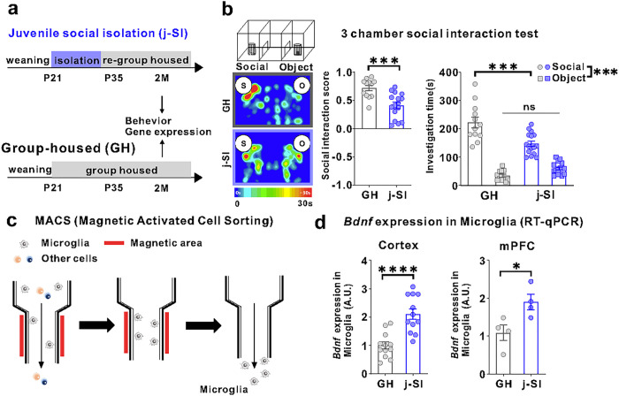
Our single housing condition started at P21 and lasted for two weeks, followed by re-grouped housing with age-, sex-, and strain-matched mice (Fig. 1a). While single housing conditions induce varying alterations in the brain and behavioral consequences in mice, depending on the strain, sex, and age [37, 38], our procedure has been shown to induce agitation and defective social behaviors and alter the plasticity in the mPFC [12, 14, 15, 39, 40]. In this study, C57BL/6J male mice isolated during p21–p35 also exhibited impaired sociability in a three-chamber social preference test at two months of age (Fig. 1a, b). In the open field test, no differences in basal activity or anxiety existed between j-SI and GH mice (Supplementary Fig. 1a, b). Bdnf mRNA expression in microglia from the cerebral and prefrontal cortexes was higher in j-SI mice than GH mice (Fig. 1c, d). However, no differences were observed in Bdnf mRNA expression in the bulk tissues of the cerebral cortex and mPFC between the j-SI and GH mice (Supplementary Fig. 1a, c). These findings indicate that microglial Bdnf expression changes are specific to microglia [29]. Moreover, the lack of social stimulation during the juvenile period or subsequent re-socialization increases microglial Bdnf expression in the cerebral cortex, including the mPFC.
Figure 1. Juvenile social isolation reduces sociability and increases MG-Bdnf expression.
(a) Schema of the GH and juvenile social isolation (j-SI) mice time series. The experiments were started at P56. (b) Sociability j-SI mice displayed reduced sociability in the three-chamber sociability test compared with GH mice. j-SI mice had a lower social interaction score than GH mice (t(26) = 3.948, p =0.0005, unpaired two-tailed Student’s t-test, GH: n = 12, j-SI: n = 16) and spent less time near novel mice (F1,26 (interaction) = 17.16, p = 0.0003, two-way ANOVA followed by Bonferroni post hoc test, Bonferroni post hoc analysis of GH S vs. j-SI S, p < 0.0001 GH: n = 12, j-SI: n = 16). S, social; O, object. (c) Image of magnetic-activated cell sorting. (d) (Left) Bdnf expression measured using real-time quantitative polymerase chain reaction (RT-qPCR) in microglia recovered from the cortex was higher in the j-SI mice than in the GH mice (t(22) = 4.876, p < 0.0001, unpaired two-tailed Student’s t-test, GH: n = 12, j-SI: n = 12). (Right) Bdnf expression measured using RT-qPCR in microglia recovered from the mPFC was higher in j-SI than in GH mice (t(6) = 2.818, p=0.0304, unpaired two-tailed Student’s t-test, GH: n = 4 (from 16 mice), j-SI: n = 4 (from 16 mice)). The value was log-transformed with a base of 10 because of the right-skewed distribution. Each dot indicates the expression of Bdnf in microglia collected from the mPFC of four mice. *p < 0.05, ***p < 0.001, ****p < 0.0001. Data are presented as the mean ± SEM. 2M: two months of age, A.U.: arbitrary unit
Persistently overexpressed MG-BDNF causes social impairment and functional changes in the mPFC pyramidal cells
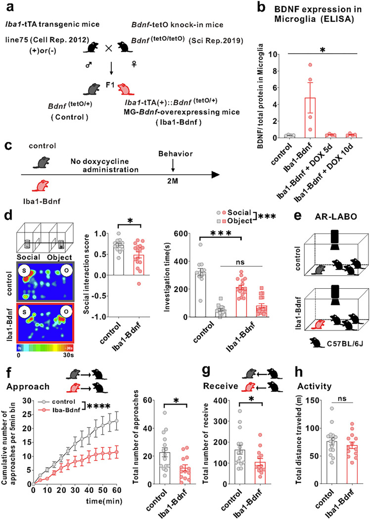
To recapitulate the microglia-specific and sustained Bdnf overexpression observed in j-SI and examine the causal relationship between microglia-specific Bdnf gene upregulation and social impairment, we exploited a tet-off system [41] to induce microglia-specific Bdnf mRNA expression (Fig. 2a). MG-BDNF overexpression was con rmed in Iba1-tTA::Bdnf(tetO/+) mice without DOX and MG-BDNF overexpression in Iba1-tTA::Bdnf(tetO/+) mice was normalized from day 5 after orally administering DOX (Fig. 2b). Bdnf mRNA expressions in the bulk tissues of the cerebral cortex and mPFC did not differ between the Iba1-tTA::Bdnf(tetO/+) and control mice, suggesting that genetically-induced MG-Bdnf overexpression in Iba1-tTA::Bdnf(tetO/+) mice does not significantly alter total Bdnf mRNA expression levels in the cerebral cortex or mPFC (Supplementary Fig. 2a, b).
Figure 2. Overexpression of MG-BDNF leads to impaired sociability in adulthood.
(a) Diagram of the generation of MG-Bdnf-overexpressing mice. The tet-off system was activated in double transgenic mice (F1), which were obtained by crossing Iba1-tTA transgenic mice with Bdnf-tetO knock-in mice. Iba1-tTA(+)::Bdnf(tetO/+) mice were the MG-Bdnf-overexpressing mice, and Bdnf(tetO/+) mice were the control mice. (b) BDNF expression in microglia recovered from the cortex, as measured using ELISA. Iba1-tTA(+)::Bdnf(tetO/+) mice had higher BDNF expression than Bdnf(tetO/+) mice; this was normalized from day 5 using doxycycline (The Kruskal-Wallis test, 9.086 , p= 0.0118; n = 4 per group). (c) Schema of the behavioral experiments without doxycycline. The experiments were started at P61. (d) The Iba1-tTA(+)::Bdnf(tetO/+) mice were less social in the three-chamber sociability test than the Bdnf(tetO/+) mice. The Iba1-tTA(+)::Bdnf(tetO/+) mice had a lower social interaction score than Bdnf(tetO/+) mice (U = 47, p = 0.0226, Mann–Whitney U test, Bdnf(tetO/+): n = 12, Iba1-tTA(+)::Bdnf(tetO/+): n = 16)(left) and spent less time near novel mice (F1,26(interaction) = 14.63, p = 0.0007, two-way ANOVA followed by Bonferroni post hoc test; Bonferroni post hoc analysis of Iba1-tTA(+)::Bdnf(tetO/+) S vs. Bdnf(tetO/+) S, p = 0.0001, Bdnf(tetO/+): n = 12, Iba1-tTA(+)::Bdnf(tetO/+): n = 16)(right). S, social; O, object (e) Schematic of the Augmented Reality-based Long-term Animal Behavior Observing system (AR-LABO). Three novel C57BL/6J mice were placed with a target mouse and observed for 1 hr. (f) (Left) The cumulative number of approaches per 5-min bin for Bdnf(tetO/+) and Iba1-tTA(+)::Bdnf(tetO/+) mice. Iba1-tTA(+)::Bdnf(tetO/+) mice accumulates significantly less cumulative approaches to other mice than Bdnf(tetO/+) mice (two-way RM ANOVA, phenotype (control or Iba1-BDNF) × time (5 min bin) interaction F(12,312) = 3.926, p <0.0001; effect of phenotype F(1,26) = 6.670, p = 0.0158; effect of time F(12,312) = 39.31, p <0.0001 , Bdnf(tetO/+): n =15, Iba1-tTA(+)::Bdnf(tetO/+): n = 13). (Right) Iba1-tTA(+)::Bdnf(tetO/+) mice approached other mice less than the Bdnf(tetO/+) mice (t(26) = 2.717, p = 0.0116, unpaired two-tailed Student’s t-test, Bdnf(tetO/+): n =15, Iba1-tTA(+)::Bdnf(tetO/+): n = 13). (g) The Iba1-tTA(+)::Bdnf(tetO/+) mice were less approached by other mice than Bdnf(tetO/+) mice (t(26) = 2.120, p = 0.0437, unpaired two-tailed Student’s t-test, Bdnf(tetO/+): n = 15, Iba1-tTA(+)::Bdnf(tetO/+): n = 13). (h) No differences in activity were observed during AR-LABO between the Iba1-tTA(+)::Bdnf(tetO/+) and Bdnf(tetO/+) mice (t(26) = 0.8059, p = 0.4276, unpaired two-tailed Student’s t-test, Bdnf(tetO/+): n = 15, Iba1-tTA(+)::Bdnf(tetO/+): n = 13). *p < 0.05, ***p < 0.001. Data are presented as the mean ± SEM. 2M: two months of age, control: Bdnf(tetO/+) mice, Iba1-BDNF: Iba1-tTA(+)::Bdnf(tetO/+) mice
Next, we evaluated whether persistent MG-BDNF overexpression impairs social behavior at two months of age such as j-SI mice using a three-chamber social preference test (Fig. 2c). The Iba1-tTA::Bdnf(tetO/+) mice had a lower social interaction score than the control mice and spent less time around the novel mice (Fig. 2d). In contrast, no differences in locomotor activity or anxiety existed between the Iba1-tTA::Bdnf(tetO/+) and control mice (Supplementary Fig. 2a, c). However, considering that the three-chamber apparatus has certain interpretative issues [42] and that social behavior through a barrier has different molecular substrates than true physical social interactions [43], we further assessed real social behavior in a naturalistic environment among multiple animals using the augmented reality-based long-term animal behavior observing system (AR-LABO). This system is an improved version of our previous system that can determine the position of each mouse under social housing conditions [44–46]. The subject mice and three age-matched male C57BL/6J mice that had never been cohoused with each other were placed in a new cage at two months of age, and their movements were observed for 1 h (Fig. 2e). Compared to the control, the Iba1-tTA::Bdnf(tetO/+) mice made fewer approaches to other mice (Fig. 2f). They were also less often approached by other mice (Fig. 2g). No difference in total activity existed between the Iba1-tTA::Bdnf(tetO/+) and control mice during the 1-h recording time (Fig. 2h). These results suggest that sustained MG-BDNF overexpression induces social deficits in adulthood.
To address whether artificial BDNF overexpression causes mPFC dysfunction, we examined the mPFC layer V pyramidal cells using whole-cell patch-clamp recordings at two months of age (Fig. 3a). Compared with the adult control mice, excitability was significantly reduced in the adult Iba1-tTA::Bdnf(tetO/+) mice in terms of spike frequency and amplitude (Fig. 3b, c). In contrast, no significant differences existed in the spike thresholds (Fig. 3c). Spontaneous excitatory postsynaptic current (sEPSC) frequency was significantly lower (Fig. 3d, e), and spontaneous inhibitory postsynaptic current (sIPSC) frequency was significantly higher (Fig. 3f, g) in the adult Iba1-tTA::Bdnf(tetO/+) mice compared with the control mice. In contrast, sEPSC and sIPSC amplitudes did not significantly differ (Fig. 3e, g). Furthermore, in Iba1-tTA::Bdnf(tetO/+) mice, miniature EPSC (mEPSC) frequency was significantly lower (Fig. 3h, i), and miniature IPSC (mIPSC) frequency and amplitude were significantly higher (Fig. 3j, k) than in the control mice (Fig. 3k). These results suggest that a sustained increase in MG-BDNF from early childhood may decrease the excitability of mPFC layer V pyramidal cells. It also increases the number of inhibitory synaptic inputs and decreases the excitatory synaptic inputs in such cells, enhancing the activity of inhibitory neuronal circuits. In summary, our results demonstrated that overexpressed MG-BDNF was sufficient to induce mPFC dysfunction and social impairment.
Figure 3. MG-BDNF overexpression affects the inhibitory synaptic inputs to mPFC pyramidal cells and the excitability of such cells.
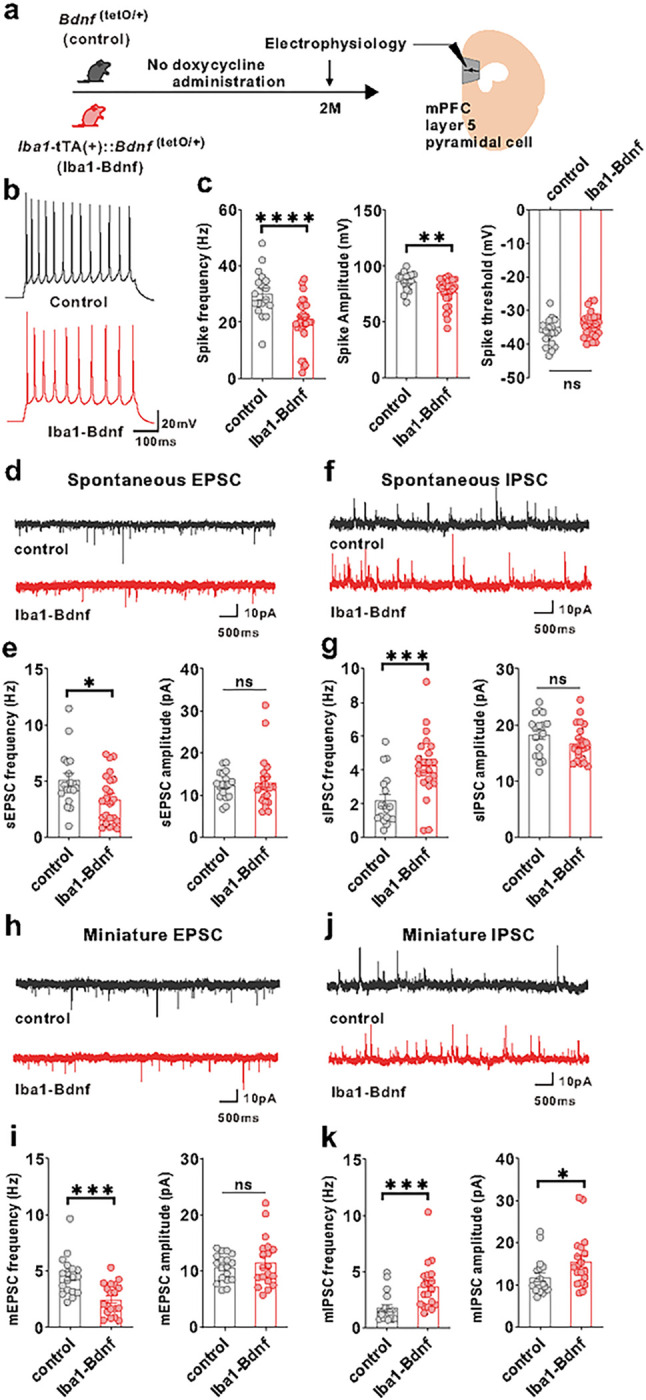
(a) Electrophysiological analysis of mPFC layer V pyramidal cells in adulthood without doxycycline administration. The experiments were started at P62. (b) Representative traces recorded from mPFC pyramidal cells at 200-pA injection. (c) Iba1-tTA(+)::Bdnf(tetO/+) mice had lower spike frequency (U = 93.50, p < 0.0001, Mann–Whitney U test) (left) and amplitude (U = 131, p = 0.0026, Mann–Whitney U test) (middle) than Bdnf(tetO/+) mice at 200-pA injection. No significant differences existed in spike threshold (t(46) =1.997, p = 0.0518, unpaired two-tailed Student’s t-test) (right). (n = 18 cells from three biologically independent Bdnf(tetO/+) mice, n = 30 cells from four biologically independent Iba1-tTA(+)::Bdnf(tetO/+) mice). (d) Representative traces of spontaneous EPSCs. (e) (Left) The Iba1-tTA(+)::Bdnf(tetO/+) mice had lower sEPSC frequency than Bdnf(tetO/+) mice (U = 130, p = 0.0436, Mann–Whitney U test). (Right) No significant difference existed in sEPSC amplitude (U = 185, p = 0.5761, Mann–Whitney U test). (f) Representative traces of spontaneous IPSCs. (g) (Left) Iba1-tTA(+)::Bdnf(tetO/+) mice showed increased sIPSC frequency compared with Bdnf(tetO/+) mice (U = 85, p = 0.0010, Mann–Whitney U test). (Right) There was no significant difference in sIPSC amplitude (t(39) = 1.440, p = 0.1579, unpaired two-tailed Student’s t-test). (e, g) n = 18 cells from three biologically independent Bdnf(tetO/+) mice, n = 23 cells from four biologically independent Iba1-tTA(+)::Bdnf(tetO/+) mice. (h) Representative traces of miniature EPSCs. (i) (Left) Iba1-tTA(+)::Bdnf(tetO/+) mice had lower mEPSC frequency than Bdnf(tetO/+) mice. (t(35) = 3.949, p = 0.0004, unpaired two-tailed Student’s t-test). (Right) There was no significant difference in mEPSC amplitude (U = 158, p = 0.7074, Mann–Whitney U test). (j) Representative traces of miniature IPSCs. (k) The Iba1-tTA(+)::Bdnf(tetO/+) mice had increased mIPSC frequency (U = 53, p = 0.0002, Mann–Whitney U test)(left) and amplitude (U = 101, p = 0.0335, Mann–Whitney U test)(right) than the Bdnf(tetI/+) mice. (i, k) n = 18 cells from three biologically independent Bdnf(tetO/+) mice, n =19 cells from three biologically independent Iba1-tTA(+)::Bdnf(tetO/+) mice. *p < 0.05, **p < 0.01, ***p < 0.001, ****p < 0.0001. Data are presented as the mean ± SEM. 2M: two months of age, control: Bdnf(tetO/+) mice, Iba1-BDNF: Iba1-tTA(+)::Bdnf(tetO/+) mice
MG-BDNF affects the complement system in the mPFC
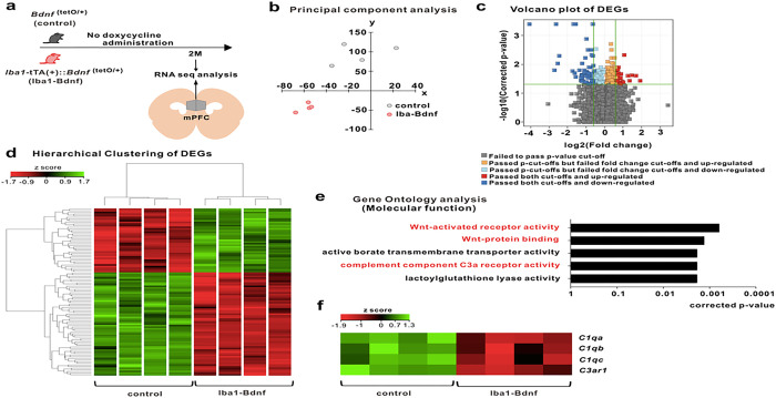
RNA-sequencing and principal component analysis revealed that MG-BDNF overexpression during the juvenile period altered mPFC distribution at two months of age (Fig. 4a, b). Compared with control mice, 63 genes were downregulated, and 39 were upregulated in the adult Iba1-tTA::Bdnf(tetO/+) mice (Fig. 4c, d). Gene Ontology molecular function analysis using differentially expressed genes downregulated in Iba1-tTA::Bdnf(tetO/+) mice revealed that sustained MG-BDNF overexpression altered gene expression related to Wnt-activated receptor activity (corrected p-value < 0.0001), Wnt-protein binding (corrected p-value < 0.0001), active borate transmembrane transporter activity (corrected p-value = 0.0018), and complement C3a receptor activity (corrected p-value = 0.0018; Fig. 4e). Heatmaps of Wnt signaling- and complement system-related gene expression were indicated with Z-scores, referencing the Kyoto Encyclopedia of Genes and Genomes pathways [47] (Supplementary Fig. 3 and Fig. 4f). This heat mapping of C1qa, C1qb, C1qc, and C3ar1 expression using Z-scores suggested reduced function of this complement cascade beginning with C1q in Iba1-tTA::Bdnf(tetO/+) mice. In particular, the Iba1-tTA::Bdnf(tetO/+) mice had significantly decreased C1qa (corrected p-value = 0.0241) and C3ar1 expression (corrected p-value = 0.0288). These results suggest that MG-BDNF overexpression decreases the complement cascade functional status in the mPFC.
Figure 4. MG-BDNF overexpression affects the complement system.
(a) RNA-seq analysis of the mPFC in adult Bdnf(tetO/+) (n = 4) and Iba1-tTA(+)::Bdnf(tetO/+) (n = 4) mice without doxycycline. The experiments was started at P64. (b) Principal component analysis revealing gene expression differences between the Bdnf(tetO/+) and Iba1-tTA(+)::Bdnf(tetO/+) mice. (c) Volcano plot of differentially expressed genes (DEGs). The thresholds are log2 fold change > 1.5 and p < 0.05. (d) Heatmap and hierarchical clustering of DEGs between Bdnf(tetO/+) and Iba1-tTA(+)::Bdnf(tetO/+) mice. The thresholds are log2 fold change > 1.5 and p < 0.05. Gene expression levels are indicated in the heatmap by the Z-scores in the legend. (e) The Gene Ontology analysis of down-regulated DEGs in the Iba1-tTA(+)::Bdnf(tetO/+) mice compared with Bdnf(tetO/+) mice suggested the involvement of the Wnt signaling pathway (Wnt-activated receptor pathway, p = 0.0006; Wnt-protein binding, p = 0.0013) and complement component C3a receptor activity (p = 0.0018) in molecular function. (f) Differences in expression levels of selected complement genes in Bdnf(tetO/+) and Iba1-tTA(+)::Bdnf(tetO/+) mice in RNA-Seq analysis. Gene expression levels are presented in the heatmap by the Z-scores in the legend. 2M: two months of age, control: Bdnf(tetO/+) mice, Iba1-BDNF: Iba1-tTA(+)::Bdnf(tetO/+) mice
Normalizing MG-BDNF overexpression during the juvenile period rescues impaired sociability and mPFC pyramidal cell dysfunction in adulthood
Given that BDNF is known to be associated with the critical period of experience-dependent neural plasticity [25], we manipulated MG-BDNF overexpression by administering DOX at different time points and measured the social behaviors and electrophysiological properties of the mPFC pyramidal neurons in adulthood to investigate whether MG-BDNF regulates social behavior and mPFC function in a time-specific manner. First, MG-BDNF overexpression during the juvenile period was suppressed by oral DOX administration from p21 in the Iba1-tTA::Bdnf(tetO/+). Similarly, the control mice were also fed DOX from p21 (Fig. 5a). In the three-chamber social preference test, no difference was detected in the social interaction score or time spent around novel mice between the adult Iba1-tTA::Bdnf(tetO/+) and control mice at two months of age (Fig. 5b), indicating that the suppressing MG-BDNF overexpression from the juvenile period normalized impaired social behavior in adulthood. No significant differences existed in locomotion or anxiety in the open field test (Supplementary Fig. 4). Regarding neuronal function, differences were not observed in spike frequency or amplitude of the excitability of the mPFC layer V pyramidal cells (Fig. 5c, d). Similar results were observed in the sEPSC and mEPSC frequencies (Fig. 5e, f, i, and j). In addition, the sIPSC or mIPSC frequency was comparable between the Iba1-tTA::Bdnf(tetO/+) and control mice (Fig. 5g, h, k, l). These results suggest that normalizing MG-BDNF expression in Iba1-tTA::Bdnf(tetO/+) from the juvenile period leads to comparable excitability of mPFC layer V pyramidal cells and its inhibitory inputs to the control mice in adulthood.
Figure 5. Normalizing MG-BDNF during the juvenile period does not impair sociability or abnormal inhibitory inputs in the mPFC.
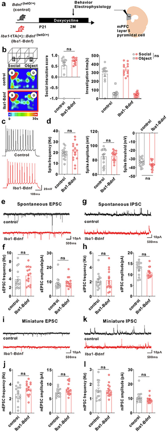
(a–l) The Iba1-tTA(+)::Bdnf(tetO/+) mice were administered doxycycline from p21 at weaning to normalize MG-BDNF. Bdnf(tetO/+) mice were also administered doxycycline from p21 as the control. Behavioral and electrophysiological experiments were started at p61. (b) Normalizing MG-BDNF from p21 did not reduce sociability in adult Iba1-tTA(+)::Bdnf(tetO/+) mice in the three-chamber social test. Both groups had no differences in the social interaction score (U = 57, p = 0.2701, Mann–Whitney U test, Bdnf(tetO/+): n = 12, Iba1-tTA(+)::Bdnf(tetO/+): n = 13)(left) or social investigation time (F1,23(interaction) = 2.626, p = 0.1187, two-way ANOVA, Bdnf(tetO/+): n = 12, Iba1-tTA(+)::Bdnf(tetO/+): n = 13)(right). S, social; O, object. (c) Representative traces recorded from mPFC pyramidal cells at 200-pA injection. (d) No differences existed between Bdnf(tetO/+) and Iba1-tTA(+)::Bdnf(tetO/+) mice treated with doxycycline from p21 in the spike frequency (U = 110, p = 0.5192, Mann–Whitney U test)(left), spike amplitude (U = 120, p = 0.7944, Mann–Whitney U test)(middle), or threshold (t(30) = 2.026, p = 0.0517, unpaired two-tailed Student’s t-test)(right) at 200-pA injection (n = 17 cells from three biologically independent Bdnf(tetO/+) mice, n = 15 cells from four biologically independent Iba1-tTA(+)::Bdnf(tetO/+) mice). (e) Representative traces of spontaneous EPSCs. (f) No differences were observed between Bdnf(tetO/+) and Iba1-tTA(+)::Bdnf(tetO/+) mice treated with doxycycline from p21 in sEPSC frequency (U = 85, p = 0.2671, Mann–Whitney U test)(left) or amplitude (U = 89, p = 0.3453, Mann–Whitney U test)(right). (g) Representative traces of spontaneous IPSCs. (h) Normalizing MG-BDNF from p21 did not increase sIPSC frequency (U = 107, p = 0.8381, Mann–Whitney U test)(left) or amplitude (U = 69, p = 0.0742, Mann–Whitney U test)(right) in Iba1-tTA(+)::Bdnf(tetO/+) mice. (f, h) n = 15 cells from ve biologically independent Bdnf(tetO/+) mice, n = 15 cells from three biologically independent Iba1-tTA(+)::Bdnf(tetO/+) mice. (i) Representative traces of miniature EPSCs. (j) No differences existed between Bdnf(tetO/+) and Iba1-tTA(+)::Bdnf(tetO/+) mice treated with doxycycline from p21 in mEPSC frequency (t(28) = 1.988, p = 0.0566, unpaired two-tailed Student’s t-test) (left) or amplitude (t(28) = 1.729, p = 0.0948, unpaired two-tailed Student’s t-test)(right). (k) Representative traces of miniature IPSCs. (l) Normalizing MG-BDNF from p21 did not increase mIPSC frequency (t(28) = 0.01783, p = 0.9859, unpaired two-tailed Student’s t-test)(left) or amplitude (U = 76, p = 0.1370, Mann–Whitney U test)(right) in Iba1-tTA(+)::Bdnf(tetO/+) mice. (j, l) n = 15 cells from five biologically independent Bdnf(tetO/+) mice, n = 15 cells from three biologically independent Iba1-tTA(+)::Bdnf(tetO/+) mice. Data are presented as the mean ± SEM. 2M: two months of age, control: Bdnf(tetO/+) mice, Iba1-BDNF: Iba1-tTA(+)::Bdnf(tetO/+) mice.
Next, we assessed the effect of delayed normalization of MG-BDNF overexpression after p45 on social behavior and mPFC layer V pyramidal cell function. DOX was orally administered to the Iba1-tTA::Bdnf(tetO/+) and control mice from p45–p50 (Supplementary Fig. 5a). The Iba1-tTA::Bdnf(tetO/+) and control mice exhibited comparable social interaction scores and time spent around the novel mice at two months of age (Supplementary Fig. 5b). Similarly, no differences existed in locomotion or anxiety during the open field test (Supplementary Fig. 5c). In contrast, normalizing MG-BDNF during adulthood did not improve the electrophysiological abnormalities of mPFC layer V pyramidal cells in the Iba1-tTA::Bdnf(tetO/+) mice at two months of age. The spike frequency of excitability remained significantly reduced (Supplementary Fig. 5d). In addition, sIPSC and mIPSC frequencies remained significantly increased in the Iba1-tTA::Bdnf(tetO/+) mice compared with control mice (Supplementary Fig. 5f, h). Furthermore, the sIPSC amplitude was significantly reduced in the Iba1-tTA::Bdnf(tetO/+) mice compared with control mice (Supplementary Fig. 5f). No significant differences existed in the frequency or amplitude of sEPSCs or mEPSCs (Supplementary Fig. 5e, g). These results suggest that MG-BDNF overexpression during the juvenile period may be critical for forming inhibitory synapses in the mPFC. However, even the MG-BDNF intervention during adulthood can improve social behavior.
BDNF expression in human M2 macrophages correlates with childhood experiences
Given the significant correlation in Bdnf expression between microglia from the brain and peripheral blood mononuclear cells in mice (Supplementary Fig. 6a), we measured BDNF expression in human peripheral macrophages, which share properties with microglia [48–50], following the juvenile experience-dependent increase of MG-Bdnf in mice. CD14-positive monocytes were collected from the peripheral blood of participants and differentiated into M1/M2 macrophages; subsequently, BDNF mRNA expression was measured (Supplementary Fig. 6b). The Japanese version of the CATS was used to assess adverse childhood experiences [51, 52]. A positive correlation existed between the total CATS scores and BDNF expression in M2 macrophages. In the CATS sub-items, neglect and punishment, among other variables, positively correlated with BDNF expression in M2 macrophages (Table 1). In contrast, no significant correlations existed between BDNF expression in M1 macrophages and the CATS total or sub-item scores (Table 1). These results indicate that adverse childhood experiences may increase BDNF expression in M2 macrophages, even in humans.
Table 1.
Correlation between human macrophage BDNF expression and adverse childhood experiences
| M1 | M2 | |||
|---|---|---|---|---|
| CATS | rs | p value | rs | p value |
| Total score | 0.069 | 0.675 | 0.409 | 0.010 * |
| Sub-item scores | ||||
| Sexual abuse | −0.114 | 0.488 | 0.293 | 0.071 |
| Punishment | 0.230 | 0.159 | 0.396 | 0.013 * |
| Neglect | 0.008 | 0.960 | 0.348 | 0.030 * |
| Emotional abuse | −0.043 | 0.795 | 0.321 | 0.046 |
| Others | 0.093 | 0.574 | 0.367 | 0.022 * |
Discussion
Little is known concerning the microglia’s influence on mPFC development and its function, such as social behavior [53]. As such, microglia are receiving significantly more attention in psychiatric research [54, 55]. In particular, understanding microglia’s role in the mPFC circuit formation is essential due to the critical implication of the mPFC in the pathobiology of neuropsychiatric disorders [53, 56–58]. In this study, we first demonstrated that j-SI mice during p21–p35 had increased MG-Bdnf expression and reduced sociability, consistent with previous studies [12, 14]. Next, we investigated the impact of these microglial changes on social behavior and mPFC function using MG-BDNF-overexpressing mice. Sustained MG-BDNF overexpression resulted in impaired social behavior, a reduced ring capacity of mPFC layer V pyramidal cells, and reduced excitatory inputs and enhanced inhibitory inputs to mPFC layer V pyramidal cells, implying an altered excitatory/inhibitory balance. Notably, the post-weaning normalization of MG-BDNF (from p21) ameliorated the impairment of social behaviors and the ring and abnormal excitatory/inhibitory balance in mPFC layer V pyramidal cells. These results suggest that MG-BDNF during the juvenile period is crucial to developing social behaviors and mPFC function. In contrast, when MG-BDNF was normalized from adulthood (p45–p50), the ring and excitatory/inhibitory balance of mPFC layer V pyramidal cells remained abnormal. These findings indicate that MG-BDNF has a critical window of effects on social behavior and mPFC function. The mPFC complement system might be implicated in a possible underlying mechanism, consistent with previous findings that microglial experience-dependent synaptic pruning depends on its related complement system [1, 2, 7].
We used BdnftetO/+ mice as the control group and Iba1-tTA::BdnftetO/+ mice as the experimental group. Our BdnftetO/+ mice were derived from ES cells of 129/SvEv mice for homologous recombination and backcrossed to C57BL/6J mice for more than five generations. While Iba1-tTA mice were originally developed in fertilized eggs of C57BL/6J, they were maintained as breeders with our BdnftetO/+ mice. Accordingly, the alleles near the transgene Iba1-tTA are expected to be enriched with those of C57BL/6J and alleles in the rest of the genome contained randomly mixed 129/SvEv and C57BL/6J alleles originating from BdnftetO/+ mice. Thus, the expected impacts of a systematic genetic background bias between the control and experimental groups are minimized, as the alleles near Iba1-tTA transgene are those of C57BL/6J in both control and experimental groups and the rest of the genome contained a random mixture of 129/SvEv and C57BL/6J alleles; the mixed genetic backgrounds of BdnftetO/+ mice were present in both the control and experimental groups [34]. Moreover, if the phenotypes reflected genetic background differences between control and experimental groups instead of or in addition to MG-BDNF overexpression, some phenotypic differences between the two groups should have remained after normalizing MG-BDNF levels; no phenotypic difference was seen in social behavior (Fig. 5b) or electrophysiological recordings (Fig. 5c–l).
Recently, Schalbetter et al. reported that microglia affect mPFC function and its relative cognition in a time-specific manner [24]. Although microglia are reportedly related to social behavior [20–22], no study has examined the relationship between sociability and time-specific development of the mPFC with microglia. The j-SI mouse with robust impairment of social behavior is a potential model for human neglect [11, 12]; however, it also makes it possible to elucidate the mechanism of social circuit formation in a limited social experience-dependent window, similar to that of sensory deprivation [59]. Social experience deprivation during the juvenile period (p21–35) has been suggested to affect the excitatory/inhibitory balance in mPFC [16–18], mPFC–pPVT neural circuits [14], and glial cells, such as oligodendrocytes [12] and microglia [13], all of which may be responsible for reduced sociability in these mice. Following the current finding that juvenile social experience deprivation elevates MG-BDNF expression in mPFC, a novel mechanism could elucidate the experience-dependent development of social behaviors. In mice overexpressing MG-BDNF, we demonstrated that higher MG-BDNF expression reduced social behaviors, suggesting that MG-BDNF may be critical in developing social abilities. In addition to a robust social assessment, i.e., the three-chamber social test, we also applied the AR-LABO, in which multiple mice were simultaneously traced under free-moving conditions to confirm their social behavior. MG-BDNF-overexpressing mice exhibited fewer approaches to other mice and received fewer approaches from others. This might be due to MG-BDNF-overexpressing mice emitting lower levels of chemical communication, such as ultrasound, urine, and pheromones [60, 61].
Furthermore, MG-BDNF modulates the excitability and excitatory/inhibitory input of mPFC layer V pyramidal cells in a limited window from the juvenile period (p21) to adulthood (p45–50). This is consistent with the critical period for social ability acquisition in mice, which is from p21 to p35 [12]. In contrast, normalizing MG-BDNF during adulthood did not improve the excitability and excitatory/inhibitory balance of mPFC layer V pyramidal cells, although impaired social behavior was ameliorated. MG-BDNF is implicated in learning-dependent neural plasticity [29] and may modify social circuit formation in brain regions other than the mPFC, even in adulthood [62]. Previous studies have also reported that microglia are related to mPFC circuit formation and cognitive maturation in adolescence [24]; thus, normalizing MG-BDNF after p45–50 may be sufficient to restore sociability.
The excitability of layer V pyramidal cells in the mPFC of MG-BDNF-overexpressing mice is similar to that observed in j-SI mice [16, 18]. This reduction in the excitability of layer V mPFC neurons may be associated with the hypoactivity of mPFC neurons that project subcortically to regulate social behavior [14], leading to reduced sociability. The relationship between MG-BDNF and the development and maturation of inhibitory neuronal circuits is poorly understood; however, enhancing inhibitory neuronal circuits, as in this study, is likely consistent with a known function of BDNF: promoting the formation and maintenance of inhibitory neural synapses during brain development [25, 63–65]. Particularly, BDNF regulates the critical visual cortex period, and overexpressed BDNF leads to the premature maturation of inhibitory neural circuits, leading to early closure of the critical visual cortex period [25]. Juvenile PFC development strengthens inhibitory neurotransmission within the brain, altering the excitatory/inhibitory balance [66, 67], which is implicated in the social function of the mPFC [19]. Overexpressed MG-BDNF might similarly close the critical social development window, disrupting mPFC development and reducing sociability by strengthening inhibitory neural circuits. In this study, normalizing MG-BDNF from adulthood (p45–p50) did not ameliorate the enhanced inhibitory neuronal circuitry. In the rodent neocortex, inhibitory synapse formation primarily occurs postnatally and rapidly (before adolescence) reaches adult-like inhibitory synapse density [68, 69]. The time course of rodent inhibitory synapse functional maturation is similar to that of inhibitory synapse formation. Specifically, IPSC frequency becomes prominent postnatally and displays adult-like properties before adolescence [70]. Enhancing inhibitory neuronal circuits via overexpressed MG-BDNF may increase the density and function of inhibitory synapses. Previous studies have also revealed increased inhibitory inputs in mPFC layer V pyramidal cells in j-SI mice and other abnormalities in inhibitory interneuron functions in the mPFC [11, 15, 17, 18]. Abnormalities in inhibitory circuits induced by juvenile isolation and changes in MG-BDNF expression might be a potential mechanism for the experience-dependent impairment of social development.
In this study, we performed RNA-seq of the mPFC; our findings suggested the involvement of the complement system as a mechanism of MG-BDNF-induced reduction of sociability. The relationship between BDNF and the complement system has not previously been reported; nevertheless, complement C3 signaling starting at C1q is crucial for the experience-dependent synaptic pruning of microglia [2, 7]. Thus, decreased C1q and C3ar1 [71–74] expression may reduce microglial pruning and inhibit mPFC circuit purification. In addition, the relationship between the complement system and psychiatric disorders is gradually becoming more evident [75, 76], indicating that further investigations are needed. Our experiments with mice have a limitation. The duration during which MG-BDNF overexpression remained suppressed may be critical rather than the timing of the DOX administration initiation. However, we have shown that resocialization after P35 in j-SI mice does not improve either sociability or mPFC function [12, 16–18], which may support the timing specificity in the current study.
Childhood experiences were also associated with BDNF expression in human peripheral M2 macrophages in this study. Microglia and macrophages should be considered separately [48] as primitive myeloid progenitor cells (microglia’s origin) migrate from the yolk sac into the brain from the embryonic period [77]. Thus, results in mice and humans cannot be directly compared; however, a relationship exists between childhood experiences and BDNF expression in macrophages that share similarities in CD11b expression and phagocytic capacity with microglia [48, 49] (the resident macrophages in the brain [49, 50]). M1 macrophages have high antigen-presenting activity and pro-inflammatory cytokine-releasing capacity. In contrast, M2 macrophages have multiple roles aside from inflammation, including anti-inflammatory responses and tissue remodeling, and secrete numerous growth and neurotrophic factors [78–80]. M2 macrophages are also implicated in the pathobiology of neuropsychiatric and neurodegenerative disorders [81–83]. Whether high levels of BDNF in M2 macrophages are associated with reduced sociability remains unclear; however, BDNF abnormalities have been identified in humans with autism spectrum and posttraumatic stress disorders [84–86]. They may also be associated with the reduced sociability of these disorders [87, 88].
In conclusion, these findings indicate that MG-BDNF is critical in developing social behaviors and mPFC function in a time-specific manner, potentially related to juvenile social experience-dependent social development. Our results provide new insights into experience-dependent social behavior formation and mPFC development.
Acknowledgments:
We acknowledge the participants that contributed to the clinical study. Research reported in this publication was supported by JSPS KAKENHI (20K16674 to TK), AMED-PRIME (21gm6310015h0002 to MM), AMED-CREST (1510009h0001 to MM), and AMED (21wm04250XXs0101 and 21uk1024002h0002 to MM). Additionally, this work was supported by the National Institutes of Health (R01MH099660, R01DC015776, and R21HD053114 to NH). Furthermore, this work was also supported by the Osaka Medical Research Foundation for Intractable Diseases.
Footnotes
Conflict of interest: N/A
Contributor Information
Manabu Makinodan, Nara Medical University.
Takashi Komori, Nara Medical University School of Medicine.
Kazuya Okamura, Nara Medical University School of Medicine.
Minobu Ikehara, Nara Medical University School of Medicine.
Kazuhiko Yamamuro, Nara Medical University School of Medicine.
Nozomi Endo, Nara Medical University School of Medicine.
Kazuki Okumura, Nara Medical University School of Medicine.
Takahira Yamauchi, Nara Medical University School of Medicine.
Daisuke Ikawa, Nara Medical University School of Medicine.
Noriko Ouji-Sageshima, Nara Medical University School of Medicine.
Michihiro Toritsuka, Nara Medical University School of Medicine.
Ryohei Takada, Keio University School of Medicine.
Yoshinori Kayashima, Keio University School of Medicine.
Rio Ishida, Keio University School of Medicine.
Yuki Mori, Keio University School of Medicine.
Kohei Kamikawa, Keio University School of Medicine.
Yuki Noriyama, Keio University School of Medicine.
Yuki Nishi, Keio University School of Medicine.
T Ito, Keio University School of Medicine.
Yasuhiko Saito, Keio University School of Medicine.
Mayumi Nishi, Keio University School of Medicine.
Toshifumi Kishimoto, Keio University School of Medicine.
Kenji Tanaka, Keio University School of Medicine.
Noboru Hiroi, University of Texas Health Science Center at San Antonio.
References
- 1.Tremblay MÈ, Lowery RL, Majewska AK. Microglial interactions with synapses are modulated by visual experience. PLoS Biol 2010; 8: e1000527. [DOI] [PMC free article] [PubMed] [Google Scholar]
- 2.Schafer DP, Lehrman EK, Kautzman AG, Koyama R, Mardinly AR, Yamasaki R et al. Microglia sculpt postnatal neural circuits in an activity and complement-dependent manner. Neuron 2012; 74: 691–705. [DOI] [PMC free article] [PubMed] [Google Scholar]
- 3.Paolicelli RC, Bolasco G, Pagani F, Maggi L, Scianni M, Panzanelli P et al. Synaptic pruning by microglia is necessary for normal brain development. Science 2011; 333: 1456–1458. [DOI] [PubMed] [Google Scholar]
- 4.Hensch TK. Critical period regulation. Annu Rev Neurosci 2004; 27: 549–579. [DOI] [PubMed] [Google Scholar]
- 5.Hensch TK. Critical period plasticity in local cortical circuits. Nat Rev Neurosci 2005; 6: 877–888. [DOI] [PubMed] [Google Scholar]
- 6.Miyamoto A, Wake H, Ishikawa AW, Eto K, Shibata K, Murakoshi H et al. Microglia contact induces synapse formation in developing somatosensory cortex. Nat Commun 2016; 7: 12540. [DOI] [PMC free article] [PubMed] [Google Scholar]
- 7.Dejanovic B, Wu T, Tsai M-C, Graykowski D, Gandham VD, Rose CM et al. Complement C1q-dependent excitatory and inhibitory synapse elimination by astrocytes and microglia in Alzheimer’s disease mouse models. Nat Aging 2022, 2: 837–850. [DOI] [PMC free article] [PubMed] [Google Scholar]
- 8.Gibel-Russo R, Benacom D, Di Nardo AA. Non-cell-autonomous factors implicated in parvalbumin interneuron maturation and critical periods. Front Neural Circuits 2022; 16: 875873. [DOI] [PMC free article] [PubMed] [Google Scholar]
- 9.Sipe GO, Lowery RL, Tremblay MÈ, Kelly EA, Lamantia CE, Majewska AK. Microglial P2Y12 is necessary for synaptic plasticity in mouse visual cortex. Nat Commun 2016; 7: 10905. [DOI] [PMC free article] [PubMed] [Google Scholar]
- 10.Kalish BT, Barkat TR, Diel EE, Zhang EJ, Greenberg ME, Hensch TK. Single-nucleus RNA sequencing of mouse auditory cortex reveals critical period triggers and brakes. Proc Natl Acad Sci U S A 2020; 117: 11744–11752. [DOI] [PMC free article] [PubMed] [Google Scholar]
- 11.Xiong Y, Hong H, Liu C, Zhang YQ. Social isolation and the brain: effects and mechanisms. Mol Psychiatry 2023; 28: 191–201. [DOI] [PMC free article] [PubMed] [Google Scholar]
- 12.Makinodan M, Rosen KM, Ito S, Corfas G. A critical period for social experience-dependent oligodendrocyte maturation and myelination. Science 2012; 337: 1357–1360. [DOI] [PMC free article] [PubMed] [Google Scholar]
- 13.Ikawa D, Makinodan M, Iwata K, Ohgidani M, Kato TA, Yamashita Y et al. Microglia-derived neuregulin expression in psychiatric disorders. Brain Behav Immun 2017; 61: 375–385. [DOI] [PubMed] [Google Scholar]
- 14.Yamamuro K, Bicks LK, Leventhal MB, Kato D, Im S, Flanigan ME et al. A prefrontal-paraventricular thalamus circuit requires juvenile social experience to regulate adult sociability in mice. Nat Neurosci 2020; 23: 1240–1252. [DOI] [PMC free article] [PubMed] [Google Scholar]
- 15.Bicks LK, Yamamuro K, Flanigan ME, Kim JM, Kato D, Lucas EK et al. Prefrontal parvalbumin interneurons require juvenile social experience to establish adult social behavior. Nat Commun 2020; 11: 1003. [DOI] [PMC free article] [PubMed] [Google Scholar]
- 16.Yamamuro K, Yoshino H, Ogawa Y, Makinodan M, Toritsuka M, Yamashita M et al. Social Isolation During the Critical Period Reduces Synaptic and Intrinsic Excitability of a Subtype of Pyramidal Cell in Mouse Prefrontal Cortex. Cereb Cortex 2018; 28: 998–1010. [DOI] [PubMed] [Google Scholar]
- 17.Yamamuro K, Yoshino H, Ogawa Y, Okamura K, Nishihata Y, Makinodan M et al. Juvenile social isolation enhances the activity of inhibitory neuronal circuits in the medial prefrontal cortex. Front Cell Neurosci 2020; 14: 105. [DOI] [PMC free article] [PubMed] [Google Scholar]
- 18.Okamura K, Yoshino H, Ogawa Y, Yamamuro K, Kimoto S, Yamaguchi Y et al. Juvenile social isolation immediately affects the synaptic activity and ring property of fast-spiking parvalbumin-expressing interneuron subtype in mouse medial prefrontal cortex. Cereb Cortex 2023; 33: 3591–3606. [DOI] [PubMed] [Google Scholar]
- 19.Bicks LK, Koike H, Akbarian S, Morishita H. Prefrontal cortex and social cognition in mouse and man. Front Psychol 2015; 6: 1805. [DOI] [PMC free article] [PubMed] [Google Scholar]
- 20.Zhan Y, Paolicelli RC, Sforazzini F, Weinhard L, Bolasco G, Pagani F et al. Deficient neuron-microglia signaling results in impaired functional brain connectivity and social behavior. Nat Neurosci 2014; 17: 400–406. [DOI] [PubMed] [Google Scholar]
- 21.Xu ZX, Kim GH, Tan JW, Riso AE, Sun Y, Xu EY et al. Elevated protein synthesis in microglia causes autism-like synaptic and behavioral aberrations. Nat Commun 2020; 11: 1797. [DOI] [PMC free article] [PubMed] [Google Scholar]
- 22.Lowery RL, Mendes MS, Sanders BT, Murphy AJ, Whitelaw BS, Lamantia CE et al. Loss of P2Y12 has behavioral effects in the adult mouse. Int J Mol Sci 2021; 22: 1868. [DOI] [PMC free article] [PubMed] [Google Scholar]
- 23.Mallya AP, Wang HD, Lee HNR, Deutch AY. Microglial pruning of synapses in the prefrontal cortex during adolescence. Cereb Cortex 2019; 29: 1634–1643. [DOI] [PMC free article] [PubMed] [Google Scholar]
- 24.Schalbetter SM, von Arx AS, Cruz-Ochoa N, Dawson K, Ivanov A, Mueller FS et al. Adolescence is a sensitive period for prefrontal microglia to act on cognitive development. Sci Adv 2022; 8: eabi6672. [DOI] [PMC free article] [PubMed] [Google Scholar]
- 25.Huang ZJ, Kirkwood A, Pizzorusso T, Porciatti V, Morales B, Bear MF et al. BDNF regulates the maturation of inhibition and the critical period of plasticity in mouse visual cortex. Cell 1999; 98: 739–755. [DOI] [PubMed] [Google Scholar]
- 26.Coull JA, Beggs S, Boudreau D, Boivin D, Tsuda M, Inoue K et al. BDNF from microglia causes the shift in neuronal anion gradient underlying neuropathic pain. Nature 2005; 438: 1017–1021. [DOI] [PubMed] [Google Scholar]
- 27.Nakajima K, Honda S, Tohyama Y, Imai Y, Kohsaka S, Kurihara T. Neurotrophin secretion from cultured microglia. J Neurosci Res 2001; 65: 322–331. [DOI] [PubMed] [Google Scholar]
- 28.Trang T, Beggs S, Salter MW. Brain-derived neurotrophic factor from microglia: a molecular substrate for neuropathic pain. Neuron Glia Biol 2011; 7: 99–108. [DOI] [PMC free article] [PubMed] [Google Scholar]
- 29.Parkhurst CN, Yang G, Ninan I, Savas JN, Yates JR, Lafaille JJ et al. Microglia promote learning-dependent synapse formation through brain-derived neurotrophic factor. Cell 2013; 155: 1596–1609. [DOI] [PMC free article] [PubMed] [Google Scholar]
- 30.Song M, Martinowich K, Lee FS. BDNF at the synapse: why location matters. Mol Psychiatry 2017; 22: 1370–1375. [DOI] [PMC free article] [PubMed] [Google Scholar]
- 31.Tanaka KF, Matsui K, Sasaki T, Sano H, Sugio S, Fan K et al. Expanding the repertoire of optogenetically targeted cells with an enhanced gene expression system. Cell Rep 2012; 2: 397–406. [DOI] [PubMed] [Google Scholar]
- 32.Sano K, Kawashima M, Imada T, Suzuki T, Nakamura S, Mimura M et al. Enriched environment alleviates stress-induced dry-eye through the BDNF axis. Sci Rep 2019; 9: 3422. [DOI] [PMC free article] [PubMed] [Google Scholar]
- 33.Suzuki T, Tanaka KF. Downregulation of Bdnf expression in adult mice causes body weight gain. Neurochem Res 2022; 47: 2645–2655. [DOI] [PubMed] [Google Scholar]
- 34.Hiroi N. Critical reappraisal of mechanistic links of copy number variants to dimensional constructs of neuropsychiatric disorders in mouse models. Psychiatry Clin Neurosci 2018, 72:301–321. [DOI] [PMC free article] [PubMed] [Google Scholar]
- 35.Rein B, Ma K, Yan Z. A standardized social preference protocol for measuring social deficits in mouse models of autism. Nat Protoc 2020; 15: 3464–3477. [DOI] [PMC free article] [PubMed] [Google Scholar]
- 36.Benjamini Y, Drai D, Elmer G, Kafkafi N, Golani I. Controlling the false discovery rate in behavior genetics research. Behav Brain Res 2001; 125: 279–284. [DOI] [PubMed] [Google Scholar]
- 37.Arakawa H. Ethological approach to social isolation effects in behavioral studies of laboratory rodents. Behav Brain Res 2018, 341:98–108. [DOI] [PubMed] [Google Scholar]
- 38.Harper KM, Hiramoto T, Tanigaki K, Kang G, Suzuki G, Trimble W et al. Alterations of social interaction through genetic and environmental manipulation of the 22q11.2 gene Sept5 in the mouse brain. Hum Mol Genet 2012, 21:3489–3499. [DOI] [PMC free article] [PubMed] [Google Scholar]
- 39.Liu J, Dietz K, DeLoyht JM, Pedre X, Kelkar D, Kaur J et al. Impaired adult myelination in the prefrontal cortex of socially isolated mice. Nat Neurosci 2012, 15:1621–1623. [DOI] [PMC free article] [PubMed] [Google Scholar]
- 40.Himmler BT, Pellis SM, Kolb B. Juvenile play experience primes neurons in the medial prefrontal cortex to be more responsive to later experiences. Neurosci Lett 2013, 556:42–45. [DOI] [PubMed] [Google Scholar]
- 41.Gossen M, Bujard H. Tight control of gene expression in mammalian cells by tetracycline-responsive promoters. Proc Natl Acad Sci U S A 1992; 89: 5547–5551. [DOI] [PMC free article] [PubMed] [Google Scholar]
- 42.Jabarin R, Netser S, Wagner S. Beyond the three-chamber test: toward a multimodal and objective assessment of social behavior in rodents. Mol Autism 2022, 13: 41. [DOI] [PMC free article] [PubMed] [Google Scholar]
- 43.Fukumitsu K, Kaneko M, Maruyama T, Yoshihara C, Huang AJ, McHugh TJ et al. Amylin-Calcitonin receptor signaling in the medial preoptic area mediates a liative social behaviors in female mice. Nat Commun 2022, 13: 709. [DOI] [PMC free article] [PubMed] [Google Scholar]
- 44.Endo N., Ujita W., Fujiwara M., Miyauchi H., Mishima H., Makino Y et al. Multiple animal positioning system shows that socially-reared mice influence the social proximity of isolation-reared cagemates. Commun Biol 2018; 1: 225. [DOI] [PMC free article] [PubMed] [Google Scholar]
- 45.Endo N, Makinodan M, Somayama N, Komori T, Kishimoto T, Nishi M. Characterization of behavioral phenotypes in the BTBR T + Itpr3tf/J mouse model of autism spectrum disorder under social housing conditions using the multiple animal positioning system. Exp Anim 2019; 68: 319–330. [DOI] [PMC free article] [PubMed] [Google Scholar]
- 46.Endo N., Makinodan M., Mannari-Sasagawa T., Horii-Hayashi N., Somayama N., Komori T et al. The effects of maternal separation on behaviours under social-housing environments in adult male C57BL/6 mice. Sci Rep 2021; 11: 527. [DOI] [PMC free article] [PubMed] [Google Scholar]
- 47.Kanehisa M, Goto S. KEGG: kyoto encyclopedia of genes and genomes. Nucleic Acids Res 2000; 28: 27–30. [DOI] [PMC free article] [PubMed] [Google Scholar]
- 48.Cuadros MA, Sepulveda MR, Martin-Oliva D, Marín-Teva JL, Neubrand VE. Microglia and microglia-like cells: similar but different. Front Cell Neurosci 2022; 16: 816439. [DOI] [PMC free article] [PubMed] [Google Scholar]
- 49.Li Q, Barres BA. Microglia and macrophages in brain homeostasis and disease. Nat Rev Immunol 2018; 18: 225–242. [DOI] [PubMed] [Google Scholar]
- 50.Bennett ML, Bennett FC. The influence of environment and origin on brain resident macrophages and implications for therapy. Nat Neurosci 2020; 23: 157–166. [DOI] [PubMed] [Google Scholar]
- 51.Sanders B, Becker-Lausen E. The measurement of psychological maltreatment: early data on the Child Abuse and Trauma Scale. Child Abuse Negl 1995; 19: 315–323. [DOI] [PubMed] [Google Scholar]
- 52.Tanabe H, Ozawa S, Goto K. Psychometric properties of the Japanese version of the Child Abuse and Trauma Scale (CATS). The 9th Annual Meeting of the Japanese Society for Traumatic Stress Studies. 2010. [Google Scholar]
- 53.Blagburn-Blanco SV, Chappell MS, De Biase LM, DeNardo LA. Synapse-specific roles for microglia in development: New horizons in the prefrontal cortex. Front Mol Neurosci 2022; 15: 965756. [DOI] [PMC free article] [PubMed] [Google Scholar]
- 54.Tay TL, Béchade C, D’Andrea I, St-Pierre MK, Henry MS, Roumier A et al. Microglia gone rogue: impacts on psychiatric disorders across the lifespan. Front Mol Neurosci 2017; 10: 421. [DOI] [PMC free article] [PubMed] [Google Scholar]
- 55.Zengeler KE, Lukens JR. Innate immunity at the crossroads of healthy brain maturation and neurodevelopmental disorders. Nat Rev Immunol 2021; 21: 454–468. [DOI] [PMC free article] [PubMed] [Google Scholar]
- 56.Schubert D, Martens GJ, Kolk SM. Molecular underpinnings of prefrontal cortex development in rodents provide insights into the etiology of neurodevelopmental disorders. Mol Psychiatry 2015; 20: 795–809. [DOI] [PMC free article] [PubMed] [Google Scholar]
- 57.Arnsten AF, Rubia K. Neurobiological circuits regulating attention, cognitive control, motivation, and emotion: disruptions in neurodevelopmental psychiatric disorders. J Am Acad Child Adolesc Psychiatry 2012; 51: 356–367. [DOI] [PubMed] [Google Scholar]
- 58.Hare BD, Duman RS. Prefrontal cortex circuits in depression and anxiety: contribution of discrete neuronal populations and target regions. Mol Psychiatry 2020; 25: 2742–2758. [DOI] [PMC free article] [PubMed] [Google Scholar]
- 59.Erzurumlu RS, Gaspar P. Development and critical period plasticity of the barrel cortex. Eur J Neurosci 2012; 35: 1540–1553. [DOI] [PMC free article] [PubMed] [Google Scholar]
- 60.Portfors CV, Perkel DJ. The role of ultrasonic vocalizations in mouse communication. Curr Opin Neurobiol 2014; 28: 115–120. [DOI] [PMC free article] [PubMed] [Google Scholar]
- 61.Silverman JL, Yang M, Lord C, Crawley JN. Behavioural phenotyping assays for mouse models of autism. Nat Rev Neurosci 2010; 11: 490–502. [DOI] [PMC free article] [PubMed] [Google Scholar]
- 62.Xu S, Jiang M, Liu X, Sun Y, Yang L, Yang Q et al. Neural circuits for social interactions: From microcircuits to input-output circuits. Front Neural Circuits 2021; 15: 768294. [DOI] [PMC free article] [PubMed] [Google Scholar]
- 63.Abidin I, Eysel UT, Lessmann V, Mittmann T. Impaired GABAergic inhibition in the visual cortex of brain-derived neurotrophic factor heterozygous knockout mice. J Physiol 2008; 586: 1885–1901. [DOI] [PMC free article] [PubMed] [Google Scholar]
- 64.Palizvan MR, Sohya K, Kohara K, Maruyama A, Yasuda H, Kimura F et al. Brain-derived neurotrophic factor increases inhibitory synapses, revealed in solitary neurons cultured from rat visual cortex. Neuroscience 2004; 126: 955–966. [DOI] [PubMed] [Google Scholar]
- 65.Berghuis P, Dobszay MB, Sousa KM, Schulte G, Mager PP, Härtig W et al. Brain-derived neurotrophic factor controls functional differentiation and microcircuit formation of selectively isolated fast-spiking GABAergic interneurons. Eur J Neurosci 2004; 20: 1290–1306. [DOI] [PubMed] [Google Scholar]
- 66.Lewis DA, Hashimoto T, Volk DW. Cortical inhibitory neurons and schizophrenia. Nat Rev Neurosci 2005; 6: 312–324. [DOI] [PubMed] [Google Scholar]
- 67.Caballero A, Tseng KY. GABAergic function as a limiting factor for prefrontal maturation during adolescence. Trends Neurosci 2016; 39: 441–448. [DOI] [PMC free article] [PubMed] [Google Scholar]
- 68.Micheva KD, Beaulieu C. Quantitative aspects of synaptogenesis in the rat barrel eld cortex with special reference to GABA circuitry. J Comp Neurol 1996; 373: 340–354. [DOI] [PubMed] [Google Scholar]
- 69.De Felipe J, Marco P, Fairén A, Jones EG. Inhibitory synaptogenesis in mouse somatosensory cortex. Cereb Cortex 1997; 7: 619–634. [DOI] [PubMed] [Google Scholar]
- 70.Le Magueresse C, Monyer H. GABAergic interneurons shape the functional maturation of the cortex. Neuron 2013; 77: 388–405. [DOI] [PubMed] [Google Scholar]
- 71.Litvinchuk A, Wan YW, Swartzlander DB, Chen F, Cole A, Propson NE et al. Complement C3aR inactivation attenuates tau pathology and reverses an immune network deregulated in tauopathy models and Alzheimer’s disease. Neuron 2018; 100: 1337–1353.e5. [DOI] [PMC free article] [PubMed] [Google Scholar]
- 72.Vasek MJ, Garber C, Dorsey D, Durrant DM, Bollman B, Soung A et al. A complement-microglial axis drives synapse loss during virus-induced memory impairment. Nature 2016; 534: 538–543. [DOI] [PMC free article] [PubMed] [Google Scholar]
- 73.Surugiu R, Catalin B, Dumbrava D, Gresita A, Olaru DG, Hermann DM et al. Intracortical administration of the complement C3 receptor antagonist tri uoroacetate modulates microglia reaction after brain injury. Neural Plast 2019; 2019: 1071036. [DOI] [PMC free article] [PubMed] [Google Scholar]
- 74.Butler CA, Popescu AS, Kitchener EJA, Allendorf DH, Puigdellívol M, Brown GC. Microglial phagocytosis of neurons in neurodegeneration, and its regulation. J Neurochem 2021; 158: 621–639. [DOI] [PubMed] [Google Scholar]
- 75.Perez Sierra D, Tripathi A, Pillai A. Dysregulation of complement system in neuropsychiatric disorders: A mini review. Biomark Neuropsychiatry 2022; 7: 100056. [DOI] [PMC free article] [PubMed] [Google Scholar]
- 76.Druart M, Le Magueresse C. Emerging roles of complement in psychiatric disorders. Front Psychiatry 2019; 10: 573. [DOI] [PMC free article] [PubMed] [Google Scholar]
- 77.Ginhoux F, Greter M, Leboeuf M, Nandi S, See P, Gokhan S et al. Fate mapping analysis reveals that adult microglia derive from primitive macrophages. Science 2010; 330: 841–845. [DOI] [PMC free article] [PubMed] [Google Scholar]
- 78.Sica A, Mantovani A. Macrophage plasticity and polarization: in vivo veritas. J Clin Invest 2012; 122: 787–795. [DOI] [PMC free article] [PubMed] [Google Scholar]
- 79.Martinez FO, Gordon S. The M1 and M2 paradigm of macrophage activation: time for reassessment. F1000Prime Rep 2014; 6: 13. [DOI] [PMC free article] [PubMed] [Google Scholar]
- 80.Rőszer T. Understanding the mysterious M2 macrophage through activation markers and effector mechanisms. Mediators Inamm 2015; 2015: 816460. [DOI] [PMC free article] [PubMed] [Google Scholar]
- 81.Ascoli BM, Parisi MM, Bristot G, Antqueviezc B, Géa LP, Colombo R et al. Attenuated in ammatory response of monocyte-derived macrophage from patients with BD: a preliminary report. Int J Bipolar Disord 2019; 7: 13. [DOI] [PMC free article] [PubMed] [Google Scholar]
- 82.Chu F, Shi M, Zheng C, Shen D, Zhu J, Zheng X et al. The roles of macrophages and microglia in multiple sclerosis and experimental autoimmune encephalomyelitis. J Neuroimmunol 2018; 318: 1–7. [DOI] [PubMed] [Google Scholar]
- 83.Moehle MS, West AB. M1 and M2 immune activation in Parkinson’s Disease: Foe and ally. Neuroscience 2015; 302: 59–73. [DOI] [PMC free article] [PubMed] [Google Scholar]
- 84.Notaras M, van den Buuse M. Neurobiology of BDNF in fear memory, sensitivity to stress, and stress-related disorders. Mol Psychiatry 2020; 25: 2251–2274. [DOI] [PubMed] [Google Scholar]
- 85.Notaras M, Hill R, van den Buuse M. The BDNF gene Val66Met polymorphism as a modi er of psychiatric disorder susceptibility: progress and controversy. Mol Psychiatry 2015; 20: 916–930. [DOI] [PubMed] [Google Scholar]
- 86.Liu SH, Shi XJ, Fan FC, Cheng Y. Peripheral blood neurotrophic factor levels in children with autism spectrum disorder: a meta-analysis. Sci Rep 2021; 11: 15. [DOI] [PMC free article] [PubMed] [Google Scholar]
- 87.Gyawali S, Patra BN. Autism spectrum disorder: Trends in research exploring etiopathogenesis. Psychiatry Clin Neurosci 2019; 73: 466–475. [DOI] [PubMed] [Google Scholar]
- 88.Bjornsson AS, Hardarson JP, Valdimarsdottir AG, Gudmundsdottir K, Tryggvadottir A, Thorarinsdottir K et al. Social trauma and its association with posttraumatic stress disorder and social anxiety disorder. J Anxiety Disord 2020; 72: 102228. [DOI] [PubMed] [Google Scholar]


