Abstract
Background
Chronic kidney disease (CKD) is a worldwide public health problem affecting millions of people. Probiotics and postbiotics are associated with valuable compounds with antibacterial, anti-inflammatory, and immunomodulatory effects, preserving renal function in CKD patients. The current study is aimed to evaluate the efficacy of Limosilactobacillus fermentum (L. fermentum) and its postbiotic in an animal model of cisplatin-induced CKD.
Methods
The animals were divided into four experimental groups (normal mice, CKD mice with no treatment, CKD mice with probiotic treatment, and CKD mice with postbiotic treatment). CKD mice were induced by a single dose of cisplatin 10 mg/kg, intraperitoneally. For 28 days, the cultured probiotic bacteria and its supernatant (postbiotic) were delivered freshly to the related groups through their daily water. Then, blood urea nitrogen (BUN) and creatinine (Cr) of plasma samples as well as glutathione (GSH), lipid peroxidation, reactive oxygen species, and total antioxidant capacity of kidneys were assessed in the experimental mice groups. In addition, histopathological studies were performed on the kidneys.
Results
Application of L. fermentum probiotic, and especially postbiotics, significantly decreased BUN and Cr (P < 0.0001) as well as ROS formation and lipid peroxidation levels (P < 0.0001) along with increased total antioxidant capacity and GSH levels (P < 0.001). The histopathologic images also confirmed their renal protection effect. Interestingly, the postbiotic displayed more effectiveness than the probiotic in some assays. The improvement effect on renal function in the current model is mainly mediated by oxidative stress markers in the renal tissue.
Conclusions
In conclusion, it was found that the administration of L. fermentum probiotic, and particularly its postbiotic in cisplatin-induced CKD mice, showed promising effects and could successfully improve renal function in the animal model of CKD. Therefore, probiotics and postbiotics are considered as probably promising alternative supplements to be used for CKD.
Keywords: Probiotic, Postbiotic, Limosilactobacillus fermentum, Renoprotection, CKD mice model, In-vivo study, Integrative medicine
Introduction
Chronic kidney disease (CKD) is characterized by irreversible and progressive alteration in the function and structure of the kidney during months or years [1, 2]. It is considered one of the fastest-growing causes of death and is estimated to become the fifth global cause by 2040 [3]. The progression of the disease might be influenced by several factors, such as dietary intake, mental stress, and medications [4]. Current approaches for CKD management include low protein and sodium intake, blood pressure control, and glycemic control [5]. However, no effective therapy exists for this health issue, and innovative strategies are necessary to manage, control, and even treat the disease [5, 6].
Limosilactobacillus fermentum (L. fermentum) is one of the common probiotic strains in nature [7, 8], usually isolated from fermenting plant material, bread, dairy products, naturally fermented sausages, saliva, and breast milk [8, 9]. Apart from extensive applications in the food industry, there have been many studies regarding the effectiveness of L. fermentum [8, 10, 11], for example, prevention and treatment of gastrointestinal diseases, prevention of alcoholic liver disorder, alleviating colorectal cancer risk and a lot more [12, 13], which are summarized in Table 1.
Table 1.
Various in vivo studies regarding the effectiveness of L. fermentum
| Study | Finding |
|---|---|
| The effect of L. fermentum in animal models of ethanol-induced liver disease | Considerable decrease in ethanol-induced liver tissue damage [14, 15]. |
| The effect of L. fermentum on hypercholesterolemia | Amelioration of hypercholesterolemia by the probiotic’s antioxidant effect, anti-inflammatory effect, and gut barrier function [16, 17]. |
| The effect of L. fermentum on colitis | Effective reduction of the symptoms of colitis in mice through different mechanisms, such as modulating the nuclear factor-κB (NF-κB) signaling pathway and ameliorating the inflammation and/or antioxidant properties [18–20]. |
| The effect of L. fermentum on sleep disturbance | Efficient amelioration of sleep disturbance produced by the first night effect (FNE) and promotion of non-rapid eye movement (NREM) sleep in mice [21]. |
| The effect of L. fermentum on Helicobacter. Pylori | Inhibition of the Helicobacter pylori colonization [22]. |
| The effect of L. fermentum following local administration on vaginal infection | The antimicrobial preventative as well as curative effects against Escherichia coli [23]. |
| The effect of L. fermentum on colorectal cancer | Attenuation of the risk of colorectal cancer [24, 25]. |
| The effect of L. fermentum on aging | Potential decrease of aging symptoms in rats and mice via its various properties, such as antioxidant effects [26, 27]. |
| The effect of L. fermentum on renal damage in a systemic lupus erythematosus mouse model | Prevention of the impairment of kidney function and damage through various mechanisms such as reducing blood lipopolysaccharides, reduction of inflammation and oxidative stress, as well as immune complex deposition [28]. |
The International Scientific Association of Probiotics and Prebiotics (ISAPP) defines postbiotics as the preparation of inanimate microorganisms and/or their components, which induces a health benefit on the host [29]. Different components are considered postbiotics, such as cell-free supernatant, functional proteins, extracellular polysaccharides (EPS), enzymes, cell wall fragments, short-chain fatty acid, and bacterial lysate [30]. Since postbiotics are free of living microorganisms, the possible risks associated with postbiotic use might be fewer than the probiotics while maintaining their effectiveness [30, 31]. Postbiotics could be an attractive alternative for other biotic members. Postbiotics can be absorbed and appropriately metabolized and have shown higher stability, facile transportation, and essential signaling potential with different organs and tissues [32–34].
Furthermore, postbiotics have other favorable properties, including anti-inflammatory, immuno-modulatory, antioxidant, antitumor, anti-hypertensive, infection prevention, anti-atherosclerotic, autophagy, and antiproliferative properties [35–37]. However, there have been fewer investigations that studied the efficacy of postbiotics. To the best of our knowledge, no study has been conducted about postbiotics in kidney diseases.
The present study is aimed to examine the potential efficacy of L. fermentum and its postbiotic in cisplatin‑induced CKD in an animal model. Therefore, we assessed various factors, including blood urea nitrogen (BUN) and plasma creatinine (Cr) of serum samples, glutathione (GSH), lipid peroxidation, reactive oxygen species (ROS), and total antioxidant capacity of kidneys. Also, we examined the histopathological consequences of the probiotic and its bioactive metabolites to investigate their efficacy.
Materials and methods
Materials
De Man, rogosa & sharpe (MRS) broth medium was prepared from Himedia (India). Cisplatin was obtained from Ebewe Pharma (Austria). Sodium thiopental, tris, potassium chloride (KCl), dichlorofluorescein (DCF), phosphoric acid, thiobarbituric acid, n-butanol, trichloroacetic acid (TCA), ethylenediaminetetraacetic acid (EDTA), and dithio-bis-(2-nitrobenzoic acid) (DTNB, Ellman’s reagent) were purchased from Merck (Germany). Acetic acid, sodium acetate, ferric chloride dihydrate, and 2,4,6-tris(2-pyridyl)-s-triazine (TPTZ) were also from Merck (Germany). All other solvents and reagents utilized to prepare various buffer solutions were of analytical grade and purchased from Merck (Germany). All the preparations were made using deionized water (Direct Q UV3, Millipore, USA).
Probiotic and postbiotic preparation
Limosilactobacillus fermentum PTCC No. 1744 as the probiotic strain was purchased from Persian Type Culture Collection in Iranian Research Organization for Science and Technology (IROST). The bacteria were cultured in MRS broth medium for 48 h at 37 °C under microaerophilic conditions until the stationary phase was achieved. The pH of 7.2 was adjusted for the media. Afterward, the number of viable bacteria was counted by plate counts using MRS agar, and an inoculum of bacteria containing an approximate density of 109 CFU/ml was prepared.
Preparation of postbiotic was done according to the method previously described by Montazeri-Najafabady et al. [38]. Briefly, the bacteria were centrifuged at 4000 g and 4 °C for 20 min using a refrigerated centrifuge (Eppendorf 5804 R Germany), and the biomass (including probiotic cells) was collected. The supernatant postbiotic was filtered through a 0.2 μm membrane filter to remove any remaining probiotic bacteria. The filtered supernatant (containing postbiotic) was then lyophilized by a freeze-drier (Alpha 1-2LD Plus, Martin Christ, Germany) and kept at − 20 °C until further use. The final product was held for a maximum of 12 days.
Experimental design
Twenty male BALB/c mice with an average weight of 22.5 ± 2.5 g and an age of 8 weeks were supplied from the comparative and experimental medicine center of Shiraz University of Medical Sciences. All the animals were housed in standard cages where water and standard food were easily accessible. The mice were kept in suitable conditions under the average temperature of 22 ± 2 °C and humidity of 44 ± 5% with a 12-h light-dark cycle. To inhibit any stress effects on animals, they were allowed to adapt with the new situation for two weeks [39, 40]. All animal procedures were performed under the supervision of the institutional ethics committee of Shiraz University of Medical Sciences, Shiraz, Iran (Ethics committee code: IR.SUMS.REC.1400.057). The ARRIVE guidelines for the care and use of laboratory animals were also followed.
The mice were divided into four experimental groups: normal mice received distilled water (no treatment, Group 1), CKD mice received distilled water (no treatment, Group 2), CKD mice received L. fermentum probiotic bacteria (Group 3), and CKD mice received postbiotic (Group 4), each group contained five mice. To induce CKD in the animals, the mice received a single dose of cisplatin (10 mg/kg) through intraperitoneal (i.p.) injection and studied five days later [41]. The probiotic (for group 3) and postbiotic (for group 4) were freshly prepared every day and added to the animals’ water at a proportion of 1:4 probiotic/postbiotic: daily water (15 ml daily water for each mouse). The experiment was performed for 28 days. Sample collections (serum and kidney pieces) were collected for biochemical/histological assessment following deep anesthesia/scarification on day 28.
Biochemical assay
Blood urea nitrogen (BUN) and creatinine (cr) assay
The mice were anesthetized by intra-peritoneal sodium thiopental at 70 mg/kg, and blood samples (5 ml for each piece) were collected carefully from the abdominal vein. Then, the blood samples were clotted at room temperature and centrifuged at 1000 rpm for 25 min to separate serum parts. The obtained serum was kept at -20 °C for further experiments. The blood serum was utilized to assess the experimental animals’ BUN and creatinine as biomarkers of renal injury using an automated biochemistry analyzer (BM/Hitachi 747, Tokyo, Japan).
Renal oxidative stress (ROS) assay
ROS formation
250 mg of the kidney tissue for each mouse was weighed and poured into 2.5 ml of cooled tris buffer (40 mM, pH = 7.4). Then, the tissue was homogenized with a homogenizer (IKA T 25 digital ULTRA-TURRAX®). 100 µl of the homogeneous mixture was added to 1ml of cooled tris buffer (40 mM, pH = 7.4), and then 5 µl of DCF solution (1 µM) was added to the obtained mixture. After 30 min incubation at 37 ° C, the fluorescence intensity of the samples was measured at the excitation and emission wavelengths of 485 and 525 nm, respectively, using a fluorimeter (FLUOstar Omega® multifunctional microplate reader, BMG LABTECH, Germany) [42].
Tissue lipid peroxidation
The kidney tissue (500 mg) was mixed with 5 ml of KCl solution (1.15% w: v) and homogenized at the temperature of 4 °C. Then, 0.5 ml of the homogenized tissue was blended with 3 mL of 1% m-phosphoric acid and 1 ml of 0.6% w: v of thiobarbituric acid and mixed gently for 5 min. Next, the mixture was heated at 100 °C for 45 min in a water bath. After that, 2 mL of n-butanol was added to the cooled mixture and vortexed for 5 min. After centrifuging the samples, 100 µL of the upper phase (n-butanol phase) was added to a 96-well plate. The absorbance was measured by a spectrophotometer (EPOCH® plate reader, BioTek®, USA) at the wavelength of 532 nm [42].
Total antioxidant capacity
Ferric reducing antioxidant power or ferric-reducing ability of plasma (FRAP) assay was used to assess the total antioxidant capacity, which is based on the reduction of the complex of ferric tripyridyltriazine (Fe3+-TPTZ) to ferrous tripyridyltriazine (Fe2+-TPTZ) via the antioxidants of a sample at low pH [42]. The end product, Fe2+-TPTZ, shows a blue color in which absorption is measured [43]. Accordingly, 500 mg of the kidney tissue was weighed and poured into 5 ml of cooled KCl solution (1.15% W/V). The tissue was homogenized using the homogenizer. After that, 100 µL of the homogeneous mixture was mixed with 3 mL of FRAP reagent (2.5 ml acetate buffer (300 mM, pH = 3) and 0.25 ml ferric chloride dihydrate (20 mM)) and 0.25 mL of TPTZ solution. Following incubation for 4 min, the mixture was centrifuged at 10,000 rpm for 1 min. Finally, the absorption was read using a cuvette spectrophotometer (BioTEK® Instruments, Highland Park, USA) at the wavelength of 593 nm.
Glutathione (GSH) level
The kidney tissue (500 mg) was homogenized in 5 mL of 40 mM EDTA solution at 4 °C to measure the GSH level. Then, 5 ml of the homogenized mixture was mixed with 4 ml of distilled water and 1 ml of TCA (50% W/V) and stirred vigorously. The resulting mixture was centrifuged at 3000 g and 4 °C for 15 min. 2 ml of the supernatant was added to 4 ml of 0.4 M Tris buffer and 0.1 ml of 10 mM DTNB (Ellman’s reagent) followed by shaking well. Finally, the absorbance of each sample was measured using the spectrophotometer at the wavelength of 412 nm in less than 5 min [42].
Histopathological study
As previously mentioned, the kidney samples were preserved in a 10% formalin solution for histopathological assay [44]. The paraffin-embedded kidney tissue sections were prepared and stained with the hematoxylin-eosin dye. The destruction in kidney sections (damage %) was reported by investigating glomerular atrophy, tubular change, interstitial nephritis, and vascular change [44]. A semi-quantitative grading method was applied. The degree of nephropathy changes was calculated compared to the control group.
Statistical analysis
Statistical analysis was performed using GraphPad software version 8 (v8.4.0, GraphPad Software Inc., San Diego, CA). Comparisons were carried out via a one-way analysis of variance followed by Tukey’s post hoc test. A P-value less than 0.05 was considered statistically significant. Data were expressed as means ± Standard Deviation (n = 5) [45].
Results
BUN assay
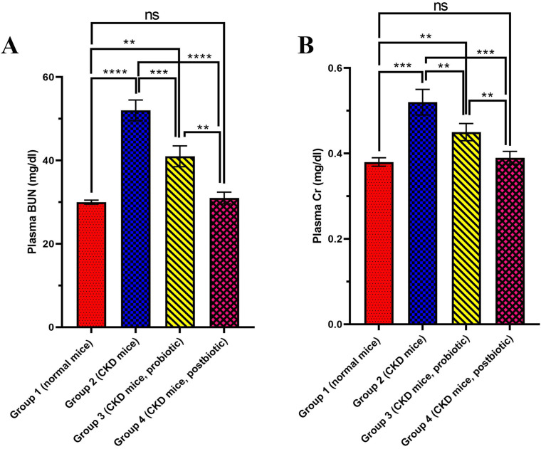
Various CKD-induced mice groups were orally treated by L. fermentum, postbiotic or nothing, and then evaluated for the BUN level. As the results showed (Fig. 1A), cisplatin treatment (Group 2) significantly elevated the BUN level to 50.0 ± 3.0 mg/dl (P < 0.0001) compared to the control group (Group 1, 30.0 ± 1.3 mg/dl), which is healthy mice with normal renal function. Among all the groups, the value of BUN was the highest for the cisplatin-treated group that received no treatment (Group 2). Treatment of cisplatin-induced CKD groups with probiotic bacteria (Group 3) and the supernatant (postbiotic, Group 4) significantly decreased BUN level (P < 0.001 and P < 0.0001, respectively) in comparison to the cisplatin-treated group that received no treatment (Group 2). Among all the groups, the BUN level was the least (P < 0.0001) for the control group (Group 1, 30.0 ± 1.3 mg/dl) as well as the group that received the postbiotic (Group 4, 31.0 ± 1.4 mg/dl), the levels were not significantly different from each other (P > 0.05), followed by the group that received the probiotic (Group 3, 41.0 ± 2.5 mg/dl).
Fig. 1.
(A) Blood urea nitrogen (BUN) and (B) creatinine (Cr) levels, in different mice groups of Group 1 (normal mice, no treatment), Group 2 (Chronic kidney disease (CKD) mice, no treatment, Group 3 (CKD mice, L. fermentum probiotic treatment) and Group 4 (CKD mice, L. fermentum postbiotic treatment) at the end of Day 28. Data are expressed as Mean ± Standard deviation (SD) for five replicates. * (P < 0.05), ** (P < 0.01), *** (P < 0.001), **** (P < 0.0001), ***** (P < 0.00001) and ****** (P < 0.000001) and ns (not significant) are different levels of significance
Creatinine (Cr) assay
The effects of L. fermentum and postbiotic on Cr levels in cisplatin-induced CKD are demonstrated in Fig. 1B. Accordingly, cisplatin treatment (Group 2) led to a significant increase (P < 0.001) of Cr level to 0.52 ± 0.03 mg/dl compared to 0.38 ± 0.01 mg/dl for the control group (Group 1). Among all the groups, the Cr level was the maximum for the cisplatin-treated group without treatment (Group 2). It was shown that treatment of mice groups with probiotic bacteria (Group 3) and the supernatant (Group 4, postbiotic) significantly reduced the Cr level (P < 0.01 and P < 0.001, respectively) in comparison to the cisplatin-treated group, which received no treatment (Group 2). The Cr level was 0.45 ± 0.02 mg/dl for the group that received probiotics (Group 3). The Cr level was the minimum for the control group (Group 1, 0.38 ± 0.01 mg/dl, P > 0.001), which was as much as the group that received postbiotic (Group 4) (0.39 ± 0.01 mg/dl, P > 0.05).
ROS formation
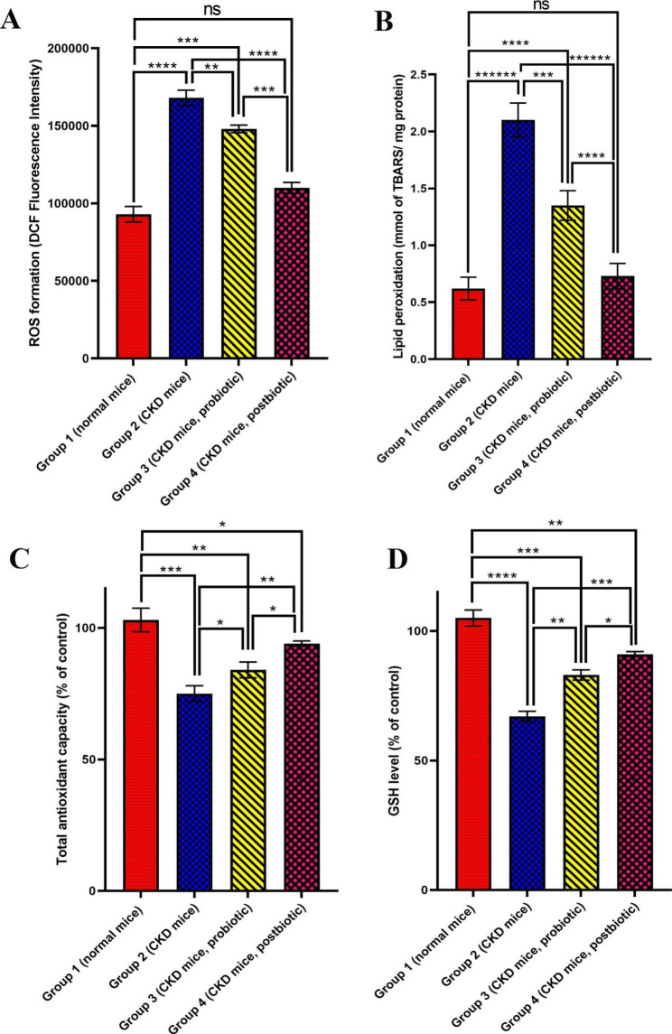
As presented in Fig. 2A, the ROS increased after mice were treated with cisplatin alone (Group 2, P < 0.0001) in comparison to the control group. Probiotics (Group 3) and postbiotics (Group 4) significantly attenuated ROS formation (P < 0.01 and P < 0.0001, respectively) in comparison to the cisplatin-treated group without treatment (Group 2). The amount of ROS in kidney tissue in the control group (Group 1) is the lowest of all groups (P < 0.0001). Among the two groups of 3 and 4, the postbiotic (Group 4) displayed the most suppressive effect against ROS formation in cisplatin-induced CKD (P < 0.001).
Fig. 2.
(A) Renal oxidative species (ROS) formation, (B) lipid peroxidation, (C) total antioxidant capacity, and (D) glutathione (GSH) level in kidney tissue in different mice groups of Group 1 (normal mice, no treatment), Group 2 (Chronic kidney disease (CKD) mice, no treatment, Group 3 (CKD mice, L. fermentum probiotic treatment) and Group 4 (CKD mice, L.fermentum postbiotic treatment) at the end of day 28. Data are expressed as Mean ± Standard deviation (SD) for five replicates. * (P < 0.05), ** (P < 0.01), *** (P < 0.001), **** (P < 0.0001), ***** (P < 0.00001) and ****** (P < 0.000001) and ns (not significant) are different levels of significance
Tissue lipid peroxidation
The effects of probiotics and postbiotics on lipid peroxidation are shown in Fig. 2B. The antineoplastic agent, cisplatin, significantly augmented lipid peroxidation amount (Group 2, 2.10 ± 0.2 mmol TBARS/mg protein) compared to the control group (Group 1, 0.60 ± 0.07 mmol TBARS/mg protein, P < 0.000001). Treatment with the probiotic (Group 3) and postbiotic (Group 4) significantly declined lipid peroxidation compared to the cisplatin-treated mice group (Group 2) (P < 0.0001 and P < 0.000001, respectively). The result indicates that the postbiotic-treated mice group (Group 4) showed high efficacy in lipid peroxidation decrease (0.73 mmol ± 0.09 TBARS/mg protein, P < 0.00001), whose value was near the control group (Group 1) (P > 0.05).
Total antioxidant capacity
Total antioxidant capacity (% of control) following various treatments of mice groups with L. fermentum probiotic and postbiotics is shown in Fig. 2C. It was revealed that total antioxidant capacity significantly decreased after administration of cisplatin (Group 2, 75% ± 3, P < 0.001) compared to the control group (Group 1, 102% ± 5). Supplementation with L. fermentum probiotic (Group 3) and postbiotic (Group 4) could improve the total antioxidant capacity. However, the postbiotic (Group 4) displayed the more ameliorative effect in comparison to the probiotic (Group 3) (P < 0.05).
GSH level
Figure 2D presents the GSH level (% control) in different cisplatin-induced CKD mice groups treated with L.fermentum probiotic and postbiotic. The GSH level significantly decreased after cisplatin administration (Group 2, 67% ± 2, P < 0.0001) compared to the control group (Group 1, 104% ± 3). Supplementation with probiotics (Group 3) and postbiotics (Group 4) considerably enhanced the GSH levels in comparison to the cisplatin-treated group without treatment (P < 0.01 and P < 0.001). However, the postbiotic (Group 4) presented the better effect than the probiotic (Group 3)(P < 0.05).
Histopathological study
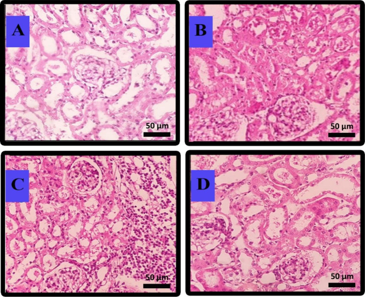
The damage percentage of kidney tissues and the histological sections of the kidneys related to different cisplatin-induced CKD mice following treatment with L. fermentum probiotics and postbiotics are shown in Table 2; Fig. 3. The results indicated that 19% of tubular and 10% of interstitial cells were damaged after cisplatin administration (Group 2). At the same time, no cytotoxic effects were detected in glomerular and vascular cells (Table 2; Fig. 3B). The tubular cell damage decreased after the administration of probiotic (Group 3, Table 2; Fig. 3C), and postbiotic (Group 4, Table 2; Fig. 3D) to 13% and 1%, respectively. In the case of interstitial cells, the cell injury was attenuated to 1% following postbiotic administration (Group 4).
Table 2.
Kidney histopathological changes at the end of Day 28 in different mice groups
| Changes | Group 1 Control mice |
Group 2 Cisplatin-induced CKD group |
Group 3 Cisplatin-induced CKD mice group receiving L. fermentum probiotic bacteria |
Group 4 Cisplatin-induced CKD mice group receiving L. fermentum postbiotic |
|---|---|---|---|---|
| Glomerular atrophy | No noticeable change | No noticeable change | No noticeable change | No noticeable change |
| Tubular atrophy | No noticeable change | 19% | 13% | 1% |
| Interstitial nephritis | No noticeablechange | 10% | 10% | 1% |
| Vascular changes | No noticeable change | No noticeable change | No noticeable change | No noticeable change |
Fig. 3.
Histopathological assessment of kidney tissue in different mice groups at Day 28 through hematoxylin–eosin staining. (A) Normal mice group receiving no treatment (Group 1), (B) chronic kidney disease (CKD) induced mice group receiving no treatment (Group 2), (C) chronic kidney disease (CKD) induced mice group receiving L. fermentum probiotic bacteria (Group 3) and (D) chronic kidney disease (CKD) induced mice group receiving postbiotic (Group 4). Magnification, X 400
Discussion
CKD is a universal public health problem affecting millions, and its prevalence has recently increased. Discovering new therapeutic agents can be considered essential to slow the progression of CKD. Interestingly, there has been growing use of nutritional and natural remedies for managing various chronic diseases, which have shown promising effects [46–48]. Probiotics, particularly their bioactive metabolites, called bacterial supernatant postbiotics, have recently attracted considerable interest in various research areas due to their beneficial characteristics. The present study investigated the possible effects of L. fermentum probiotic and postbiotic on a cisplatin-induced CKD mice model through multiple assays.
Cisplatin was utilized to induce CKD in mice because of its ability to cause nephrotoxicity as one of its determining side effects [49]. This study compared the mice group receiving cisplatin (CKD-induced mice) without post-treatment with the standard mice group receiving no treatment in all assays (Figs. 1, 2 and 3). The results revealed that cisplatin administration could lead to a significant increase of BUN, Cr, ROS, and lipid peroxidation while a significant reduction of antioxidant capacity and GSH, which indicates the efficacy of the CKD induction cisplatin in the mice is observed.
CKD is usually linked with BUN or serum creatinine [50], and their elevations are considered nephrotoxicity indices [51]. The results of BUN (Fig. 1A) and Cr (Fig. 1B) assays suggest the high efficacy of the probiotic and the better efficacyof its metabolites (postbiotic) in reducing BUN and Cr levels towards the normal ones in CKD. Similar to our results, several in-vivo studies and clinical trials have shown the benefits of probiotic supplementation in reducing BUN and creatinine levels. An in vivo study reported that using L. fermentum decreased BUN and Cr levels significantly near normal conditions in lead-induced oxidative damage model in rats [52]. In another study, administering Lactobacillus casei Shirota to CKD-induced rats reduced BUN and Cr levels [53]. Another study observed that the BUN level was decreased in nephrectomized animals after being fed with a probiotic of Lactobacilli, Bifidobacteria, and Streptococcus thermophilus [54]. In addition, it was revealed that treatment with probiotic Sporosarcina pasteurii strain 6452 could significantly reduce the BUN level and increase the life spans of the nephrectomy rats [55]. Also, it was revealed that the BUN levels decreased and the life quality improved significantly in patients with CKD stages 3 and 4 after treatment with various probiotics such as Lactobacillus acidophilus, Streptococcus thermophilus, and Bifidobacterium longum [56] and also Lactobacillus casei Shirota [51].
The administration of the probiotic and postbiotic led to a significant reduction of the ROS level, indicating that these supplements can improve the disease conditions to normal. However, the postbiotic could result in the highest decrease of ROS to a level like the normal one. Regarding lipid peroxidation assay (Fig. 2B), treatment with probiotics and postbiotics demonstrated a significant reduction in lipid peroxidation, indicating their ability to decrease the CKD severity. The postbiotics were the most effective treatment among all tested groups. According to Fig. 2C, it was demonstrated that treatment with a probiotic and postbiotic could significantly increase the total antioxidant capacity, which was attenuated in cisplatin-induced CKD. The increase in TAC following treatment can be an indicator of CKD improvement.
Interestingly, the highest rise of antioxidant capacity was attributed to the postbiotic treatment. The obtained results agreed with several studies in which the antioxidant properties of probiotics have been confirmed and reported [57]. Also, as presented in Fig. 2D, the administration of probiotics and postbiotics significantly attenuated the GSH level decline in cisplatin-induced CKD. The maximum increase was related to the postbiotic, which suggests its highest efficacy in ameliorating CKD regarding GSH level. GSH could protect the cisplatin damage to the kidney through different mechanisms, such as improving the kidney’s function in the clearance of BUN and Cr and decreasing the renal production of malondialdehyde (MDA) as an index of lipid peroxidation [58]. L. fermentum can attenuate lipid peroxidation and oxidative damage through efficient scavenging of active free radicals of oxygen near cells and regulate the signal pathways associated with antioxidation in host cells, protecting the body from oxidative stress [52, 59]. It was shown that L. fermentum could decrease ROS levels while increase GSH in different body tissues such as serum, liver, and kidney, resulting in high antioxidant capacity [52]. In addition, it was demonstrated that L. fermentum could decrease inflammation.
Furthermore, L. fermentum could activate the response of a signaling pathway named Keap1/Nrf2/ARE to secrete more antioxidant molecules. Besides, it can stimulate the expression of several genes to produce HO-1, NQO1, and γ-GCS, which have antioxidant capacity. HO-1 enhances the body’s ability to resist oxidative stress and cell damage. NQO1, a soluble flavone ubiquitous in almost all animal species, avoids the one-electron reduction of some toxic free radicals and preserves the reduced form of fat-soluble antioxidants to protect the body from oxidative stress. γ-GCS is an antioxidant factor of the Keap1/Nrf2/ARE signaling pathway. The rate-limiting enzyme in GSH biosynthesis scavenges many free radicals to decrease cell oxidative damage. Following the increase of these chemical productions, HO-1, NQO1, and γ-GCS, the oxidative stress response induced by a health problem would be reduced.
The histopathological results of kidney tissue showed that its use could lead to fewer glomeruli and tubular damage and less inflammation following the administration of L. fermentum. Interestingly, the liver and kidneys treated with L. fermentum were similar to the morphology obtained for the control group [52]. In the current study, probiotic therapy attenuated cisplatin-induced damage to the renal cells (19% in tubular and 10% in interstitial). However, according to results of different assays performed in the present study, the postbiotic treatment could show a complete recovery of the tubular and interstitial damages (approximately 1% of injuries remained). These findings are in agreement with the histopathological analysis of previous studies using probiotics in CKD, in which inflammation and damage of various parts of the kidney tissue decreased significantly [60–62]. The deterioration of kidney tissues revealed that the L. fermentum derivative’s administration attenuated CKD progression, and the postbiotic was much more effective. No detrimental effects of cisplatin were observed in glomeruli and vascular cells. These results demonstrate that the 4-week evaluation period in the present study needs to be revised, and the follow-up time should be prolonged to assess these factors.
The ameliorative effects of L. fermentum and particularly postbiotics against cisplatin-induced CKD in the current study may be related to various properties of the probiotics and postbiotics, including anti-inflammatory as well as antioxidant properties and their impact on increasing the antioxidant capacity and GSH levels [63]. One of the leading causes of chronic inflammation in CKD is dysbiosis. This situation is evident in the early stages of CKD, which creates a pro-inflammatory environment in the host. In this situation, bacteria with destructive enzymes such as urease, indole, and p-cresol increase and cause uremic toxins to accumulate in body fluids and help inflammation. Under conditions of microbial imbalance in the gut, epithelial tight junctions are damaged, the permeability of the gut barrier increases, and pathogenic bacterial products such as lipopolysaccharides leak into the circulation, leading to inflammation. Probiotics help reduce uremic toxins by replacing the composition of the gastrointestinal microbiota and positively competing with pathogens for nutrients and receptor binding sites.
Few studies investigate the efficacy of postbiotics in various health problems. Also, there have not been any studies on kidney diseases because it is recognized as a novel member of the -biotics family. However, some studies revealed that probiotic metabolites (and maybe postbiotics) directly attenuate the activation of pro-inflammatory nuclear factor (NF-κB) due to reduced lipo-polysaccharides (LPS). Also, they may modulate the gut microbiota and, potentially, the inflammatory state. In our study, probiotic and prebiotic inflammation status was independent of all these dietary factors. Recent research suggests that postbiotics plays a significant role in preserving and improving kidney health in preclinical studies. Lee et al. showed a beneficial effect of lacto-GABA-salt and postbiotics-GABA-salt (obtained from the fermentation of Lactobacillus plantarum BJ21) against cisplatin-induced renal histological changes [64]. Unfortunately, they do not provide details on the compounds obtained or the postbiotic composition. The reason behind the more efficacy of postbiotics than probiotics in the present study might be due to the concentration of these secreted bioactive substances in the postbiotic formulation. When postbiotics are used, responsible bioactive substances, such as functional proteins, extracellular polysaccharides, enzymes, cell wall fragments, and short-chain fatty acids, may exist in higher concentrations than the probiotics that have been utilized. Postbiotics present their therapeutic activities via different mechanisms, including modulation of the systemic and local immune response, helping the proper microbiome balance, and, therefore, modification of their metabolites. The postbiotics can also augment the epithelial barrier, modulate the immune system response, and modify metabolic activities and system signaling via the peripheral and central nervous systems [65].
In a review study, Favero et al. acknowledged that the therapeutic strategy of postbiotics was still defined as targeting downstream signaling pathways of the gastrointestinal microbiome. They proposed a roadmap for the clinical application of postbiotics for kidney disease [66]. It is worth noting that postbiotics have potent therapeutic effects on cisplatin-induced CKD caused by other factors. Further research is needed to verify the beneficial effects of certain ingredients of postbiotics on humans and other animals with kidney diseases to elucidate the detailed mechanisms of the renoprotective effects. To achieve the desired protective effect against nephrotoxicity, researchers should consider all aspects of the relevant mechanisms and take comprehensive measures or combinations of drugs. Furthermore, advancements of molecular biology technology has led to performing researches that rely on targeted therapy using postbiotics or derivatives highly selective for the kidney as carriers, chemically coupling these factors into biological treatments.
Postbiotics may have multiple active targets rather than only one unique target. Therefore, a postbiotic product may play various roles, exhibit wide use, and even have increased potential toxicity or side effects. Since some pathways of cisplatin-induced kidney injury are also involved in the antitumor effects of cisplatin, postbiotics may also affect cisplatin-mediated antitumor efficacy. In the future perspective of postbiotics, it is critical to identify the most effective probiotics in the development and progression of kidney health and define the mechanisms underlying their beneficial function, as not all bacterial components may be involved. For the comprehensive application, it is also necessary to overcome the limitations of postbiotic formulations, especially the problem of their instability due to time and temperature. Postbiotics should be designed and formulated to be more effective so that they do not undergo degradation and denaturation until the desired target in the body is reached. The production, route of administration, and definite characterization are the other problems of using postbiotics in kidney diseases.
The batch-to-batch variability of the manufactured postbiotics and scalability are other concerns that must be resolved. Moreover, more complex postbiotics consisting of diverse bacteria encoding beneficial metabolites in CKD conditions may be explored. Finally, randomized clinical trials in patients with kidney diseases should be designed. Short-term clinical trials should establish their safety in humans and examine biomarkers of their biological activity (e.g., oxaluria in patients with hyperoxaluria). Performing clinical trials focusing on broader efficacy endpoints may be more challenging in terms of methodology and financial resources, but they are required to gather the datasets necessary for approval by the medicine’s regulatory agencies. Finally, utilizing multiple dosing models (e.g., two doses of 15 mg/kg cisplatin, 2 weeks apart) may be more reflective of CKD caused by cisplatin in real preclinical setting than that we used in the current study (single dose of 10 mg/kg cisplatin).
Conclusion
The efficacy of L. fermentum probiotic and postbiotic administration in a CKD mice model was evaluated through various biological assays as well as histopathological study. L. fermentum probiotic and postbiotic treatment of cisplatin-induced CKD mice resulted in protective effects by decreasing BUN, Cr, ROS formation, and lipid peroxidation levels while increasing TAC (total antioxidant capacity) and GSH levels, in which the postbiotic effects were higher. The histopathologic findings also confirmed the results of the biological assays. These results indicated that the tubular and interstitial cell damages decreased significantly following probiotic and postbiotic administration. It can be pointed out that treatment with probiotics and postbiotics could improve CKD in all the studied factors. However, the postbiotic-treated mice group (Group 4) had more significant improvement than the probiotic-treated mice group (Group 3).
Acknowledgements
The support and facilities provided by the vice-chancellery for research affairs, Shiraz University of Medical Sciences, are acknowledged.
Authors’ contributions
Ahmad Gholami and Seyyedeh Narjes Abootalebi designed the study. Data was collected by Yousef Ashoori, Reza Heidari, Kimia Kazemi, Navid Omidifar and Mohammad Mehdi ommati. Data statistical analysis was carried out by Nasim Golkar and Reza Heidari. The approximate manuscript draft was prepared by Nima Montazeri-Najafabady and Yousef Ashoori. Manuscript was written by Nasim Golkar and reviewed by Nasim Golkar and Ahmad Gholami. All authors approved the final manuscript.
Funding
The financial support for the present article was obtained from the vice chancellery for research affairs, Shiraz University of Medical Sciences, under grant No.23876.
Data Availability
All data generated or analyzed during this study are included in this published article.
Declarations
Ethics approval and consent to participate
The animal experiment was carried out based on animal management and welfare by the Helsinki University of Finland. All the performed protocols and procedures were approved by the Animal Ethics Committee of Shiraz University of Medical Sciences under the code number IR.SUMS.REC.1399.238. All the study methods are reported in accordance with ARRIVE guidelines (https://arriveguidelines.org).
Consent for publication
Not applicable.
Competing interests
The authors declare that they have no conflicts of interest.
Footnotes
Publisher’s Note
Springer Nature remains neutral with regard to jurisdictional claims in published maps and institutional affiliations.
Contributor Information
Reza Heidari, Email: rheidari@sums.ac.ir.
Nasim Golkar, Email: nassim_golkar@yahoo.com.
References
- 1.Askari H, Sanadgol N, Azarnezhad A, Tajbakhsh A, Rafiei H, Safarpour AR, et al. Kidney diseases and COVID-19 infection: causes and effect, supportive therapeutics and nutritional perspectives. Heliyon. 2021;7(1):e06008. doi: 10.1016/j.heliyon.2021.e06008. [DOI] [PMC free article] [PubMed] [Google Scholar]
- 2.Sadeghi H, Karimizadeh E, Sadeghi H, Mansourian M, Abbaszadeh-Goudarzi K, Shokripour M et al. Protective effects of hydroalcoholic extract of rosa canina fruit on vancomycin-induced nephrotoxicity in rats. Journal of Toxicology. 2021;2021. [DOI] [PMC free article] [PubMed]
- 3.Zheng HJ, Guo J, Wang Q, Wang L, Wang Y, Zhang F, et al. Probiotics, prebiotics, and synbiotics for the improvement of metabolic profiles in patients with chronic kidney disease: a systematic review and meta-analysis of randomized controlled trials. Crit Rev Food Sci Nutr. 2021;61(4):577–98. doi: 10.1080/10408398.2020.1740645. [DOI] [PubMed] [Google Scholar]
- 4.Webster AC, Nagler EV, Morton RL, Masson P. Chronic Kidney Disease The Lancet. 2017;389(10075):1238–52. doi: 10.1016/S0140-6736(16)32064-5. [DOI] [PubMed] [Google Scholar]
- 5.Liu T, Wang X, Li R, Zhang ZY, Fang J, Zhang X. Effects of probiotic preparations on inflammatory cytokines in chronic kidney disease patients: a systematic review and meta-analysis. Curr Pharm Biotechnol. 2021;22(10):1338–49. doi: 10.2174/1389201021666201119124058. [DOI] [PubMed] [Google Scholar]
- 6.Gheitasi I, Azizi A, Omidifar N, Doustimotlagh AH. Renoprotective effects of Origanum majorana methanolic L and carvacrol on ischemia/reperfusion-induced kidney injury in male rats. Evidence-Based Complement Altern Med. 2020;2020:1–9. doi: 10.1155/2020/9785932. [DOI] [Google Scholar]
- 7.Calasso M, Gobbetti M. Lactic Acid Bacteria | Lactobacillus spp.: Other Species. In: Fuquay JW, editor. Encyclopedia of Dairy Sciences (Second Edition). San Diego: Academic Press; 2011. p. 125 – 31.
- 8.Naghmouchi K, Belguesmia Y, Bendali F, Spano G, Seal BS, Drider D. Lactobacillus fermentum: a bacterial species with potential for food preservation and biomedical applications. Crit Rev Food Sci Nutr. 2020;60(20):3387–99. doi: 10.1080/10408398.2019.1688250. [DOI] [PubMed] [Google Scholar]
- 9.Ashoori Y, Mohkam M, Heidari R, Abootalebi SN, Mousavi SM, Hashemi SA, et al. Development and in vivo characterization of probiotic lysate-treated chitosan nanogel as a novel biocompatible formulation for wound healing. Biomed Res Int. 2020;2020:1–9. doi: 10.1155/2020/8868618. [DOI] [PMC free article] [PubMed] [Google Scholar]
- 10.Golkar N, Ashoori Y, Heidari R, Omidifar N, Abootalebi SN, Mohkam M et al. A novel effective formulation of bioactive compounds for wound healing: preparation, in vivo characterization, and comparison of various postbiotics cold creams in a rat model. Evidence-Based Complementary and Alternative Medicine. 2021;2021. [DOI] [PMC free article] [PubMed]
- 11.Montazeri-Najafabady N, Kazemi K, Gholami A. Recent advances in antiviral effects of probiotics: potential mechanism study in prevention and treatment of SARS-CoV-2. Biologia. 2022;77(11):3211–28. doi: 10.1007/s11756-022-01147-y. [DOI] [PMC free article] [PubMed] [Google Scholar]
- 12.Liu B, Zhang J, Yi R, Zhou X, Long X, Pan Y et al. Preventive effect of Lactobacillus fermentum CQPC08 on 4-Nitroquineline-1-Oxide Induced Tongue Cancer in C57BL/6 mice. Foods (Basel, Switzerland). 2019;8(3). [DOI] [PMC free article] [PubMed]
- 13.Bond DM, Morris JM, Nassar N. Study protocol: evaluation of the probiotic Lactobacillus Fermentum CECT5716 for the prevention of mastitis in breastfeeding women: a randomised controlled trial. BMC Pregnancy Childbirth. 2017;17(1):1–8. doi: 10.1186/s12884-017-1330-8. [DOI] [PMC free article] [PubMed] [Google Scholar]
- 14.Barone R, Rappa F, Macaluso F, Caruso Bavisotto C, Sangiorgi C, Di Paola G, et al. Alcoholic liver disease: a mouse model reveals protection by Lactobacillus fermentum. Clin translational Gastroenterol. 2016;7(1):e138. doi: 10.1038/ctg.2015.66. [DOI] [PMC free article] [PubMed] [Google Scholar]
- 15.Kim B-K, Lee I-O, Tan P-L, Eor J-Y, Hwang J-K, Kim S-H. Protective effect of Lactobacillus fermentum LA12 in an alcohol-induced rat model of alcoholic steatohepatitis. Korean J Food Sci Anim Resour. 2017;37(6):931. doi: 10.5851/kosfa.2017.37.6.931. [DOI] [PMC free article] [PubMed] [Google Scholar]
- 16.Palani Kumar MK, Halami PM, Serva Peddha M. Effect of Lactobacillus fermentum MCC2760-Based probiotic curd on hypercholesterolemic C57BL6 mice. ACS Omega. 2021;6(11):7701–10. doi: 10.1021/acsomega.1c00045. [DOI] [PMC free article] [PubMed] [Google Scholar]
- 17.Thumu SCR, Halami PM. In vivo safety assessment of Lactobacillus fermentum strains, evaluation of their cholesterol-lowering ability and intestinal microbial modulation. J Sci Food Agric. 2020;100(2):705–13. doi: 10.1002/jsfa.10071. [DOI] [PubMed] [Google Scholar]
- 18.Zhou X, Liu H, Zhang J, Mu J, Zalan Z, Hegyi F, et al. Protective effect of Lactobacillus fermentum CQPC04 on dextran sulfate sodium–induced colitis in mice is associated with modulation of the nuclear factor-κB signaling pathway. J Dairy Sci. 2019;102(11):9570–85. doi: 10.3168/jds.2019-16840. [DOI] [PubMed] [Google Scholar]
- 19.Chen Z, Yi L, Pan Y, Long X, Mu J, Yi R, et al. Lactobacillus fermentum ZS40 ameliorates inflammation in mice with Ulcerative Colitis Induced by Dextran Sulfate Sodium. Front Pharmacol. 2021;12:700217. doi: 10.3389/fphar.2021.700217. [DOI] [PMC free article] [PubMed] [Google Scholar]
- 20.Chauhan R, Sudhakaran Vasanthakumari A, Panwar H, Mallapa RH, Duary RK, Batish VK, et al. Amelioration of colitis in mouse model by exploring antioxidative potentials of an indigenous probiotic strain of < i > Lactobacillus fermentum Lf1. Biomed Res Int. 2014;2014:206732. doi: 10.1155/2014/206732. [DOI] [PMC free article] [PubMed] [Google Scholar]
- 21.Lin A, Shih C-T, Chu H-F, Chen C-W, Cheng Y-T, Wu C-C, et al. Lactobacillus fermentum PS150 promotes non-rapid eye movement sleep in the first night effect of mice. Sci Rep. 2021;11(1):16313. doi: 10.1038/s41598-021-95659-3. [DOI] [PMC free article] [PubMed] [Google Scholar]
- 22.Merino J, García A, Pastene E, Salas A, Saez K, González C. Lactobacillus fermentum UCO-979 C strongly inhibited Helicobacter pylori SS1 in Meriones unguiculatus. Beneficial Microbes. 2018;9(4):625–7. doi: 10.3920/BM2017.0160. [DOI] [PubMed] [Google Scholar]
- 23.Pascual L, Ruiz F, Giordano W, Barberis IL. Vaginal colonization and activity of the probiotic bacterium Lactobacillus fermentum L23 in a murine model of vaginal tract infection. J Med Microbiol. 2010;59(3):360–4. doi: 10.1099/jmm.0.012583-0. [DOI] [PubMed] [Google Scholar]
- 24.Kahouli I, Malhotra M, Westfall S, Alaoui-Jamali MA, Prakash S. Design and validation of an orally administrated active L. fermentum-L. acidophilus probiotic formulation using colorectal cancer apc Min/+ mouse model. Appl Microbiol Biotechnol. 2017;101(5):1999–2019. doi: 10.1007/s00253-016-7885-x. [DOI] [PubMed] [Google Scholar]
- 25.Kahouli I, Malhotra M, Tomaro-Duchesneau C, Rodes LS, Aloui-Jamali MA, Prakash S. Identification of lactobacillus fermentum strains with potential against colorectal cancer by characterizing short chain fatty acids production, anti-proliferative activity and survival in an intestinal fluid: in vitro analysis. J Bioanalysis Biomed. 2015;7(4):104. [Google Scholar]
- 26.Hor Y-Y, Ooi C-H, Khoo B-Y, Choi S-B, Seeni A, Shamsuddin S, et al. Lactobacillus strains alleviated aging symptoms and aging-induced metabolic disorders in aged rats. J Med Food. 2019;22(1):1–13. doi: 10.1089/jmf.2018.4229. [DOI] [PubMed] [Google Scholar]
- 27.Sharma R, Kapila R, Kapasiya M, Saliganti V, Dass G, Kapila S. Dietary supplementation of milk fermented with probiotic Lactobacillus fermentum enhances systemic immune response and antioxidant capacity in aging mice. Nutr Res. 2014;34(11):968–81. doi: 10.1016/j.nutres.2014.09.006. [DOI] [PubMed] [Google Scholar]
- 28.de la Visitación N, Robles-Vera I, Toral M, O’Valle F, Moleon J, Gómez-Guzmán M, et al. Lactobacillus fermentum CECT5716 prevents renal damage in the NZBWF1 mouse model of systemic lupus erythematosus. Food Funct. 2020;11(6):5266–74. doi: 10.1039/D0FO00578A. [DOI] [PubMed] [Google Scholar]
- 29.Salminen S, Collado MC, Endo A, Hill C, Lebeer S, Quigley EM, et al. The International Scientific Association of Probiotics and Prebiotics (ISAPP) consensus statement on the definition and scope of postbiotics. Nat Reviews Gastroenterol Hepatol. 2021;18(9):649–67. doi: 10.1038/s41575-021-00440-6. [DOI] [PMC free article] [PubMed] [Google Scholar]
- 30.Żółkiewicz J, Marzec A, Ruszczyński M, Feleszko W. Postbiotics-A step beyond pre- and Probiotics. Nutrients. 2020;12(8):2189. doi: 10.3390/nu12082189. [DOI] [PMC free article] [PubMed] [Google Scholar]
- 31.Mohkam M, Rasoul-Amini S, Shokri D, Berenjian A, Rahimi F, Sadraeian M, et al. Characterization and in vitro probiotic assessment of potential indigenous Bacillus strains isolated from soil rhizosphere. Minerva Biotecnologica. 2016;28(1):19–28. [Google Scholar]
- 32.Rad AH, Aghebati-Maleki L, Kafil HS, Gilani N, Abbasi A, Khani N. Postbiotics, as dynamic biomolecules, and their promising role in promoting food safety. Biointerface Res Appl Chem. 2021;11(6):14529–44. doi: 10.33263/BRIAC116.1452914544. [DOI] [Google Scholar]
- 33.Shigwedha N. Probiotical cell fragments (PCFs) as “novel nutraceutical ingredients. J Biosci Med. 2014;2(03):43. [Google Scholar]
- 34.Tomar SK, Anand S, Sharma P, Sangwan V, Mandal S. Role of probiotics, prebiotics, synbiotics and postbiotics in inhibition of pathogens. The Battle against Microbial Pathogens: Basic Science, Technological Advances and Educational Programs; Méndez-Vilas, A, Ed. 2015:717 – 32.
- 35.Peluzio MdCG, Martinez JA, Milagro FI, Postbiotics Metabolites and mechanisms involved in microbiota-host interactions. Trends Food Sci Technol. 2021;108:11–26. doi: 10.1016/j.tifs.2020.12.004. [DOI] [Google Scholar]
- 36.Rad AH, Aghebati-Maleki L, Kafil HS, Gilani N, Abbasi A, Khani N. Postbiotics, as dynamic biomolecules, and their promising role in promoting food safety. 2021.
- 37.Wegh CAM, Geerlings SY, Knol J, Roeselers G, Belzer C. Postbiotics and their potential applications in Early Life Nutrition and Beyond. Int J Mol Sci. 2019;20(19):4673. doi: 10.3390/ijms20194673. [DOI] [PMC free article] [PubMed] [Google Scholar]
- 38.Montazeri-Najafabady N, Ghasemi Y, Dabbaghmanesh MH, Ashoori Y, Talezadeh P, Koohpeyma F, et al. Exploring the bone sparing effects of postbiotics in the post-menopausal rat model. BMC Complement Med Ther. 2021;21(1):155. doi: 10.1186/s12906-021-03327-w. [DOI] [PMC free article] [PubMed] [Google Scholar]
- 39.Azarang A, Farshad O, Ommati MM, Jamshidzadeh A, Heidari R, Abootalebi SN et al. Protective role of probiotic supplements in hepatic steatosis: a rat model study. BioMed Research International. 2020;2020. [DOI] [PMC free article] [PubMed]
- 40.Ommati MM, Li H, Jamshidzadeh A, Khoshghadam F, Retana-Márquez S, Lu Y, et al. The crucial role of oxidative stress in non-alcoholic fatty liver disease-induced male reproductive toxicity: the ameliorative effects of iranian indigenous probiotics. Naunyn Schmiedebergs Arch Pharmacol. 2022;395(2):247–65. doi: 10.1007/s00210-021-02177-0. [DOI] [PubMed] [Google Scholar]
- 41.Sears SM, Orwick A, Siskind LJ. Modeling cisplatin-induced kidney injury to increase translational potential. Nephron. 2023;147(1):13–6. [DOI] [PMC free article] [PubMed]
- 42.Heidari R, Niknahad H. The role and study of mitochondrial impairment and oxidative stress in cholestasis. In: Vinken M, editor. Experimental Cholestasis Research. Methods in Molecular Biology. 1981. New York, NY: Springer; 2019. p. 117 – 32. [DOI] [PubMed]
- 43.Omidifar N, Nili-Ahmadabadi A, Gholami A, Dastan D, Ahmadimoghaddam D, Nili-Ahmadabadi H. Biochemical and histological evidence on the Protective Effects of < i > Allium hirtifolium Boiss (Persian Shallot) as an Herbal supplement in Cadmium-Induced Hepatotoxicity. Evidence-Based Complement Altern Med. 2020;2020:7457504. doi: 10.1155/2020/7457504. [DOI] [PMC free article] [PubMed] [Google Scholar]
- 44.Jamshidzadeh A, Heidari R, Golzar T, Derakhshanfar A. Effect of Eisenia foetida extract against cisplatin-induced kidney injury in rats. J Diet Suppl. 2016;13(5):551–9. doi: 10.3109/19390211.2015.1124163. [DOI] [PubMed] [Google Scholar]
- 45.Gholami A, Dabbaghmanesh MH, Ghasemi Y, Koohpeyma F, Talezadeh P, Montazeri-Najafabady N. The ameliorative role of specific probiotic combinations on bone loss in the ovariectomized rat model. BMC Complement Med Ther. 2022;22(1):241. doi: 10.1186/s12906-022-03713-y. [DOI] [PMC free article] [PubMed] [Google Scholar]
- 46.Chen D-Q, Hu H-H, Wang Y-N, Feng Y-L, Cao G, Zhao Y-Y. Natural products for the prevention and treatment of kidney disease. Phytomedicine. 2018;50:50–60. doi: 10.1016/j.phymed.2018.09.182. [DOI] [PubMed] [Google Scholar]
- 47.Hashemi MS, Namiranian N, Tavahen H, Dehghanpour A, Rad MH, Jam-Ashkezari S, et al. Efficacy of pomegranate seed powder on glucose and lipid metabolism in patients with type 2 diabetes: a prospective randomized double-blind placebo-controlled clinical trial. Complement Med Res. 2021;28(3):226–33. doi: 10.1159/000510986. [DOI] [PubMed] [Google Scholar]
- 48.Zareie E, Mansouri P, Hosseini H, Sadeghpour O, Shirbeigi L, Hejazi S, et al. Effect of oral administration of Triphala, a polyphenol-rich prebiotic, on scalp sebum in patients with scalp seborrhea a randomized clinical trial. J Dermatological Treat. 2022;33(2):1011–6. doi: 10.1080/09546634.2020.1800568. [DOI] [PubMed] [Google Scholar]
- 49.Pabla N, Dong Z. Cisplatin nephrotoxicity: mechanisms and renoprotective strategies. Kidney Int. 2008;73(9):994–1007. doi: 10.1038/sj.ki.5002786. [DOI] [PubMed] [Google Scholar]
- 50.Saggi SJ, Mercier K, Gooding JR, Friedman E, Vyas U, Ranganathan N, et al. Metabolic profiling of a chronic kidney disease cohort reveals metabolic phenotype more likely to benefit from a probiotic. Int J probiotics prebiotics. 2017;12(1):43–54. [PMC free article] [PubMed] [Google Scholar]
- 51.Miranda Alatriste PV, Urbina Arronte R, Gómez Espinosa CO, Espinosa Cuevas Mde L. Effect of probiotics on human blood urea levels in patients with chronic renal failure. Nutr Hosp. 2014;29(3):582–90. doi: 10.3305/nh.2014.29.3.7179. [DOI] [PubMed] [Google Scholar]
- 52.Long X, Sun F, Wang Z, Liu T, Gong J, Kan X, et al. Lactobacillus fermentum CQPC08 protects rats from lead-induced oxidative damage by regulating the Keap1/Nrf2/ARE pathway. Food Funct. 2021;12(13):6029–44. doi: 10.1039/D1FO00589H. [DOI] [PubMed] [Google Scholar]
- 53.Alla F, Sadeek EA, editors. Effect of Arabic Gum as Prebiotics and Lactobacillus casei Shirota (LcS) as Probiotic on Oxidative Stress and Renal Function in Adenin… Induced Chronic Renal Failure in Rats2018.
- 54.Ranganathan N, Ranganathan P, Friedman EA, Joseph A, Delano B, Goldfarb DS, et al. Pilot study of probiotic dietary supplementation for promoting healthy kidney function in patients with chronic kidney disease. Adv therapy. 2010;27(9):634–47. doi: 10.1007/s12325-010-0059-9. [DOI] [PubMed] [Google Scholar]
- 55.Ranganathan N, Patel BG, Ranganathan P, Marczely J, Dheer R, Pechenyak B et al. In vitro and in vivo assessment of intraintestinal bacteriotherapy in chronic kidney disease. ASAIO journal (American Society for Artificial Internal Organs: 1992). 2006;52(1):70–9. [DOI] [PubMed]
- 56.Natarajan R, Pechenyak B, Vyas U, Ranganathan P, Weinberg A, Liang P, et al. Randomized controlled trial of strain-specific probiotic formulation (renadyl) in dialysis patients. Biomed Res Int. 2014;2014:568571. doi: 10.1155/2014/568571. [DOI] [PMC free article] [PubMed] [Google Scholar]
- 57.Mishra V, Shah C, Mokashe N, Chavan R, Yadav H, Prajapati J. Probiotics as potential antioxidants: a systematic review. J Agric Food Chem. 2015;63(14):3615–26. doi: 10.1021/jf506326t. [DOI] [PubMed] [Google Scholar]
- 58.Xu Y-Y, Jiang N, Liu T-S, Qu H-Q, Wang T. Evaluation of the effect of glutathione on cisplatin antitumor activity and kidney injury at different administration times. Mol Med Rep. 2012;6(5):1075–80. doi: 10.3892/mmr.2012.1033. [DOI] [PubMed] [Google Scholar]
- 59.Vinusha KS, Deepika K, Johnson TS, Agrawal GK, Rakwal R. Proteomic studies on lactic acid bacteria: a review. Biochem Biophys Rep. 2018;14:140–8. doi: 10.1016/j.bbrep.2018.04.009. [DOI] [PMC free article] [PubMed] [Google Scholar] [Retracted]
- 60.Wang IK, Yen T-H, Hsieh P-S, Ho H-H, Kuo Y-W, Huang Y-Y, et al. Effect of a probiotic combination in an experimental mouse model and clinical patients with chronic kidney disease: a pilot study. Front Nutr. 2021;8:661794. doi: 10.3389/fnut.2021.661794. [DOI] [PMC free article] [PubMed] [Google Scholar]
- 61.Hsiao Y-P, Chen H-L, Tsai J-N, Lin M-Y, Liao J-W, Wei M-S, et al. Administration of Lactobacillus reuteri combined with Clostridium butyricum attenuates Cisplatin-Induced Renal damage by gut microbiota reconstitution, increasing butyric acid production, and suppressing renal inflammation. Nutrients. 2021;13(8):2792. doi: 10.3390/nu13082792. [DOI] [PMC free article] [PubMed] [Google Scholar]
- 62.Lee Y-J, Li K-Y, Wang P-J, Huang H-W, Chen M-J. Alleviating chronic kidney disease progression through modulating the critical genus of gut microbiota in a cisplatin-induced Lanyu pig model. J Food Drug Anal. 2020;28(1):103–14. doi: 10.1016/j.jfda.2019.10.001. [DOI] [PubMed] [Google Scholar]
- 63.Golkar N, Ashoori Y, Heidari R, Omidifar N, Abootalebi SN, Mohkam M, et al. A Novel Effective Formulation of Bioactive Compounds for Wound Healing: Preparation, <i > in Vivo</i > characterization, and comparison of various Postbiotics Cold Creams in a rat model. Evidence-Based Complement Altern Med. 2021;2021:8577116. doi: 10.1155/2021/8577116. [DOI] [PMC free article] [PubMed] [Google Scholar]
- 64.Lee H, Ji SY, Hwangbo H, Kim MY, Kim DH, Park BS, et al. Protective effect of Gamma Aminobutyric Acid against Aggravation of Renal Injury caused by High Salt Intake in Cisplatin-Induced Nephrotoxicity. Int J Mol Sci. 2022;23(1):502. doi: 10.3390/ijms23010502. [DOI] [PMC free article] [PubMed] [Google Scholar]
- 65.Salminen S, Collado MC, Endo A, Hill C, Lebeer S, Quigley EMM, et al. The International Scientific Association of Probiotics and Prebiotics (ISAPP) consensus statement on the definition and scope of postbiotics. Nat Reviews Gastroenterol Hepatol. 2021;18(9):649–67. doi: 10.1038/s41575-021-00440-6. [DOI] [PMC free article] [PubMed] [Google Scholar]
- 66.Favero C, Giordano L, Mihaila SM, Masereeuw R, Ortiz A, Sanchez-Niño MD. Postbiotics and Kidney Disease Toxins. 2022;14(9):623. doi: 10.3390/toxins14090623. [DOI] [PMC free article] [PubMed] [Google Scholar]
Associated Data
This section collects any data citations, data availability statements, or supplementary materials included in this article.
Data Availability Statement
All data generated or analyzed during this study are included in this published article.


