Abstract
Background
Praziquantel (PZQ) has been the first line antischistosomal drug for all species of Schistosoma, and the only available drug for schistosomiasis japonica, without any alternative drugs since the 1980s. However, PZQ cannot prevent reinfection, and cannot cure schistosomiasis thoroughly because of its poor activity against juvenile schistosomes. In addition, reliance on a single drug is extremely dangerous, the development and spread of resistance to PZQ is becoming a great concern. Therefore, development of novel drug candidates for treatment and control of schistosomiasis is urgently needed.
Methodologys/principal findings
One of the PZQ derivative christened P96 with the substitution of cyclohexyl by cyclopentyl was synthesized by School of Pharmaceutical Sciences of Shandong University. We investigated the in vitro and in vivo activities of P96 against different developmental stages of S. japonicum. Parasitological studies and scanning electron microscopy were used to study the primary action characteristics of P96 in vitro. Both mouse and rabbit models were employed to evaluate schistosomicidal efficacy of P96 in vivo. Besides calculation of worm reduction rate and egg reduction rate, quantitative real-time PCR was used to evaluate the in vivo antischistosomal activity of P96 at molecular level. In vitro, after 24h exposure, P96 demonstrated the highest activities against both juvenile and adult worm of S. japonicum in comparison to PZQ. The antischistosomal efficacy was concentration-dependent, with P96 at 50μM demonstrating the most evident schistosomicidal effect. Scanning electron microscopy demonstrated that P96 caused more severe damages to schistosomula and adult worm tegument compared to PZQ. In vivo, our results showed that P96 was effective against S. japonicum at all developmental stages. Notably, its efficacy against young stage worms was significantly improved compared to PZQ. Moreover, P96 retained the high activity comparable to PZQ against the adult worm of S. japonicum.
Conclusions
P96 is a promising drug candidate for chemotherapy of schistosomiasis japonica, which has broad spectrum of action against various developmental stage, potentially addressing the deficiency of PZQ. It might be promoted as a drug candidate for use either alone or in combination with PZQ for the treatment of schistosomiasis.
Author summary
Schistosomiasis is one of the neglected tropical diseases caused by infection of Schistosoma spp. Currently, in the absence of effective vaccines for schistosomiasis, PZQ is the first line drug chosen for the treatment and control of schistosomiasis in most developing countries. However, after long-term and large-scale administration of PZQ, drug-resistance has been a great concern. Therefore, there is a need for new therapies. In this study, we assessed the in vitro and in vivo antischistosomal effect of a PZQ derivative, named P96, with only a small modification of the cyclohexyl moiety, which was substituted by cyclopentyl group. It is this small modification that gives us a big surprise. In vitro, all the biological assessments, including viability reduction rate and morphological properties by scanning electron microscopy, demonstrate that P96 has superior anti-schistosomula activity compared to PZQ, and retains similar or even higher anti-adult S. japonicum activity to PZQ. The antischistosomal effect of P96 is dose-dependent. In vivo, P96 displays high efficacy against all developmental stages of S. japonicum, with significantly improved efficacy against young stage worms compared to PZQ. Furthermore, the quantitative detection results of specific circulatory SjR2 DNA prove that P96 at higher concentration of 300 mg/kg has similar activity to PZQ against adult schistosome at molecular level in rabbit sera with infection of schistosomiasis. In conclusion, P96 is a promising drug candidate for chemotherapy of schistosomiasis, potentially addressing the deficiency of PZQ, and might be promoted for use either alone or in combination with PZQ for treatment and control of schistosomiasis.
Introduction
Schistosomiasis is a relatively neglected tropical disease caused by blood flukes of the genus Schistosoma which afflicts more than 250 million people worldwide [1]. Globally, schistosomiasis is endemic in 78 countries, and nearly 800 million people are at risk of being infected [1–3]. Six geographically distinct species of Schistosoma, including S. mansoni, S. haematobium, S. japonicum, S. intercalatum, S. mekongi, S. guineensis, are responsible for infections in humans, resulting in significant morbidity and attributing to over 200,000 deaths per year [2–4]. The disability-adjusted life years (DALYs) caused by schistosomiasis ranges from 1.9 million to 70million according to different estimates [2–6].
To date, no efficacious schistosomal vaccine for human is available, and praziquantel (PZQ) remains the solely available drug for the treatment and control of schistosomiasis [6]. Despite its efficacy against adult worms of all schistosome species infecting humans, PZQ does not kill developing schistosomes, and cannot prevent reinfection, which is clearly the exclusive reason for the persistence of schistosomiasis [7–8]. In addition, after long-term and large-scale of mass drug administration campaigns, PZQ-resistance has been a constant concern [2–8]. In fact, PZQ-resistant isolates of S. mansoni have been firstly demonstrated by Fallon and Doenhoff in 1994 [9]. In 1995, the first case of acquired resistance to PZQ was recorded in Senegal [10]. Although the resistance of S. japonicum to PZQ has not been reported, the therapeutic dose in mainland China has increased from one 40 mg/kg dose to its current level of two 60 mg/kg doses [11]. In the following studies, more than one scientific team demonstrated the possibility of PZQ-resistance [12–17]. All the evidences indicate that reliance on a single drug is not sustainable, searching for new antischistosomal compounds is of priority for the treatment and control of schistosomiasis.
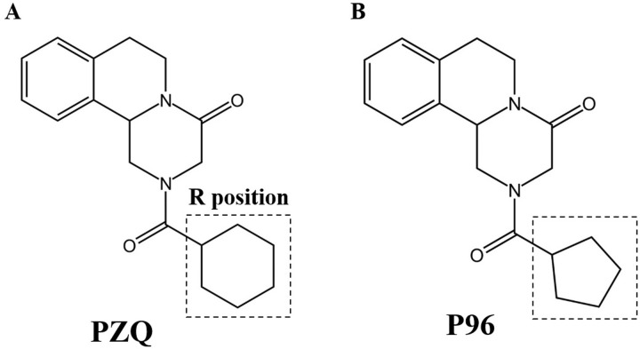
As we know, understanding the mechanism of PZQ is crucial for developing new drug candidates for chemotherapy of schistosomiasis. Many researches and designs have been conducted to try to make the mechanism of PZQ clear. Encouragingly, Park et al used ligand- and target-based methods to define a binding site for PZQ in a juxtamembrane cavity within the voltage sensor-like domain of a transient receptor potential melastatin ion channel (Sm.TRPMPZQ) in schistosomes, a broadly conserved parasitic flatworm ion channel, which could be activated by PZQ to cause calcium entry and worm paralysis [18]. Moreover, in the companion article, Le Clec’h et al used genome-wide association to map loci underlying PZQ response and identified the same transient receptor potential channel in S. mansoni determined variation in PZQ response. This channel could be activated by nanomolar concentrations of PZQ [19]. In order to characterize the pharmacological specificity of the schistosome TRP channel activated by PZQ, a series of 43 PZQ derivatives including enantiomers (R)-PZQ, (S)-PZQ and the major trans-(R)-4-OH PZQ metabolite were synthesized by Park group. The structure-activity relationships of these analogs revealed the cyclohexyl moiety (R group, Fig 1A) in PZQ is resolved as a critical determinant of efficacy. Major modifications of this moiety yielded inactive or low potency analogs [18]. The result was consistent with previous reports that structural features of the cyclohexyl group are likely related to antischistosomal activity [8]. PZQ undergoes rapid metabolism and is converted into a major trans-cyclohexanol metabolite, which is much less effective than PZQ itself [20–21]. The ketone oxidation product of the trans-cyclohexanol metabolite and other analogues with increased metabolic stability were designed and had low to modest activity against juveniles of S. japonicum and S. mansoni [18,22–23]. Although several structural changes were made in the R position (Fig 1A) with the aim of increasing the schistosomicidal activity, the vast majority of the derivatives demonstrated only low to moderate effect, their schistosomicidal activities were not comparable to PZQ [22–26]. However, in the literature reported by Park et al [18], a kind of PZQ derivative, compound 5, which was designated P96 in our group, had attracted our attention. The compound 5 with the substitution of cyclohexyl by cyclopentyl (Fig 1B), could also activate the Sm.TRPMPZQ, but had a 6-fold lower apparent affinity compared with PZQ[18].
Fig 1. Chemical structures of praziquantel (PZQ) and P96.
In this study, the derivative P96 was synthesized by School of Pharmaceutical Sciences of Shandong University, and was tested in vitro and in vivo against juvenile and adult stages of S. japonicum. Besides worm burden and egg burden, quantitative real-time PCR was employed to evaluate the antischistosomal efficacy at molecular level in vivo.
Materials and methods
Ethics statement
All the animal experiments were carried out in strict accordance with the recommendations in the Guide for the Care and Use of Laboratory Animals of the National Institutes of Health. The protocol (including mortality aspects) was approved by the Committee on the Ethics of Animal Experiments of the Soochow University (Permit Number: 2007–13).
Parasites and animals
S. japonicum infected snail (Oncomelania hupensis) were provided by the Institute of Schistosomiasis Control in Jiangsu Province (Wuxi, China). S. japonicum cercariae (Chinese mainland strain) shedding from the snails were used to infect mice models. Female ICR mice (4–6 weeks-old and weighing 15-25g) and female New Zealand rabbits (weighing 2.0–2.5kg) were provided by the Experimental Animal Center of Soochow University (Suzhou, China). All mice and rabbits were raised under specific pathogen-free conditions with controlled temperature (20 ± 2°C) and photoperiod (12 h light, 12 h dark). Each mouse was transcutaneously infected with 60±2 S. japonicum cercariae. Each rabbit was infected with 200±5 S. japonicum cercariae.
Reagents
PZQ analogue P96 was synthesized by School of Pharmaceutical Sciences of Shandong University with optimization of synthetic process reported by Park et al [18]. Briefly, 0.5 g (R)-Praziquanamine, 10 ml dichloromethane and 37.5 ml water were added into a reaction bottle on ice, adding 0.37 g anhydrous sodium carbonate into the reaction bottle. Then, 0.344 g cyclopentanoyl chloride and 2.5 ml dichloromethane were added into the reaction through a constant-pressure dropping funnel. After reaction for 30 minutes, the organic phase was separated and dried to a white powder product. PZQ powder was purchased from Sigma-Aldrich (St. Louis, MO, USA). Dulbecco’s modifed Eagle’s medium (DMEM) and penicillin/streptomycin were purchased from Life Technologies (Carlsbad, CA, USA). New-born calf serum was purchased from Biological Industries (Cromwell, CT, USA). In vitro, all chemicals were dissolved in dimethyl sulfoxide (DMSO, Fluka, Buchs, Switzerland). In vivo, all compounds were dissolved in corn oil.
In vitro treatment
Worms recovered from S. japonicum infected mice at 16 days (juvenile worms) and 35 days (adult worms) post-infection were collected through perfusion of the hepatic portal system and mesenteric veins [27]. The worms were placed in 6-well plates (Corning Costar, Corning, New York, USA) containing Dulbecco’s modified minimum Eagle’s medium (bicarbonate buffered) supplemented with 10% newborn calf serum, 100 U /ml penicillin and 100 μg/ml streptomycin, and incubated at 37°C in an atmosphere of 5% CO2 in air. Juvenile worms were divided into four groups, with five worms per well, each being tested in triplicate, as follows: group I, untreated control, incubated with complete DMEM containing 0.1% DMSO; group II, worms treated with 25 μM P96; group III, treated with 50 μM P96; group IV, treated with 100 μM PZQ. Adult worms separated by sex accepted the same treatment as juveniles. All the worms were exposed to the different compounds for about 16h, then washed three times with sterile saline, and subsequently cultured in drug-free medium. At 24, 48 and 72h post-incubation, the worms were observed under a dissecting microscope (SZX16, Olympus, Japan), and viability score was assigned as described previously [28], based on the changes of mobility and general appearance. Briefly, viability score of each worm ranged from 0 to 3: Worms with the highest score of 3, as observed in the control group during the observation period, moved more actively and softly, and the body was transparent; 2 points: Worms moved their entire bodies but stiffly and slowly, with the body translucent; 1 point: parasites moved partially and had an opaque appearance; 0 point: the worms remained contracted and did not resume movement, deemed as ‘dead’.
Scanning electron microscopy (SEM)
Ultrastructural features of tegument of schistosomes treated with P96 and PZQ were examined using SEM and were compared with control group and PZQ treatment group. For SEM, the schistosomula and male adult worms were washed three times in phosphate-buffered saline (PBS; pH 7.4) and fixed overnight at 4°C in 2.5% glutaraldehyde-PBS solution (pH 7.4). After fixation, the worms were washed again in PBS, post-fixed in 1% osmium tetroxide, dehydrated in graded ethanol, then dried for approximately 30 min. Finally, the samples were mounted on aluminum stubs, coated with gold, and examined under a Hitachi-S4700 scanning electron microscope (Chiyodaku, Japan).
In vivo treatment in mice of schistosomiasis japonica
For understanding the effect of P96 on different developmental schistosomes in vivo, female ICR mice infected with 60 ± 2 S. japonicum cercaria were randomly divided into 15 groups, with 10 mice in each group. Group 1, untreated control group, received vehicle (corn oil) only. Group 2–8, treated with an oral dose of 200 mg/kg P96 for 5 consecutive days. Treatment started at day 1(Group 2), day 3 (Group 3), day 7 (Group 4), day 14 (Group 5), day 21 (Group 6), day 28 (Group 7) and day 35 (Group 8) post-infection, respectively. Group 9–15, treated with a single dose of 200mg/kg PZQ at the same time schedule as treated with P96. In order to understand whether there was a dose-dependent effect of P96 against schistosomula, mice harbored with 14-day-old juveniles of S. japonicum were treated with P96 at a single oral dose of 100, 200, 400, 600 mg/kg for 5 consecutive days, with 8 mice in each group. At 21 days post-treatment, all mice were sacrificed to assess the worm burden and worm reduction rate.
In vivo treatment in rabbits of schistosomiasis japonica
A total of 8 female New Zealand White rabbits, weighing approximately 2.0–2.5 kg, were randomly divided into 4 groups of 2 rabbits each. Each rabbit was infected with 200 ± 5 S. japonicum cercaria. Group1, untreated control group, received vehicle (corn oil) only. Group2, treated orally with150 mg/kg P96 at 28 days post-infection. Group3, treated orally with 300 mg/kg P96 at 28 days post-infection. Group4, treated with a single oral dose of 150 mg/kg PZQ. After 3 weeks posttreatment, all the rabbits were sacrificed to recover the adult worms separated by sex for measuring the real worm burden and worm reduction rate.
Rabbit blood sample collection
Pre-infection blood samples of rabbits were collected as negative control before infection. For all the groups, S. japonicum infected blood samples were collected on the 3rd day and then weekly until 4 weeks post-infection. Blood from control group, PZQ-treated and P96-treated rabbits were collected once a day in the first week, and then weekly until 6 weeks posttreatment. Serum of each blood sample was separated by centrifugation (2000g for 10 min) after storage at 37°C for 1 h. The sera were stored at -20°C until DNA extraction.
DNA extraction
DNA from all the collected serum samples was extracted using the method described previously [29], with slight modifications. Briefly, 200 μL of infected rabbit serum were dissolved in 400 μL serum lysis buffer containing 150 mM NaCl, 10 mM EDTA, 10 mM Tris-HCl (pH 7.6), 2% SDS, 5 μg/mL salmon sperm DNA, and 250 μg/mL proteinase K (Takara, Dalian), incubated at 55°C for 1h, then extracted twice with phenol-chloroform-isoamyl alcohol (25:24:1) and precipitated with dehydrated alcohol. The DNA pellet was air-dried and dissolved in 25 μL of TE buffer (10 mM Tris-HCl, 1 mM EDTA, pH 8.0).
Design of primers
As shown in Table 1, primers were designed targeting SjR2 retrotransposon of S. japonicum. Probes were designed with 5’ terminal reporter dye FAM and 3’ terminal quencher dye TAMRA. The specificity of primers and probes were tested using a BLAST search against the Genbank database.
Table 1. Primers and probes for SjR2 quantitative real-time PCR.
| Target sequence | Forward/Reverse primer (5′→3′) | Probe (5′FAM→3′TAM) |
|---|---|---|
| SjR2 | CAGGCTTCCTTAGCTACGACTCTA | ATCCCGCTCCATCGATATCTGCTGC |
| GGATCCTGTATACGCGTTTCAGA |
Quantitative real-time PCR
The 25 μL reaction mix contained 4 μL DNA, 12.5 μL 2×Platinum qPCR Supermix-UDG (Invitrogen by life technologies), 1 μL 50 mM MgCl2, 1 μL ROX Reference Dye (1:10), 200 nM of each primer, 100 nM of probe, and distilled water to give the final volum of 25 μL. The program consisted of incubation at 50°C for 2 minutes, followed by 95°C for 2 minutes, then 45 cycles at 95°C for 15 seconds and 60°C for 45 seconds. Ten-fold serial dilutions of standard plasmid with targeting sequence of SjR2 were used to generate the standard curve to calculate the copy numbers of SjR2 DNA (S1 Fig).
Statistical analysis
All data sets were analyzed using the SPSS26.0 software package. Data of viability score were expressed as the mean value ± standard error (SE). Data of worm number and egg burden were expressed as the mean value ± standard deviation (SD). Differences between groups were analyzed by one-way ANOVA followed by Dunnett’s test. Statistical significance of the difference of the sample rates was determined by the chi-square test. A P-value<0.05 was considered to be statistically significant.
Results
P96 exhibits potent schistosomicidal effect against both juvenile and adult worm in vitro
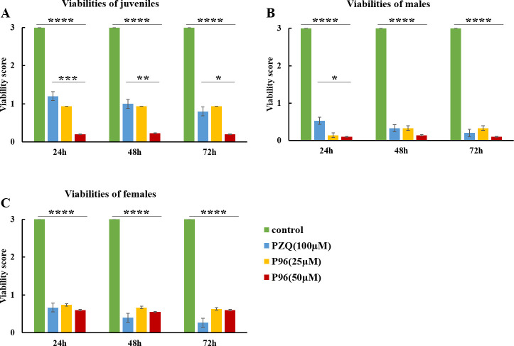
In vitro, after 24h exposure to different concentrations of P96 and PZQ, the mean viability score of males, females and juveniles was significantly decreased compared to the control group (males: F(3,68) = 260.245, P<0.0001; females: F(3,73) = 91.866, P<0.0001; juveniles: F(3,94) = 86.821, P<0.0001;). The antischistosomal effect of P96 was concentration-dependent, with P96 at 50 μM demonstrated the most obvious schistosomicidal effect against male, female and juvenile worms. The viability reduction rate of P96 at concentration of 50 μM was 96.7%, 80% and 93.3%, respectively (Table 2), which was similar (females: P = 1.000) as or even higher (males: P<0.05; juveniles: P<0.0001) than 100 μM PZQ-treated group. Unlike PZQ, the lethal effect of P96 was not time-dependent. As shown in Fig 2, from 24 h to 72 h incubation period, the viability score of P96 treated group sustained at the same level.
Table 2. In vitro effect of P96 at different concentrations against juveniles, males and females of Schistosoma japonicum.
| Concentration (μmol/L) | 24h | 48h | 72h | ||||
|---|---|---|---|---|---|---|---|
| Mortality rate (%) | Viability score (Mean ± SE)/ Viability reduction rate (%) | Mortality rate (%) | Viability score (Mean ± SE)/ Viability reduction rate (%) | Mortality rate (%) | Viability score (Mean ± SE)/ Viability reduction rate (%) | ||
| Juvenile | P96[25] | 61.1 | 0.94±0.30/68.7 | 61.1 | 0.94±0.30/68.7 | 61.1 | 0.94±0.30/68.7 |
| P96[50] | 82.2 | 0.20±0.07/93.3 | 82.2 | 0.23±0.08/92.3 | 82.2 | 0.20±0.07/93.3 | |
| PZQ[100] | 25.0 | 1.20±0.19/60.0 | 35.0 | 1.00±0.19/66.7 | 40.0 | 0.80±0.17/73.3 | |
| Control | 0 | 3.00±0.00/0.0 | 0 | 3.00±0.00/0.0 | 0 | 3.00±0.00/0.0 | |
| Male | P96[25] | 85.7 | 0.14±0.08/95.3 | 66.7 | 0.33±0.11/89.0 | 71.4 | 0.33±0.13/89.0 |
| P96[50] | 90.5 | 0.10±0.07/96.7 | 90.5 | 0.14±0.10/95.3 | 90.5 | 0.10±0.07/96.7 | |
| PZQ[100] | 46.7 | 0.53±0.13/82.3 | 66.7 | 0.33±0.13/89.0 | 80.0 | 0.2±0.11/93.3 | |
| Control | 0 | 3.00±0.00/0.0 | 0 | 3.00±0.00/0.0 | 0 | 3.00±0.00/0.0 | |
| Female | P96[25] | 29.6 | 0.74±0.10/75.3 | 33.3 | 0.67±0.09/77.6 | 37.7 | 0.63±0.09/79.0 |
| P96[50] | 40.0 | 0.60±0.11/80.0 | 50.0 | 0.55±0.14/81.7 | 40.0 | 0.60±0.11/80.0 | |
| PZQ[100] | 40.0 | 0.67±0.16/77.7 | 66.7 | 0.40±0.11/86.7 | 73.3 | 0.27±0.12/91.0 | |
| Control | 0 | 3.00±0.00/0.0 | 0 | 3.00±0.00/0.0 | 0 | 3.00±0.00/0.0 | |
Fig 2. In vitro activity of P96 against males, females and juveniles of S. japonicum.
Juvenile (A), male (B), and female (C) worms were incubated with 25 μM P96, 50 μM P96 and 100 μM PZQ for 24h, 48h and 72h of incubation in drug-free medium, following the initial exposure of parasites for 16 h to each drug and subsequent washing for drug removal. The viability was assigned using a viability score of 0–3. The control group was incubated with complete DMEM with 0.1% DMSO. ****represents significant differences compared to the control group, P<0.0001. ***represents significant differences of 50 μM P96 treatment group compared to the PZQ treatment group, P<0.0001. **represents significant differences of 50 μM P96 treatment group compared to the PZQ treatment group, P<0.01. *represents significant differences of 50 μM P96 treatment group compared to the PZQ treatment group, P<0.05.
Morphological properties by scanning electron microscopy (SEM)
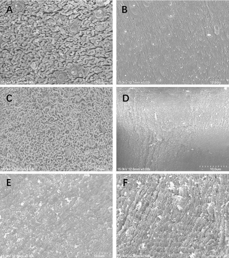
SEM studies revealed that schistosomula from control group demonstrated normal tegumental ultrastructure features (Fig 3A). Numerous ridges were uniformly arranged along the mid-body of the schistosomula (Fig 3A). After treatment with 100 μM PZQ, the ridges became swollen, however, the integrity of the tegument was not compromised (Fig 3B). In contrast, the juveniles exposed to 50 μM P96 showed significant changes in the tegument. Extensive sloughing of the tegument and severe swelling were recorded (Fig 3C). Under SEM, male S. japonicum worms from control group showed normal tegument ultrastructures (Fig 4A). The tegument of the mid-body was intact, the crests with sensory papillae were uniformly arranged along the body (Fig 4A). The inner wall of gynecophoral canal and typical ridges were preserved (Fig 4B). Males exposed to 100 μM PZQ demonstrated disarrangement of crests with swelling sensory papillae in the tegument (Fig 4C). The ridges in the inner wall of gynecophoral canal were shallow or even disappeared. Pronounced oedema, collapsed papillae and shallow peeling were observed in this area (Fig 4D). Alterations in the tegument treated by 50 μM P96 were different from that of PZQ. The tegumental structures were destroyed. The normal crests in the tegument of mid-body disappeared and fused into trabeculae. The sensory papillae were swollen, disformed, collapsed or even disappeared (Fig 4E). Severe swelling and extensive peeling of the tegument were detected in the gynecophoral canal inner wall (Fig 4F).
Fig 3. Scanning electron microscopy (SEM, ×3000) observation on the tegument of schistosomula.
(A) mid-portion of the control schistosomula in the medium with DMEM for incubation of 72h; (B) mid-portion of the worm exposed to 100 μM PZQ after 72 h of incubation with drug-free medium; (C) mid-portion of the worm exposed to 50 μM P96 after 72 h of incubation with drug-free medium.
Fig 4. Scanning electron microscopy (SEM, ×3000) observation on the tegument of male adult S.
japonicum. (A) mid-portion of the control worm; (B) inner wall of gynecophoral canal of control worm; (C) mid-portion of the worm exposed to 100 μM PZQ after 72 h of incubation with drug-free medium; (D) inner wall of gynecophoral canal of the worm exposed to 100 μM PZQ after 72 h of incubation with drug-free medium; (E) mid-portion of the worm exposed to 50 μM P96 after 72 h of incubation with drug-free medium; (F) inner wall of gynecophoral canal of the worm exposed to 50 μM P96 after 72 h of incubation with drug-free medium.
Stage-sensitivity of P96 in vivo exhibits prominent worm killing efficacy against both juveniles and adult worms
As shown in Table 3, the worm reduction rate caused by 200 mg/kg P96 in mice harbored with1-day, 3-day, 7-day and 14-day juvenile of S. japonicum ranged from 43.5% to 58.2%, which was significantly higher than 200 mg/kg PZQ treated group (9.0–27.5%), the P value was all lower than 0.05. On day 21, the effect of P96 was 45.9%, while 42.7% worm burden reduction for PZQ was observed. On day 28, adult worm stage, the worm reduction rate of P96 was 53.6%, which was similar to that of PZQ (67.1%, P = 0.157>0.05). On day 35, adult worm pairing and spawning stage, PZQ exerted the most outstanding activity, the worm reduction rate was 96.7%, however, not significantly higher than the reduction of 86.9% for P96 (P = 0.163>0.05).
Table 3. In vivo activity of P96 at a single oral dose of 200 mg/kg for 5 consecutive days against different developmental stages of S. japonicum in mice.
| Worm number (mean±SD)/worm reduction (%) | Chi-square test | P value | ||
|---|---|---|---|---|
| P96 (200 mg/kg) | PZQ (200 mg/kg) | |||
| Control a | 51.0 ± 2.1/0.0 | 51.0 ± 2.1/0.0 | / | / |
| 1-day-pi | 23.5 ± 3.5/53.9 | 37.0 ± 1.9/27.5 | χ2 = 7.428 | 0.006 |
| 3-day-pi | 28.8 ± 4.6/43.5 | 46.3 ± 4.6/9.8 | χ2 = 14.557 | <0.0001 |
| 7-day-pi | 28.5 ± 3.5/44.2 | 46.4 ± 3.4/9.0 | χ2 = 15.417 | <0.0001 |
| 14-day-pi | 21.3 ± 3.2/58.2 | 41.3 ± 2.7/19.9 | χ2 = 16.452 | <0.0001 |
| 21-day-pi | 27.6 ± 5.0/45.9 | 29.2 ± 3.3/42.7 | χ2 = 0.04 | 0.842 |
| 28-day-pi | 23.6 ± 2.3/53.6 | 16.7 ± 2.9/67.1 | χ2 = 1.998 | 0.157 |
| 35-day-pi | 6.7±0.58/86.9 | 1.7±0.6/96.7 | χ2 = 1.950 | 0.163 |
a: Mice were given an equal volume of corn oil, pi: post-infection.
The dose-response of P96 against S. japonicum juveniles in mice
Considering the promising antischistosomal efficacy of P96, we further assessed its effect against 14-day-old juveniles in S. japonicum infected mice. As shown in Table 4, with the ascending dose of P96 from 100 mg/kg to 600 mg/kg, the mean worm burden decreased, and the worm reduction rate increased from 48.1% to 68.4%, indicating that there was a dose-dependent effect of P96 against juveniles. Meanwhile, with the increasing dose of P96, the mortality of the mice declined remarkably. Fifty percent mice died in 100 mg/kg treated-group, 12.5% mice died in 200 mg/kg treated-group, no mice died in 400 mg/kg and 600 mg/kg treated-group.
Table 4. The dose-dependent effect of P96 against 14-day-old Schistosoma japonicum juveniles in mice with a daily oral dose 100–600 mg/kg for 5 consecutive days.
| Dose (mg/kg) | Worm burden of each mouse | Mean worm burden (Mean±SD) | Worm reduction (%) | |||||||
|---|---|---|---|---|---|---|---|---|---|---|
| No.1 | No.2 | No.3 | No.4 | No.5 | No.6 | No.7 | No.8 | |||
| Control | 48 | 52 | 47 | 46 | 53 | 51 | 50 | 49 | 49.5±2.4 | / |
| 100 | 30 | 27 | 22 | 26 | — | — | — | — | 25.7 ± 3.3 | 48.1 |
| 200 | 22 | 25 | 20 | 24 | 26 | 14 | 21 | — | 21.7 ± 4.0 | 56.2 |
| 400 | 23 | 18 | 17 | 23 | 19 | 20 | 18 | 21 | 19.8 ± 2.4 | 60.0 |
| 600 | 17 | 14 | 15 | 16 | 17 | 14 | 15 | 17 | 15.6 ± 1.3 | 68.4 |
—: refers to death.
The dose-response of P96 against S. japonicum adults in rabbits
Table 5 summarized the activity of P96 at different doses against 28-day-old adult worm in S. japonicum infected rabbits. Rabbits treated with 150 mg/kg P96, resulted in a statistically significant reduction in the mean total worm burden and egg burden compared with the control group (worm burden: F(3,6) = 139.655, P<0.0001, egg burden: F(3,50) = 47.399, P<0.0001). The worm reduction rate and the egg reduction rate were 65.2% and 80.1%. With the dose of P96 increasing to 300 mg/kg, the worm reduction rate was enhanced to 91.7%, which was very close to that of PZQ (98.5%) at the dose of 150 mg/kg (P = 0.661>0.05).
Table 5. Effect of P96 at different oral doses against 28-day-old Schistosoma japonicum adults in rabbit models.
| Compounds | Worm burden (Mean±SD) | Worm reduction rate(%) | Egg burden/g liver tissue (Mean±SD) | Egg reduction rate(%) | ||
|---|---|---|---|---|---|---|
| total | male | female | ||||
| P96 (150mg/kg) | 58.5±11.2* | 41.8±11.0 | 16.8±8.3 | 65.2 | 3257.8±1618.9* | 80.1 |
| P96 (300mg/kg) | 14.0±9.9* | 10.5±7.8 | 3.5±2.1 | 91.7 | 812.9±463.1* | 95.0 |
| PZQ (150mg/kg) | 2.5±0.7* | 1.5±0.7 | 1.0±0.0 | 98.5 | 400.0±228.9* | 97.6 |
| Control (corn oil) | 168.0±4.2 | 90.0±1.4 | 78.0±2.8 | / | 16378.3±8836.6 | / |
*P<0.0001, significant difference compared with control group.
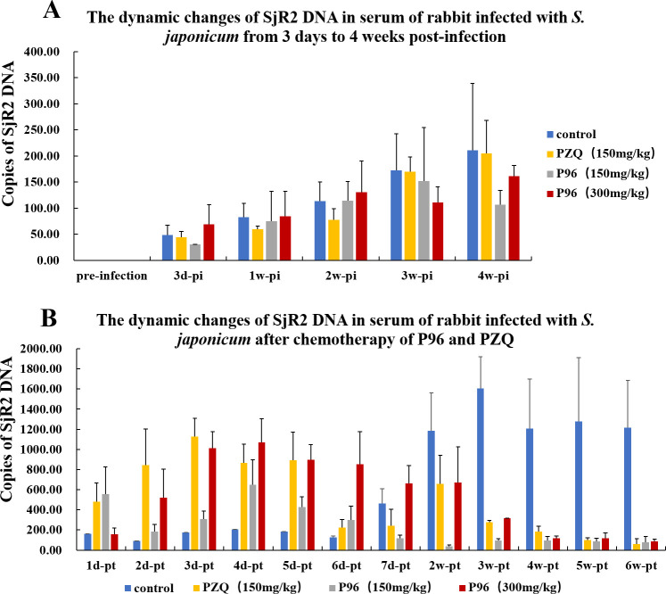
As shown in Fig 5, among the three groups of rabbits treated by different doses of PZQ and P96 at 28 days post-infection (adult worm stage), the detection results of quantitative real-time PCR showed that the copies of SjR2 DNA were very low at the beginning of the infection, the numbers were less than 300 copies (Fig 5A and S1–S4 Tables). After treatment of 150 mg/kg PZQ, the content of SjR2 DNA reached the peak at 3 days posttreatment (Fig 5B and S4 Table), which was consistent with the fact that PZQ was the most effective against schistosome adult worm. In rabbits with chemotherapy of P96 at an oral dose of 150mg/kg, the content of SjR2 DNA reached the highest on the 4rd day posttreatment, but was lower than PZQ treatment group (Fig 5B and S2 Table). With the oral dose of P96 increasing to 300mg/kg, the peak of SjR2 DNA after 4 days posttreatment was higher than that of 150mg/kg P96 treatment group, indicating that there was a dose-dependent effect of P96 against schistosome adult worm. Moreover, the content of SjR2 DNA copies in sera of rabbit with administration of 300mg/kg P96 was close to that of 150mg/kg PZQ treatment group (Fig 5B and S3 Table). As the treatment time prolonged, the content of SjR2 DNA declined quickly. At the 6th week posttreatment, the amount of SjR2 gene decreased to a very low level in rabbit with chemotherapy of 150 mg/kg PZQ, 150 mg/kg P96 and 300mg/kg P96. While in the control group, the copy numbers sustained at high level (Fig 5B and S1–S4 Tables).
Fig 5. Dynamic changes of SjR2 DNA in sera of rabbits of schistosomiasis before and after chemotherapy of different concentrations of P96 and PZQ.
(A) The copies of SjR2 DNA in rabbit sera from different treatment groups increased gradually from the 3rd day to 4 weeks post-infection (pi), but all sustained at a low level. The DNA numbers were less than 300 copies. (B) The rabbit of each group was treated with different doses of p96 and PZQ at 4weeks pi. After treatment of 150 mg/kg PZQ, the SjR2 DNA copies reached the peak at the 3rd day posttreatment (pt), and declined to a low level after 6 weeks pt. In sera of rabbit treated with 300 mg/kg P96, the SjR2 DNA copies were the highest at the 4th day pt, also declined to a low level at 6 weeks pt. The fluctuation trend of SjR2 DNA content in rabbits with treatment of 150 mg/kg P96 is consistent with the 300 mg/kg P96 treated group, but the content was lower than high concentration treated group.
Discussion
The large-scale and mono-therapeutic use of PZQ has raised many concerns for the treatment and control of schistosomiasis. The concerns mainly focused on the lack of activity against immature schistosome and the growing inclination to resistance of PZQ, which could be explanations to the poor cure rates and treatment failures in residents of high-risk regions [30]. Thus, it is of increasing importance to develop new drugs in the face of potential resistance to PZQ for the treatment of schistosomiasis, and the absence of an effective vaccine [31]. It is well known that PZQ has been the first treatment for several decades, the mechanism of action of PZQ in schistosomes has been extensively studied and discussed by many groups. However, Park team has made an important breakthrough in the field of discovering the mechanism of action of PZQ. In 2019, they demonstrated that PZQ activated a schistosome transient receptor potential (TRP) channel and defined the properties of Sm.TRPMPZQ [32]. In their following study, the authors have confirmed that Sm.TRPMPZQ is the binding site for (R)-PZQ in a juxtamembrane cavity within the voltage sensor–like domain (VSLD) of Sm.TRPMPZQ, a broadly conserved ion channel in parasitic schistosomes and other PZQ-sensitive parasites [18]. Coincidentally, Le Clec’h et al [19] used genome-wide association to map loci underlying PZQ response and also identified a transient receptor potential (Sm.TRPMPZQ) channel within the major chromosome 3 peak that could be activated by nanomolar concentrations of PZQ, and they demonstrated the genetic basis of variation in the response to PZQ in a PZQ-selected S. mansoni population, which identified that Sm.TRPMPZQ SNP underly variation in PZQ responses in S. mansoni [19]. It is exciting to see that different groups have used different methods to prove the same target that the Sm.TRPMPZQ channel is an important druggable target for schistosomiasis caused by Schistosoma spp. In order to determine the pharmacological specificity of Sm.TRPMPZQ channel, a series of 43 PZQ derivatives including one with the substitution of cyclohexyl by cyclopentyl (compound 5), christened P96 in this study, were synthesized and tested by Park et al.[18]. The ability of the analogs to activate Sm.TRPMPZQ channel and to contract adult S. mansoni worms, showed similar structure-activity relationships (SARs), with only small structural modifications of PZQ preserving activity at Sm.TRPMPZQ [18]. There was only a small change in structure of P96, but the schistosomicidal effect was significantly improved. It can cause obviously contraction in S. mansoni adult worms [18], and can also cause rigidity contraction and reduction of viability in S. japonicum worms (Fig 2 and Table 2).
It is popular and thought to be a good strategy to develop novel antischistosomal agents to synthesize new PZQ derivatives [33]. Substantial research has been conducted to the design and development of PZQ derivatives. The chemical modifications are mainly concentrated on the cyclohexane ring and aromatic ring of praziquantel [34–35]. Unfortunately, vast majority of the analogues are not promising compounds, with low or moderate antischistosomal activity, being not comparable to PZQ [22,26,33–37]. Although no compelling agents have gone to clinical testing and trials, the results are of major importance for the analysis of its chemical structure for its activity of PZQ. As mentioned by Patra et al [36], whose studies demonstrated that the C10 aromatic ring of PZQ is not suitable for structural modification [36]. To impede the metabolism and increase metabolic stability, three different cycloalkyls substituents (cyclobutyl, cyclopentyl and cyclohexyl) with a carbonyl group in its structure were used for the synthesis of the derivatives. The results exhibited as the size of ring with carbonyl group enlarged, antischistosomal activity of R-isomers increased from 41.0% to 60.0%, indicating the size of the ring is important for the activity, with the cyclohexyl has the most effective activity [22]. The available results indicated that the derivatives structurally linked to PZQ through the metabolically liable cyclohexyl ring position might not afford active derivatives [22]. These results are consistent with the findings of Park et al that the cyclohexyl moiety in PZQ is resolved as a critical determinant of efficacy. Major modifications of this moiety yielded inactive or low potency analogs, only minor alterations, such as P96, preserved comparable potency with (R)-PZQ [18].
The derivative P96 with substitution of cyclohexyl by cyclopentyl, could this small change bring us a big surprise? Unlike previously reported derivatives, most of them demonstrating less activity than PZQ, P96 exerted potent antischistosmal efficacy against both juveniles and adults of S. japonicum in vitro. The schistosomicidal effect of P96 in vitro was dose-dependent, with a concentration at 50μM demonstrating the best activity (Fig 2 and Table 2). Male adult worms seemed to be more sensitive than females of the same age (Table 2). This result was supported by the research of Le Clec’h et al [19], which demonstrated that expression of Sm.TRPMPZQ determined the response of schistosomal worms to PZQ. The expression of Sm.TRPMPZQ was 11.94-fold lower in female than in male worms, consistent with females being naturally resistant to PZQ. Due to the similar structure of P96 compared with PZQ, the female worms of S. japonicum were not as sensitive as male adult worms. As we know that PZQ is highly effective against adults, but has poor activity against juveniles. This phenomenon may partially be attributed to the lower expression level of isoform 6 of Sm.TRPMPZQ [19]. However, in our current study, P96 exhibited the most prominent activity against 16-day-old juveniles (82.2% of mortality, Table 2) compared to PZQ (25.0% of mortality, P<0.0001, Fig 2), and still retained high efficacy against adult worms, with a significant reduction of 96.7% in male worm viability compared to 82.3% of PZQ after 24h exposure (P<0.05, Fig 2). Our in vitro experiments confirmed that this small change of the structure of PZQ, substitution of cyclohexyl with cyclopentyl, did significantly improved the schistosomicidal efficacy of P96, especially for the effect against young stages of S. japonicum.
Furthermore, integrality of the tegument plays a key role for worm survival, the parasite is vulnerable to the host immune system because of the surface antigens exposure [8,33]. Our SEM observation revealed that P96 caused severe damage to the schistosomula tegument, including sloughing of the tegument with a disordered surface. Whereas PZQ only caused very light damage to the juvenile tegument, the morphological change might partially explain the poor activity of PZQ against immature schistosomes (Fig 3). The ultrastructural alterations of male adult worm treated by P96 were similar to that of PZQ (Fig 4), indicating that the potent antischistosomal activities of P96 might be correlated with its effects on worm tegument.
Although our in vitro results showed that P96 had promising antischistosomal activity, it was worth noting that potential action in vitro did not translate to impressive killing in vivo. As reported by Patra et al [38], upon alteration of the organometallic moiety to Cr (CO)3, the derivatives exhibited marked activity against S. mansoni in vitro, however, they exerted low activity in vivo [38]. In this study, two kinds of animal models were employed to evaluate the efficacy of P96 in vivo. In S. japonicum infected mouse model, P96 demonstrated outstanding antichistosomal activity against all worm developmental stage. The most prominent was it presented remarkable schistosomicidal activity against young stages superior to PZQ. Moreover, it retained the high efficacy against the 35-day-old adult S. japonicum with a reduction rate of 86.9%, which was similar to PZQ (Table 3). Our in vivo results also confirmed that the efficacy of PZQ was stage-dependent, as the worm getting mature, demonstrating the most severe worm killing effect (Table 3). In addition, the dose-response study in mice revealed that there was a dose-dependent effect of P96 against juveniles, with the highest oral dose at 600mg/kg achieved the maximum worm reduction rate (68.4%, Table 4). Thereafter, the activity of P96 was further tested in a large size rabbit model of schistosomiasis. The results also demonstrated a dose-dependent effect of P96 against adult worm. With the ascending dose of P96, the more promising reduction of worm burden was observed. At concentration of 300mg/kg, the worm reduction rate reached 91.7%, which was very close to 98.5% of PZQ (Table 5). However, further experiments are needed to identify the optimal dose for P96 in vivo.
Our quantitative real-time PCR detection results confirmed the prominent antischistosomal activity of P96. In our previous study, we have demonstrated that the specific SjR2 DNA of S. japonicum in rabbit sera mainly came from the residual body of dead worms and the disintegration of inactive eggs after chemotherapy of PZQ [39]. In this study, the SjR2 DNA detection results revealed that the content of SjR2 DNA achieved at the highest level on the 3rd day posttreatment of 150 mg/kg PZQ, indicating the higher copies of SjR2 DNA might represent better worm killing effect. In sera of rabbits with oral administration of 150 mg/kg P96, the content of SjR2 DNA reached its peak at 4 days posttreatment, but lower than that of PZQ treatment. With the dose of P96 increasing to 300 mg/kg, the copy numbers of SjR2 DNA were close to 150 mg/kg PZQ after 4 days treatment, which further verified that P96 has comparable activity to PZQ when tested against adult S. japonicum.
Conclusion
Our in vitro and in vivo results demonstrated that P96 with a small structure change, cyclohexyl substituted by cyclopentyl, had broad-spectrum antischistosomal activity, especially against immature stages. Its remarkable schistosomicidal efficacy against both young stage and adult worm of S. japonicum enabled it could serve as the promising drug candidate for treatment and control of schistosomiasis. Further studies were needed to elucidate the in vivo metabolism of P96, as well as its mechanism of action against schistosomes.
Supporting information
The target DNA sequence of SjR2 was cloned into plasmids using pMD20-T II cloning reagent Kit (Tiangen, Beijing, China), and the plasmids were purified by TIAN pure Mini Plasmid Kits (Tiangen, Beijing, China). Sequencing of the cloned amplification product confirmed that it was identical to part of the Schistosoma japonicum retrotransposon SjR2. The plasmid was tested in a series of 10-fold dilutions by quantitative real-time quantitative PCR. A standard curve of SjR2 DNA was constructed, resulting in a detection limit of 4.0 copies of SjR2 DNA. The correlation coefficient (R2) was 0.9982 and the slope was -3.2983.
(TIF)
Detection of SjR2 DNA in serum of each rabbit at different time points was repeated three times. “/” represented undetected by quantitative real-time PCR, “pi” represented post-infection, “pt” represented posttreatment. This data was demonstrated in Fig 5.
(XLSX)
Detection of SjR2 DNA in serum of each rabbit at different time points was repeated three times. “/” represented undetected by quantitative real-time PCR, “pi” represented post-infection, “pt” represented posttreatment. This data was demonstrated in Fig 5.
(XLSX)
Detection of SjR2 DNA in serum of each rabbit at different time points was repeated three times. “/” represented undetected by quantitative real-time PCR, “pi” represented post-infection, “pt” represented posttreatment. This data was demonstrated in Fig 5.
(XLSX)
Detection of SjR2 DNA in serum of each rabbit at different time points was repeated three times. “/” represented undetected by quantitative real-time PCR, “pi” represented post-infection, “pt” represented posttreatment. This data was demonstrated in Fig 5.
(XLSX)
Acknowledgments
The authors would like to thank the Marine College of Shandong University for synthesis of the compound P96 for this study.
Data Availability
All relevant data are within the manuscript.
Funding Statement
This work was financially supported by the National High-Tech Program of China (863 Project No. 2012AA020306 to DQS and CMX), National Natural Science Foundation of China (No. 82172294 to CMX, No.81902083 to JX), Priority Academic Program Development of Jiangsu Higher Education Institutions (No. YX13400214 to Bin Li, Dean of School of Biology & Basic Medical Sciences, Suzhou Medical College of Soochow University).
References
- 1.World Health Organization. Schistosomiasis (WHO, 2021). https://apps.who.int/neglected_diseases/ntddata/sch/sch.html.
- 2.Molehin AJ, McManus DP, You H. Vaccines for Human Schistosomiasis: Recent Progress, New Developments and Future Prospects. Int J Mol Sci. 2022; 23(4):2255. doi: 10.3390/ijms23042255 [DOI] [PMC free article] [PubMed] [Google Scholar]
- 3.McManus DP, Dunne DW, Sacko M, Utzinger J, Vennervald BJ, Zhou XN. Schistosomiasis. Nat Rev Dis Primers. 2018; 4(1): 13. doi: 10.1038/s41572-018-0013-8 [DOI] [PubMed] [Google Scholar]
- 4.Mohamud A Verjee. Schistosomiasis: Still a Cause of Significant Morbidity and Mortality. Res Rep Trop Med. 2019; 10:153–163. doi: 10.2147/RRTM.S204345 [DOI] [PMC free article] [PubMed] [Google Scholar]
- 5.Colley D.G. Morbidity control of schistosomiasis by mass drug administration: How can we do it best and what will it take to move on to elimination? Trop Med Health. 2014; 42 (2 Suppl): 25–32. [DOI] [PMC free article] [PubMed] [Google Scholar]
- 6.Colley DG, Bustinduy AL, Secor WE, King CH. Human schistosomiasis. Lancet. 2014; 383(9936): 2253–2264. doi: 10.1016/S0140-6736(13)61949-2 [DOI] [PMC free article] [PubMed] [Google Scholar]
- 7.Ogongo P, Nyakundi RK, Chege GK, Ochola L. The Road to Elimination: Current State of Schistosomiasis Research and Progress Towards the End Game. Front Immunol. 2022; 13:846108. doi: 10.3389/fimmu.2022.846108 [DOI] [PMC free article] [PubMed] [Google Scholar]
- 8.da Silva VBR, Campos BRKL, de Oliveira JF, Decout JL, do Carmo Alves de Lima M. Medicinal chemistry of antischistosomal drugs: Praziquantel and oxamniquine. Bioorg Med Chem. 2017; 25(13):3259–3277. doi: 10.1016/j.bmc.2017.04.031 [DOI] [PubMed] [Google Scholar]
- 9.Fallon PG, Doenhoff MJ. Drug-resistant schistosomiasis: resistance to praziquantel and oxamniquine induced in Schistosoma mansoni in mice is drug specific. Am J Trop Med Hyg. 1994; 51(1):83–88. doi: 10.4269/ajtmh.1994.51.83 [DOI] [PubMed] [Google Scholar]
- 10.Stelma FF, Talla I, Sow S, Kongs A, Niang M, Polman K, et al. Efficacy and side effects of praziquantel in an epidemic focus of Schistosoma mansoni. Am J Trop Med Hyg. 1995; 53(2):167–170. doi: 10.4269/ajtmh.1995.53.167 [DOI] [PubMed] [Google Scholar]
- 11.Fu S, Zhou XZ, Fan Y, Wu HM, Zhou XL. Comparative study on various dose schedules of praziquantel in the treatment of schistosomiasis japonica. Ji Sheng Chong Xue Yu Ji Sheng Chong Bing Za Zhi. 1983;1(1):32–36. Chinese. [PubMed] [Google Scholar]
- 12.Liang YS, Coles GC, Doenhoff MJ, Southgate VR. In vitro responses of praziquantel-resistant and -susceptible Schistosoma mansoni to praziquantel. Int J Parasitol. 2001; 31(11):1227–1235. doi: 10.1016/s0020-7519(01)00246-6 [DOI] [PubMed] [Google Scholar]
- 13.Pica-Mattoccia L, Cioli D. Sex- and stage-related sensitivity of Schistosoma mansoni to in vivo and in vitro praziquantel treatment. Int J Parasitol. 2004; 34(4): 527–533. doi: 10.1016/j.ijpara.2003.12.003 [DOI] [PubMed] [Google Scholar]
- 14.Ismail M, Botros S, Metwally A, William S, Farghally A, Tao LF, et al. Resistance to praziquantel: direct evidence from Schistosoma mansoni isolated from Egyptian villagers. Am J Trop Med Hyg. 1999; 60(6):932–935. doi: 10.4269/ajtmh.1999.60.932 [DOI] [PubMed] [Google Scholar]
- 15.Couto FF, Coelho PM, Araújo N, Kusel JR, Katz N, Jannotti-Passos LK, et al. Schistosoma mansoni: a method for inducing resistance to praziquantel using infected Biomphalaria glabrata snails. Mem Inst Oswaldo Cruz. 2011;106(2):153–157. doi: 10.1590/s0074-02762011000200006 [DOI] [PubMed] [Google Scholar]
- 16.Qian K, Liang Y, Wang W, Qu G, Li H, Yang Z, et al. Effect of praziquantel treatment on hepatic egg granulomas in mice infected with praziquantel-susceptible and resistance Schistosoma japonicum isolates. Southeast Asian J Trop Med Public Health. 2018;49(2):208–216. [Google Scholar]
- 17.Melman SD, Steinauer ML, Cunningham C, Kubatko LS, Mwangi IN, Wynn NB, et al. Reduced susceptibility to praziquantel among naturally occurring Kenyan isolates of Schistosoma mansoni. PLoS Negl Trop Dis. 2009; 3(8): e504. doi: 10.1371/journal.pntd.0000504 [DOI] [PMC free article] [PubMed] [Google Scholar]
- 18.Park SK, Friedrich L, Yahya NA, Rohr CM, Chulkov EG, Maillard D, et al. Mechanism of praziquantel action at a parasitic flatworm ion channel. Sci Transl Med. 2021; 13(625):eabj5832. doi: 10.1126/scitranslmed.abj5832 [DOI] [PMC free article] [PubMed] [Google Scholar]
- 19.Le Clec’h W, Chevalier FD, Mattos ACA, Strickland A, Diaz R, McDew-White M, et al. Genetic analysis of praziquantel response in schistosome parasites implicates a transient receptor potential channel. Sci Transl Med. 2021;13(625):eabj9114. doi: 10.1126/scitranslmed.abj9114 [DOI] [PMC free article] [PubMed] [Google Scholar]
- 20.Lima RM, Ferreira MAD, Ponte TMJ, Marques MP, Takayanagui OM, Garcia HH, et al. Enantioselective analysis of praziquantel and trans-4-hydroxypraziquantel in human plasma by chiral LC-MS/MS: Application to pharmacokinetics. J Chromatogr B Analyt Technol Biomed Life Sci. 2009; 877(27):3083–3088. doi: 10.1016/j.jchromb.2009.07.036 [DOI] [PubMed] [Google Scholar]
- 21.Andrews P. Praziquantel: mechanisms of anti-schistosomal activity. Pharmacol Ther. 1985; 29(1):129–156. doi: 10.1016/0163-7258(85)90020-8 [DOI] [PubMed] [Google Scholar]
- 22.Zheng Y, Dong LL, Hu CY, Zhao B, Yang CH, Xia CM, et al. Development of chiral praziquantel analogues as potential drug candidates with activity to juvenile Schistosoma japonicum. Bioorg Med Chem Lett. 2014; 24(13):4223–4226. doi: 10.1016/j.bmcl.2014.07.039 [DOI] [PubMed] [Google Scholar]
- 23.Dong YX, Chollet J, Vargas M, Mansour NR, Bickle Q, Alnouti Y, et al. Praziquantel analogs with activity against juvenile Schistosoma mansoni. Bioorg Med Chem Lett. 2010; 20(8):2481–2484. doi: 10.1016/j.bmcl.2010.03.001 [DOI] [PubMed] [Google Scholar]
- 24.Ronketti F, Ramana AV, Chao-Ming X, Pica-Mattoccia L, Cioli D, Todd MH. Praziquantel derivatives I: modification of the aromatic ring. Bioorg Med Chem Lett. 2007; 17(15):4154–4157. doi: 10.1016/j.bmcl.2007.05.063 [DOI] [PubMed] [Google Scholar]
- 25.Guglielmo S, Cortese D, Vottero F, Rolando B, Kommer VP, Williams DL, et al. New praziquantel derivatives containing NO-donor furoxans and related furazans as active agents against Schistosoma mansoni. Eur J Med Chem. 2014; 84:135–145. doi: 10.1016/j.ejmech.2014.07.007 [DOI] [PMC free article] [PubMed] [Google Scholar]
- 26.Wang Z, Chen J, Qiao C. Praziquantel derivatives with antischistosomal activity: aromatic ring modification. Chem Biol Drug Des. 2013; 82(2):216–225. doi: 10.1111/cbdd.12153 [DOI] [PubMed] [Google Scholar]
- 27.Duvall RH, DeWitt WB. An improved perfusion technique for recovering adult schistosomes from laboratory animals. Am J Trop Med Hyg. 1967; 16(4):483–486. doi: 10.4269/ajtmh.1967.16.483 [DOI] [PubMed] [Google Scholar]
- 28.Santiago Ede F, de Oliveira SA, de Oliveira Filho GB, Moreira DR, Gomes PA, da Silva AL, et al. Evaluation of the anti-Schistosoma mansoni activity of thiosemicarbazones and thiazoles. Antimicrob Agents Chemother. 2014; 58(1):352–363. doi: 10.1128/AAC.01900-13 [DOI] [PMC free article] [PubMed] [Google Scholar]
- 29.Xia CM, Rong R, Lu ZX, Shi CJ, Xu J, Zhang HQ, et al. Schistosoma japonicum: A PCR assay for the early detection and evaluation of treatment in a rabbit model. Exp Parasitol. 2009; 121(2): 175–179. doi: 10.1016/j.exppara.2008.10.017 [DOI] [PubMed] [Google Scholar]
- 30.Ndamse CC, Masamba P, Kappo AP. Bioorganometallic Compounds as Novel Drug Targets against Schistosomiasis in Sub-Saharan Africa: An alternative to Praziquantel? Adv Pharm Bull. 2022; 12(2): 283–297. doi: 10.34172/apb.2022.029 [DOI] [PMC free article] [PubMed] [Google Scholar]
- 31.Lee EF, Young ND, Lim NT, Gasser RB, Fairlie WD. Apoptosis in schistosomes: toward novel targets for the treatment of schistosomiasis. Trends Parasitol. 2014; 30(2):75–84. doi: 10.1016/j.pt.2013.12.005 [DOI] [PubMed] [Google Scholar]
- 32.Park SK, Gunaratne GS, Chulkov EG, Moehring F, McCusker P, Dosa PI, et al. The anthelmintic drug praziquantel activates a schistosome transient receptor potential channel. J Biol Chem. 2019; 294(49):18873–18880. doi: 10.1074/jbc.AC119.011093 [DOI] [PMC free article] [PubMed] [Google Scholar]
- 33.Vale N, Gouveia MJ, Rinaldi G, Brindley PJ, Gärtner F, Correia da Costa JM. Praziquantel for Schistosomiasis: Single-Drug Metabolism Revisited, Mode of Action, and Resistance. Antimicrob Agents Chemother. 2017; 61(5): e02582–16. doi: 10.1128/AAC.02582-16 [DOI] [PMC free article] [PubMed] [Google Scholar]
- 34.Ong YC, Roy S, Andrews PC, Gasser G. Metal Compounds against Neglected Tropical Diseases. Chem Rev. 2019; 119(2):730–796. doi: 10.1021/acs.chemrev.8b00338 [DOI] [PubMed] [Google Scholar]
- 35.Hess J, Keiser J, Gasser G. Toward organometallic antischistosomal drug candidates. Future Med Chem. 2015; 7(6):821–830. doi: 10.4155/fmc.15.22 [DOI] [PubMed] [Google Scholar]
- 36.Patra M, Ingram K, Pierroz V, Ferrari S, Spingler B, Keiser J, et al. Ferrocenyl derivatives of the anthelmintic praziquantel: design, synthesis, and biological evaluation. J Med Chem. 2012; 55(20):8790–8798. doi: 10.1021/jm301077m [DOI] [PubMed] [Google Scholar]
- 37.Sadhu PS, Kumar SN, Chandrasekharam M, Pica-Mattoccia L, Cioli D, Rao VJ. Synthesis of new praziquantel analogues: potential candidates for the treatment of schistosomiasis. Bioorg Med Chem Lett. 2012; 22(2):1103–1106. doi: 10.1016/j.bmcl.2011.11.108 [DOI] [PubMed] [Google Scholar]
- 38.Patra M, Ingram K, Pierroz V, Ferrari S, Spingler B, Gasser RB, et al. [(η(6)-Praziquantel)Cr(CO)3] derivatives with remarkable in vitro anti-schistosomal activity. Chemistry. 2013; 19(7):2232–2235. [DOI] [PubMed] [Google Scholar]
- 39.Xu J, Liu AP, Guo JJ, Wang B, Qiu SJ, Sun H, et al. The sources and metabolic dynamics of Schistosoma japonicum DNA in serum of the host. Parasitol Res. 2013; 112(1):129–133. doi: 10.1007/s00436-012-3115-3 [DOI] [PubMed] [Google Scholar]
Associated Data
This section collects any data citations, data availability statements, or supplementary materials included in this article.
Supplementary Materials
The target DNA sequence of SjR2 was cloned into plasmids using pMD20-T II cloning reagent Kit (Tiangen, Beijing, China), and the plasmids were purified by TIAN pure Mini Plasmid Kits (Tiangen, Beijing, China). Sequencing of the cloned amplification product confirmed that it was identical to part of the Schistosoma japonicum retrotransposon SjR2. The plasmid was tested in a series of 10-fold dilutions by quantitative real-time quantitative PCR. A standard curve of SjR2 DNA was constructed, resulting in a detection limit of 4.0 copies of SjR2 DNA. The correlation coefficient (R2) was 0.9982 and the slope was -3.2983.
(TIF)
Detection of SjR2 DNA in serum of each rabbit at different time points was repeated three times. “/” represented undetected by quantitative real-time PCR, “pi” represented post-infection, “pt” represented posttreatment. This data was demonstrated in Fig 5.
(XLSX)
Detection of SjR2 DNA in serum of each rabbit at different time points was repeated three times. “/” represented undetected by quantitative real-time PCR, “pi” represented post-infection, “pt” represented posttreatment. This data was demonstrated in Fig 5.
(XLSX)
Detection of SjR2 DNA in serum of each rabbit at different time points was repeated three times. “/” represented undetected by quantitative real-time PCR, “pi” represented post-infection, “pt” represented posttreatment. This data was demonstrated in Fig 5.
(XLSX)
Detection of SjR2 DNA in serum of each rabbit at different time points was repeated three times. “/” represented undetected by quantitative real-time PCR, “pi” represented post-infection, “pt” represented posttreatment. This data was demonstrated in Fig 5.
(XLSX)
Data Availability Statement
All relevant data are within the manuscript.


