

Abstract
Superior semicircular canal dehiscence (SCD) repair through the middle cranial fossa approach is typically performed on a patient in the supine position and the patient’s head is turned to the contralateral side and secured with surgical pinions or supported on a headrest. However, traditional supine positioning method may place strain on the patient’s neck, limit the surgeon’s visualization of the dehiscence due to inadequate head rotation, and compromise the ergonomic positioning of the surgeon. Here, we present a novel positioning method for middle fossa SCD repair that allows for optimal head rotation in a semi-supine position, requires less set-up and patient manipulation than the park bench position, and does not require the use of surgical pins.
Keywords: middle fossa craniotomy, patient positioning, SCD, SCDS, skull base, superior semicircular canal dehiscence
INTRODUCTION
Superior semicircular canal dehiscence syndrome (SCDS) is characterized by sound- and pressure-induced vertigo that is associated with dehiscence of the temporal bone over the superior semicircular canal.1 SCDS may be surgically treated with plugging or resurfacing of the superior semicircular canal dehiscence (SCD) via a middle cranial fossa (MCF) or transmastoid approach.2,3 The MCF approach for SCD repair is traditionally performed with the patient in the supine position.
In the supine position, the patient’s head is turned to the contralateral side and secured with surgical pins, placed on a horseshoe head holder, or supported on a flat head plate on the bed. The park bench position involves placing the patient on their side and provides for further head rotation. However, the park bench position requires extensive manipulation of the patient which is time-consuming, and increases the risk of endotracheal tube dislodgement and shoulder or arm injury.4,5 Moreover, the trajectory provided by the park bench position better serves a retro-sigmoid, posterior fossa approach. Furthermore, the geometry of the superior canal does not require such a severe head turn towards the floor. Instead, a lateral approach to the arc of the superior canal requires 70–90° of head rotation: 45° to account for the angle of the canal from the sagittal plane and an additional 20– 40° to allow the surgeon to access the arc of the canal from its lateral aspect. This amount of head rotation can only be achieved in a minority of patients with their bodies in the supine position. Even then, such a head-on-neck position risks compression of the ipsilateral jugular vein, which could lead to excess intracranial pressure.
Here we present a novel method of positioning the patient for SCD repair that allows for optimal rotation of the head in a semi-supine position, requires less set-up and patient manipulation than the park bench position, and does not require the use of surgical pins. The proposed method also allows the surgeon to maintain an ergonomically-ideal posture during microdissection, avoiding tilting of the surgeon’s head on the torso.
METHODS
A review of patients who were scheduled to undergo SCD surgery via a MCF approach by the senior author (J.P.C.) was performed. A case was selected to demonstrate the novel positioning during SCD surgery using an MCF approach. Written consent was obtained from the patient for the use of photos and video.
Technique
The patient is brought to the operating room and general anesthesia is induced. Following endotracheal intubation, the bed is rotated 180° away from anesthesia and the head is marked for placement of the surgical navigation post on the side of the patient’s head closest to the navigation tower (Fig. 1). The scalp is prepped in a sterile fashion, and the navigation post is placed. The head is then placed on a horseshoe headrest, and the head is turned to the contralateral side. The skin incision is marked, injected with local anesthesia, and auditory brainstem neuromonitoring probes are placed (Fig. 2). Facial nerve monitors are placed.
Fig. 1.

Following intubation, the bed is turned 180° away from anesthesia and the head is marked (white arrow) and then prepped in the usual sterile fashion (not pictured) for placement of the surgical navigation post on the ipsilateral side of the patient’s head.
Fig. 2.
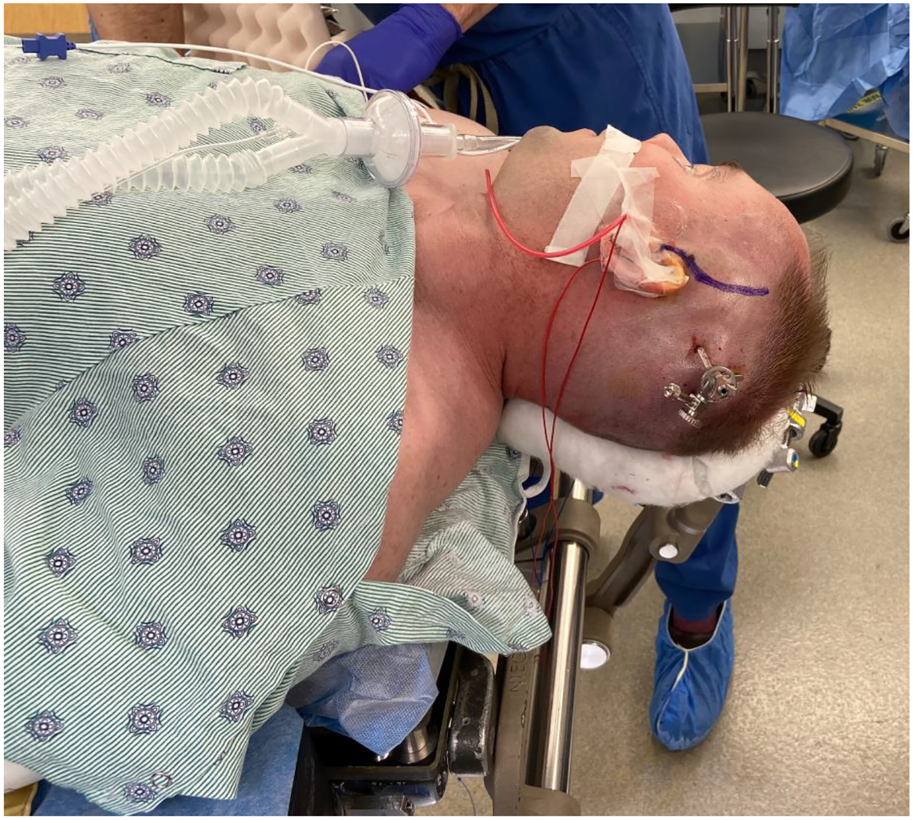
After placement of the surgical navigation post, the patient’s head is turned to the contralateral side and the incision is marked. Auditory brainstem neuromonitoring probes are then placed.
The patient is initially positioned supine on the operating table with a drawsheet beneath them. The drawsheet must be of sufficient length on either side to wrap the patient with their arms at their sides. The upper edge of the drawsheet reaches the axillae, and the torso is positioned such that the rib cage is aligned with the contralateral edge of the bed; this accounts for any tendency of the body to shift when the operating table is tilted in that direction. Padding is placed across the patient’s chest, and the patient is secured to the operating table with a circumferential wrap using three-inch silk tape around the bed and the chest (Fig. 3A). The arms are not taped within this circumfer-ential chest wrap because doing so might increase the risk of brachial plexus injury from compression should the torso shift towards the arm with bed rotation. Instead, the arms are first wrapped in foam or gel padding at the levels of the wrists and elbows and then cradled in place by tying the drawsheet across the patient’s body at the level of their chest and hips (Fig. 3B). The arms should be positioned slightly anterior to the body when secured with the bedsheet. This avoids both brachial plexus injury from scapular retraction and hyperextension of the elbows. Foam pads should be placed beneath the knots used to tie the bedsheet. Additional safety straps are placed across the hips and legs to secure the patient to the bed.
Fig. 3.

The patient is positioned with tape across the chest (white arrow), but not the arms to avoid brachial plexus injury (A). The arms are secured using a bedsheet (B). Taping the torso to the bed minimizes the risk that it will shift beyond the edge of the bed when the bed is titled towards the contralateral side.
The horseshoe headrest is positioned so that the patient’s head is turned as far as possible to the contralateral side without placing undue stress on the patient’s neck.
The operating table is then tilted towards the contralateral side to bring the patient’s temporal cranium to a plane parallel to the operating room floor (Fig. 4). The surgical site is then prepped and draped (Fig. 5). This positioning technique is further demonstrated in the Supplemental Video.
Fig. 4.
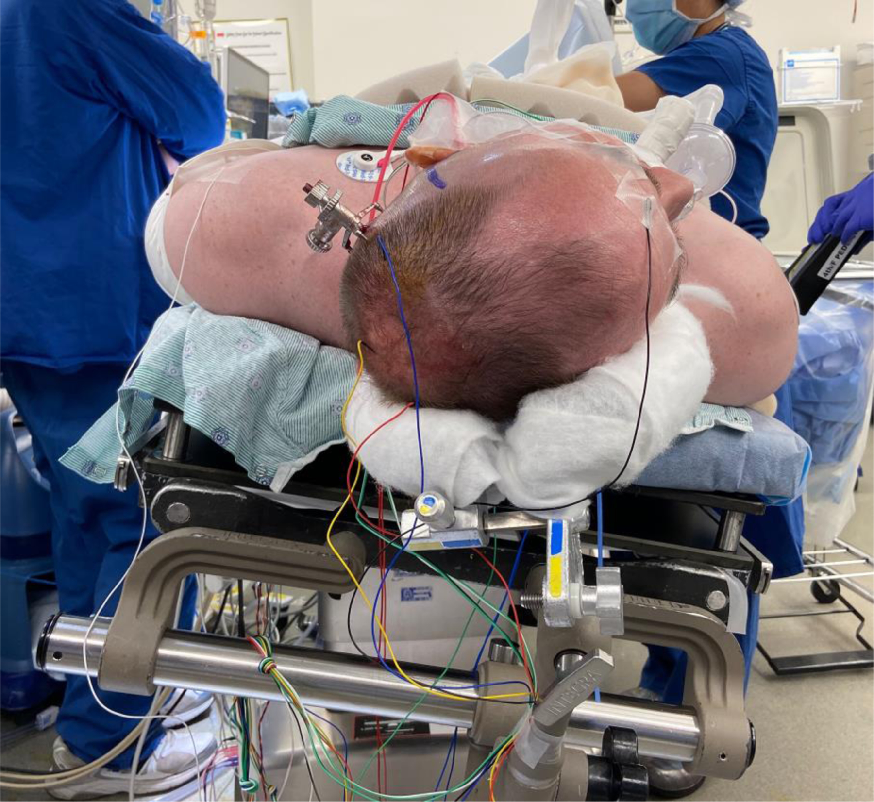
The patient is tilted to the contralateral side and the horseshoe headrest is adjusted to place the temporal cranium in a parallel plane relative to the floor.
Fig. 5.

Final view of the operative field following the placement of surgical drapes.
Assessment of Positioning Technique.
A retrospective chart review was performed for the last 100 individuals who underwent MCF SCD repair by the senior author (J.P.C.) and were positioned using this technique to evaluate its efficacy. All cases occurred between the years 2019–2022. Medical records were reviewed for the incidence of intra-operative endotracheal tube dislodgement, adequacy of surgical exposure to complete SCD repair without patient repositioning, and post-operative patient neck or shoulder discomfort. Institutional review board approval (IRB00324480) was obtained for retrospective chart review.
RESULTS
Case Example
A 43-year-old man presented with bilateral (L > R) symptoms of SCDS that included loud sound-induced vertigo and exertion-induced vertigo while running or performing other forms of exercise. His symptoms also included a distortion of his voice (autophony), pulsatile oscillopsia, and a “crackling” sound in his left ear. An audiogram showed bilateral low-frequency air-bone gaps and negative bone-conduction lines. On physical exam, positive pressure within the external auditory canal produced eye movements within the plane of the superior canal bilaterally (Hennbert sign). Ocular vestibular evoked myogenic potential (oVEMPs) amplitudes were elevated on the right and normal on the left. A computed tomography scan showed bilateral SCD and a left tegmen tympani defect with dura contacting the malleus. He was diagnosed with bilateral SCDS. Given that his symptoms were worse on the left, the decision was made to proceed with left superior semicircular canal plugging and resurfacing and tegmen repair with image guidance using an MCF approach.
The patient was positioned as described above and excellent exposure of the left superior canal and tegmen tympani defects were achieved while allowing the surgeon to maintain a favorable ergonomic position. Appropriate patient positioning was achieved with minimal manipulation of the patient following intubation. The left superior semicircular canal was plugged with fascia, bone dust, and bone chips, and a portion of the craniotomy bone flap was placed above the tegmen tympani defect. The superior canal and tegmen tympani were resurfaced using bone cement, temporalis fascia, and fibrin sealant. The bone flap was re-affixed with titanium cranioplasty hardware. Post-operatively the patient experienced an improvement in his left-side SCDS symptoms and denied any neck, arm, or shoulder discomfort. He was discharged from the hospital on postoperative day two.
Assessment of Positioning Technique.
One-hundred patients were identified who underwent MCF SCD repair by the senior author (J.P.C.) between 2019 and 2022 and were positioned using the technique described above. There were no episodes of intra-operative endotracheal tube dislodgement and adequate surgical exposure was achieved in every case without patient repositioning. One individual (38-year-old male) noted post-operative shoulder soreness on the night of surgery. This patient was examined by the orthopedics service and was not found to have any fracture or neurological deficit. His shoulder soreness improved significantly before discharge from the hospital on post-operative day three and he remained happy with his surgical outcome. No other patients reported neck or shoulder soreness post-operatively.
DISCUSSION
Here we present a novel method of surgical positioning of the patient for use in SCD repair through the MCF approach. The position approximates that of an athlete piloting a luge. This positioning combines aspects of the traditional park bench and supine positions but requires less post-intubation manipulation of the patient, does not require the use of surgical pins, and secures the head in an ergonomically favorable position for an MCF surgical approach. In our experience, the use of the horseshoe headrest in combination with this positioning technique secures the head in an ergonomically favorable and stable position without the set-up time or morbidity of surgical pins.
In the last 100 patients who have undergone MCF SCD repair using this positioning method, adequate surgical exposure has been achieved in every case. There was one instance of temporary shoulder discomfort post-operatively, without brachial plexus injury. While minor, we believe that this one case of post-operative shoulder discomfort was preventable and caused by scapular retraction. Scapular retraction may occur if the patient’s arms are positioned in a neutral or slightly posterior position relative to the shoulders, and be exacerbated by rotation of the patient’s body when the operating table is rotated in the vertical plane. Since this instance, we now pay particular attention so that the arms are slightly bent, positioned anterior to the shoulders when swaddled with the bedsheet and that the patient’s torso is aligned with the contralateral side of the operating table to limit movement with the bed rotation. Since this modification to our technique was introduced, there have been no instances of post-operative arm or shoulder discomfort over the course of 65 cases. It is unclear what the incidence of shoulder or arm injury is using the park bench position as the literature is limited to case reports,4,5 but we hypothesize it may be lower using this positioning method.
One limitation of this positioning method is that an image navigation reference must be placed on the patient because surgical pins are not used (Fig. 1). However, this reference hardware can be placed outside the surgical field and does not limit access to the surgical site. Further, image navigation should only be used as an adjuvant tool as the margin of error for image navigation is greater than the precision necessary to identify and repair canal dehiscence.
In summary, here we present a novel position for MCF SCD repair that allows for stable rotation of the head similar to that accomplished with the park bench position but requires less post-intubation manipulation of the patient and does not use surgical pins. This positioning method can also be adopted for other procedures that utilize the MCF approach.
Supplementary Material
Video 1. Video demonstration of a novel patient positioning method for middle cranial fossa repair of superior semicircular canal dehiscence.
Video content can be viewed at https://doi.org/10.1002/lary.30283
ACKNOWLEDGMENT
We would like to thank Dr. Virginia Drake for her assistance with this manuscript.
Additional supporting information may be found in the online version of this article.
This study was supported in part by funding from the National Institutes of Health (T32DC000027 to NSA) and the American Otological Society (Fellowship grant to P.S.K.).
Footnotes
Dr Brian M. Lin serves as a consultant to Akouos.
The authors have no other funding, financial relationships, or conflicts of interest to disclose.
BIBLIOGRAPHY
- 1.Minor LB, Solomon D, Zinreich JS, Zee DS. Sound- and/or pressure-induced vertigo due to bone dehiscence of the superior semicircular canal. Arch Otolaryngol Head Neck Surg. 1998;124:249–258. [DOI] [PubMed] [Google Scholar]
- 2.Walsh EM. Current management of superior semicircular canal dehiscence syndrome. Curr Opin Otolaryngol Head Neck Surg. 2020;28:340–345. [DOI] [PubMed] [Google Scholar]
- 3.Gioacchini FM, Alicandri-Ciufelli M, Kaleci S, Scarpa A, Cassandro E, Re M. Outcomes and complications in superior semicircular canal dehiscence surgery: a systematic review. Laryngoscope. 2016;126:1218–1224. [DOI] [PubMed] [Google Scholar]
- 4.Jain V, Davies M. Axillary artery compression in park bench position during a microvascular decompression. J Neurosurg Anesthesiol. 2011;23:264. [DOI] [PubMed] [Google Scholar]
- 5.Paluzzi A, Woon K, Bodkin P, Robertson IJ. ‘Scapula alata’ as a consequence of park bench position for a retro-mastoid craniectomy. Br J Neurosurg. 2007;21:522–524. [DOI] [PubMed] [Google Scholar]
Associated Data
This section collects any data citations, data availability statements, or supplementary materials included in this article.
Supplementary Materials
Video 1. Video demonstration of a novel patient positioning method for middle cranial fossa repair of superior semicircular canal dehiscence.
Video content can be viewed at https://doi.org/10.1002/lary.30283