Abstract
Mutations spanning the entire aryl hydrocarbon receptor–interacting protein (AIP) gene have been found in isolated familial cases of pituitary adenomas (PA). Missense mutations located in the N-terminus of the gene have been identified in several patients. However, the functional significance of these mutations remains a matter of controversy. In most studies, the N-terminus of AIP has been shown to regulate protein stability and subcellular localization of the AIP-AHR-HSP90 complex but not to be involved in protein–protein interactions. Other studies found that the N-terminal domain interacts directly with other proteins. The aim of this study was to analyze whether specific N-terminus AIP mutations identified in PA patients would be functionally different from wild-type (WT) AIP. In vitro analyses were used to assess the role of known N-terminus variants, a locally identified mutant, R9Q, and three other commonly genotyped N-terminus mutations R16H, V49M and K103R are found in PA patients. Given the functional effect of WT AIP on cAMP signalling alterations caused by N-terminus mutants on this pathway were also analyzed in GH3 cells. Results indicate that N-terminus mutations lead to de-regulation of the effect of WT AIP on cAMP signalling and increased cAMP thresholds in GH3 cells resulting in increased growth hormone (GH) secretion. Cycloheximide chase analysis identified a variation in protein degradation patterns between WT and N-terminus variants. Therefore, both functional and structural studies reveal that N-terminus mutations in the AIP gene alter protein behaviour significantly and hence can truly be pathogenic in nature.
Keywords: Pituitary Adenoma, Pituitary Cell, Growth Hormone Secretion, cAMP Signalling, cAMP Signalling Pathway
Introduction
Pituitary tumours are common and generally benign neoplasms of the brain. Approximately, one fourth of familial cases of pituitary adenomas are found to harbour germ-line mutations in the aryl hydrocarbon receptor- interacting protein (AIP) [1]. A number of studies have revealed that patients with AIP mutations have functional tumours with earlier onset, characteristically more aggressive tumours that do not respond well to treatment and require more surgical intervention [2]. The AIP is a co-chaperone protein with an N-terminal FKBP-like domain that lacks any PPIase activity and a C-terminal domain with three tetratricopeptide repeats (TRP). Most common gene alterations result in amino acid substitutions or a truncated AIP protein particularly within the C-terminus which is mainly responsible for protein–protein interactions [3]. AIP has been shown to act as a tumour suppressor in pituitary cells [2]. Mutations within the gene alter this ability to varying degrees, with truncating and frame-shift mutations having the most deleterious effects while point mutations leading to missense substitutions have varying degrees of effect of AIP’s tumour suppressive ability [2, 4].
AIP interacts with a number of proteins, among which are several members of the cAMP signalling pathway, including, the phosphodiesterases PDE4A5 and PDE2A, several nuclear receptors and G-proteins α - subunit 13 (Gα13) and α - subunit q (Gαq) [5–8]. The cAMP pathway plays a pivotal role in endocrine neoplasms, particularly in secretory tumours [9–12], owing to its effect on hormone production and release and hence regulating cellular physiology and proliferation [13]. Our group were the first to report the functional effect of AIP on cAMP signalling and downstream effects on cAMP targets as well as growth hormone (GH) secretion. This represented the first proposed mechanism of action by which AIP may act as a tumour suppressor in pituitary cells [14]. Since then, further evidence has been presented which supports our findings [15].
Several mutations have been identified in the AIP gene leading to truncation, frameshift, large deletions and amino acid substitutions. Mutations leading to truncation or C-terminus alterations have been considered to be the most damaging since AIP interactions were believed to occur through the C-terminus. However, there is now evidence that the N-terminus plays an important role in protein interactions [16, 17]. The N-terminus was also shown to play an important role in conserving the structure and stability of the AIP [18]. The aim of this paper was to analyse mutations located in the N-terminus of the AIP gene for serious deleterious effects on the ability of AIP to act as a tumour suppressor. Specifically, we studied the functional effect of four N-terminus missense mutations, a locally identified variant, R9Q, and another three common N-terminus variants, R16H, V49 M and K103R. The functional alteration of the wild-type AIP as a result of these missense mutations on cAMP signalling and growth hormone secretion was analysed, together with stability of the wild-type and mutant proteins in vitro settings.
Materials and Methods
Reagents, Plasmids, Cell Lines and Antibodies
Forskolin was purchased from Ascent Scientific (Bristol, UK) while 3-isobutyl-1-methylxanthine (IBMX) and cycloheximide were purchased from Sigma (Germany).The pcDNA3-WT-AIP expression vector was generously donated by Prof. Marta Korbonits and contains the human AIP cDNA which shares 97% homology to the rat Aip cDNA The Rip1-Cre-Luc plasmid [19] was generously donated by Dr. George Holz (Upstate Medical University, New York). GH3 cells were also donated by Prof. Korbonits and maintained in DMEM with 10% foetal bovine serum (Sigma) and 1% penicillin/streptomycin (Sigma). AIP and β-actin antibodies were obtained from Novus Biologicals (Colorado, USA).
Site-Directed Mutagenesis (SDM)
SDM was carried out on the pcDNA3-AIP plasmid in order to generate the four missense mutations required for the expression of the variant proteins in vitro in GH3 cells. SDM was carried out using the Phusion® Site-Direct Mutagenesis kit (Finnzymes, Finland) using the manufacturers’ instructions. Point mutations for each mutant were obtained using the following primers: R9Q (forward atcgcacgcctccaggaggacgggatc, reverse cgtcctcctggaggcgtgcgatgatatcc), R16H (forward ggacgggatccaaaaacatgtg, reverse tcccggaggcgtgcgatgat), V49 M (forward acgacgagggcaccatgctggac, reverse: cagcgtccggtagtggaacgtgg) and K103R (forward cccgctggtggccaggagtctc, reverse ccacatgcttgatgtcacagagg). Plasmids were then transformed in DH5α E.coli, grown in LB medium with ampicillin (Sigma) and extracted and purified for transfection using the AccuPrep Plasmid Mini Extraction kit (Bioneer, S Korea). SDM success was verified by sequencing prior to use.
MTT Proliferation Assays
GH3 cells were seeded in 96-well plates at 10,000 cells per well. After 24 h, the medium was changed to 5% FBS without antibiotics, and cells were transfected with 0.1 μg empty vector (pcDNA3) or wild-type/mutant AIP vector using Fugene HD (Promega, UK) transfection reagent at a 3:1 (Fugene:DNA) ratio according to manufacturer’s instructions. Cells were then left in the incubator for 48 to 96-h post-transfection, and 20 μl MTT reagent (5 ml/ml) was added to each plate at each time point, and readings were taken after 4 h incubation using the Mithras LB 940 (Berthold Technologies, USA). Five repeats per treatment were used, and experiments were carried out in triplicate.
Cycloheximide Chase and Western Blot Analyses
For the cycloheximide chase experiments, GH3 cells were transfected with either pcDNA3 empty vector (EV), wild-type AIP (WT) or one of the four variants, R9Q, R16H, V49 M and K103R-AIP. GH3 cells were seeded in 6-well plates, one plate per time point (0, 20, 24 and 28 h) at a concentration of 1 X 10⁶ cells per well. Plasmids were transfected using Fugene (Promega, UK) at a ratio of 1:3 (DNA: Fugene) with 2.5 μg of DNA per well. Two days after transfection, cells were treated with 100 μg/ml cycloheximide and cells were lysed at different time points (0, 20, 24, 28 h) from addition of cycloheximide in passive lysis buffer (Promega, UK) and stored at −80 °C.
Protein quantification was carried out using a standard Bradford assay. Thirty micrograms of proteins were then loaded on a 12% polyacrylamide gel and separated for 2 h at 100 V after which proteins were transferred onto a nitrocellulose membrane (Li-Cor Biosciences, USA) and checked for transfer with Ponceau red stain. The membrane was then blocked for 1 h with 5% low-fat Marvel milk in TBS-T, after which, the membrane was placed in a fridge overnight with both AIP and β-actin antibodies (1:5000 dilution). After washes with TBS and TBS-T, anti-mouse secondary antibody (1:10,000) was applied (Li-Cor Biosciences, USA) for 1 h at room temperature and washed again with TBS and TBS-T. The membrane was then analysed using the Odyssey® infra-red Li-Cor System. Analysis of band intensity was carried out using the ImageJ® software.
cAMP Luciferase Reporter Assay, RNA Extraction and Real-Time PCR
GH3 cells were plated in 24-well plates and transfection of plasmids, and luciferase activity was carried out as previously described [14]. Briefly, equal amounts of WT or mutant AIP and Rip1-Cre-Luc reporter plasmid with 0.0125 μg of Renilla (pSV40) plasmid were transfected per well with three replicates per treatment. After 40 h, cells were treated with vehicle (DMSO), 1 uM forskolin, 10 uM IBMX or both. After 8 h, the medium was removed, cells were washed with cold PBS, and 100 μl of passive lysis buffer were added to each well. The Dual Luciferase Assay kit (Promega, USA) was used to measure luciferase activity, read by luminometer (TD-20/20, Turner Biosystems, USA).
For RNA extraction and real-time PCR, GH3 cells were seeded in 6-well plates and transfected with either WT or mutant AIP expression vectors. After 40 h, cells were treated with vehicle, forskolin, IBMX or both, and cells were trypsinized and lyzed for RNA extraction after 3 h. RNA was extracted using the RNeasy Mini Kit (Qiagen, Germany). Reverse transcription was carried out with the GoScript kit (Promega, UK), and real-time PCR was carried out for the rat inhibitor of DNA binding-1 (Id1) gene using the ribonuclease inhibitor 1 (RI) and Gapdh genes as housekeeping genes in the ABI7300 real-time thermocycler (Applied Biosystems, USA). Primers were described earlier (14)
Intracellular cAMP and Secreted GH Quantification
GH3 cells were seeded in 24-well plates and transfected with either WT or mutant AIP expression vectors using Fugene reagent. After 48 h, the medium was changed, and cells were either treated with 1 μM forskolin only, 10 μM IBMX followed by 1 μM forskolin after 30 min or left untreated. After 1 h, the medium was collected and stored for GH quantification. The cells were washed with PBS and lysed. Endogenous cAMP was quantified using the Parameter Cyclic AMP kit (KGE002B, R & D Systems, USA) following the manufacturer’s instructions while GH in the medium was measured using the Rat Growth Hormone ELISA Assay (KRC5311, Invitrogen, USA). Cell counting was carried out using a standard haemocytometer.
In Silico Analysis and Statistical Analysis
In silico studies were carried out on the WT and mutant variants using the PolyPhen2 program (http://genetics.bwh.harvard.edu/pph2/) which predicts the impact of an amino acid change on the structure and function of protein. All data were analysed using SPSS Statistics version 17 (IBM). Kolmogorov-Smirnov test was carried out on all the raw data to determine the distribution of the data. Normally distributed data was analysed using the one-way ANOVA while non-normally distributed data was analysed with the Kruskal-Wallis analysis. Statistical significance was set at the 5% confidence level.
Results
The aim of this study was to evaluate the impact of N-terminus mutations of the AIP gene on the functional properties and stability of the AIP protein as a whole. Our previous work identified a novel mechanism of action of AIP highlighting its ability to decrease cAMP production and downstream signalling in GH3 cells. Hence, we set out to investigate whether these selected point mutations would influence the ability of AIP to reduce cAMP signalling.
Proliferation Assays
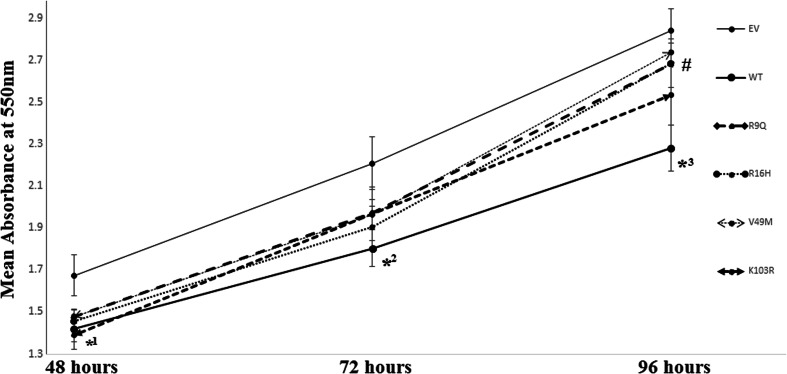
In order to analyse the effect of the various N-terminal mutants on AIP’s function as a tumour suppressor, MTT assays were carried out. Over-expression of wild-type AIP was shown to be able to reduce proliferative rates in GH3 cells in previous studies (2). As observed in Fig. 1, WT-AIP was able to reduce GH3 cell proliferation at all time points as compared to cells transfected with an empty vector. N-terminal variants of AIP were also able to reduce cell proliferation similarly as the wild-type at 48 and 72-h post-transfection. However, at 96-h post-transfection, the R9Q, R16H and V49 M variants of AIP failed to reduce GH3 cells proliferation significantly compared to the empty vector and instead had significantly higher proliferation rates as compared to WT-AIP. The K103R variant failed to alter proliferation significantly when compared to both WT-AIP and the empty vector.
Fig. 1.
Proliferation assays of WT and N-terminus variants of AIP in GH3 cells. GH3 cells were transfected with either pcDNA3 empty vector (EV) or WT or N-terminus variants, and MTT analysis was carried out at 3 time points after transfection. Points represent means of four separate experiments with five replicates per experiment. Error bars indicate standard error. (*1, p = <0.05 comparing all AIP expressing plasmids to EV at 48 h; *2, p = <0.05 comparing all AIP expressing plasmids to EV except the V49M variant at 72 h; *3, p = <0.05 comparing WT-AIP to EV at 96 h; #, p = <0.05 comparing the R9Q, R16H and V49M N-terminus variant to WT-AIP at 48 h)
WT versus N-Terminus AIP Variants: Downstream cAMP Signalling
We evaluated the impact of the selected point mutations using two methods. Firstly, a reporter gene assay in which the expression of the luciferase gene is driven by a cAMP-specific promoter or cAMP response element (CRE) was utilized. Secondly, the expression of a cAMP target gene, Id1, was also analysed to verify the effect of AIP and its mutants on downstream cAMP signalling as, in this instance, transcription of cAMP target genes. Forskolin, a cAMP–stimulating chemical and IBMX, a general phosphodiesterase inhibitor, are used in order to study the cAMP signalling pathway at maximal stimulation since differences in AIP’s effect on cAMP signalling were only observed at highly stimulated scenarios in GH3 cells (14).
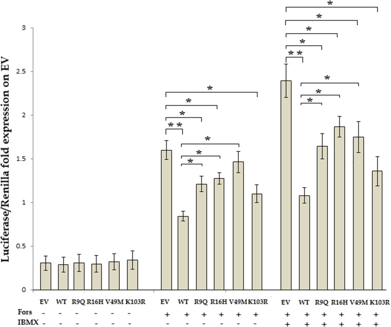
As observed in Fig. 2, as expected over-expression of WT AIP reduces CRE-driven reporter activity as compared to control (EV) in the presence of forskolin, a behaviour which is not altered by the pre-treatment with the general phosphodiesterase inhibitor IBMX. No differences were observed on basal non-stimulated levels of expression of the luciferase gene. However, stimulation with forskolin revealed statistical differences between WT and mutant AIP variants. Although the R9Q, R16H and K103R mutants were able to reduce forskolin-induced CRE-driven luciferase expression when compared to the control, the R9Q and R16H N-terminus variants were able to do so significantly less than the WT AIP. Interestingly, the V49M variant did not even alter CRE-driven luciferase activity when compared to the control in the presence of forskolin, but it did when treated with forskolin and IBMX, while the K103R mutant did not differ significantly in its activity in relation to the WT. Pre-treatment with IBMX showed the same differences between WT and mutant AIP protein activity. Furthermore, the difference between the control and V49 M variant was more apparent and reached statistically significance as observed in Fig. 2.
Fig. 2.
WT versus N-terminus variants of the AIP protein on cAMP-driven transcription. Luciferase reporter activity of GH3 cells transfected with either empty vector (EV), wild-type (WT) or N-terminus variant AIP (R9Q, R16H, V49M and K103R expression vectors) and rat Rip1 promoter-driven luciferase expression plasmid. Treatment with 10 μM IBMX and/or 1 μM forskolin 48 h after transfection is described below. Bars represent means of five separate experiments with three replicates per experiment. Error bars indicate standard error. (*p = <0.05, **p = <0.005; For, forskolin)
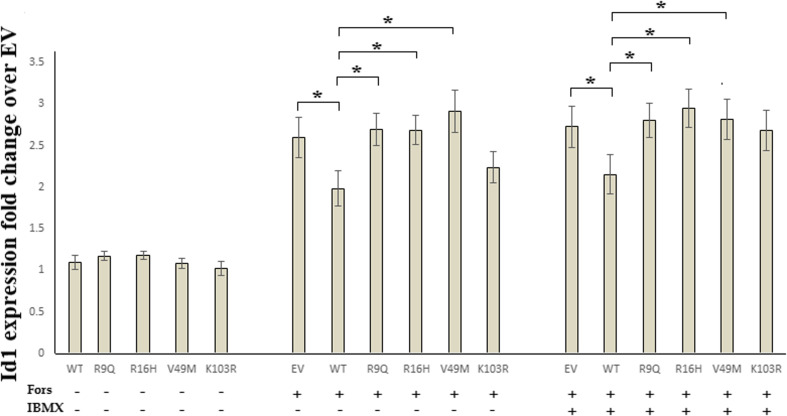
The second set of experiments on the effect of AIP over-expression on cAMP downstream signalling involved the direct measurement of a cAMP target gene by real-time PCR. The Id1 gene is a well characterized cAMP target gene with a verified CRE in its promoter region [20–23]. As observed in Fig. 3, Id1 expression is sensitive to forskolin addition which increases cAMP concentrations in all cell types. The results obtained for this indicator of cAMP signalling mirror closely those obtained for the CRE-driven-luciferase activity. Over-expression of WT and mutant AIP did not alter non-stimulated Id1 gene expression while forskolin-induced Id1 expression was significantly lower in cells over-expressing the WT AIP as compared to empty vector control. The R9Q, R16H and V49M variants failed to reduce Id1 expression in forskolin-stimulated conditions and had significantly higher expression of this gene as compared to the WT. Similarly, these variants, failed to reduce Id1 expression when pre-treated with IBMX when compared to WT AIP, while the K103R variant also failed to reduce Id1 expression significantly when compared to control but was also not significantly different from the WT behaviour. Hence the R9Q, R16H and V49 M mutant variants show significant difference in behaviour to the WT AIP but not to the control while the K103R shows no difference in behaviour when compared to both WT AIP over–expressing and control GH3 cells and under all chemical conditions.
Fig. 3.
WT versus N-terminus variants of the AIP protein on cAMP target gene expression. Id1 mRNA expression of GH3 cells transfected with empty vector (EV), wild-type (WT) or N-terminus variants of AIP and untreated or treated with 10 μM IBMX and/or 1 μM forskolin. The expression was normalized against the Ri housekeeping gene. Bars represent means of three separate experiments with two replicates per experiment. Error bars indicate standard error. (*p = <0.05, Fors, forskolin)
WT versus N-Terminus Variants: cAMP Concentrations and GH Secretion
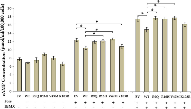
Total intracellular cAMP was measured (Fig. 4) since AIP was shown to alter cAMP production through its interaction with the Gαi proteins [14, 15]. Over-expression of EV, WT and N-terminus mutants did not alter basal levels of cAMP production. However, in the presence of forskolin cells transfected with WT and K103R variant, AIPs reduced cAMP concentrations significantly as compared to control cells while the other variants failed to do so. GH3 cells expressing the R16H and V49M AIP variant had a cAMP concentration significantly higher than WT over expressing cells under forskolin stimulation. Pre-treatment with IBMX which ablates phosphodiesterase activity, revealed even greater differences. Under PDE-inhibiting conditions, WT AIP retained the ability to reduce cAMP concentrations significantly when compared to the control while all mutant variants lost this ability. However, under these conditions, the R9Q, R16H and V49M over-expressing variant cells contained cAMP levels significantly higher than the WT over-expressing cells indicating a loss of cAMP-supressing ability under these conditions.
Fig. 4.
WT versus N-terminus variants of the AIP protein on intracellular cAMP concentration. Total intracellular cAMP of GH3 cells transfected with empty vector (EV), wild-type (WT) or N-terminus variants of the AIP. Treatment with 10 μM IBMX and/or 1 μM forskolin 48 h after transfection is described below. Bars represent means of four separate experiments with three replicates per experiment. Error bars indicate standard error. (*p = <0.05, Fors, forskolin)
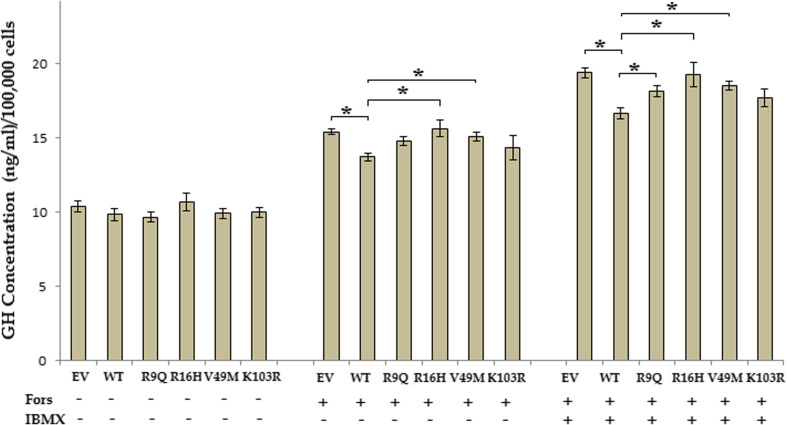
GH secretion was regulated in parallel to the cAMP concentrations (Fig. 5) as would be expected given the direct effect of cAMP level changes on GH secretion reported previously [13]. No differences were observed on basal levels of GH secretion in cells transfected with any expression vector. However, addition of forskolin led to less GH secretion in WT AIP-over-expressing cells as compared to control cells transfected with an empty vector, an ability which was not observed with any of the N-terminus variant proteins. The GH3 cells over-expressing the N-terminus variants, R16H and V49 M had significantly higher GH secretion levels than the WT AIP over-expressing cells. Pre-treatment with IBMX accentuated this difference. GH3 cells over-expressing the AIP variants R9Q, R16H and V49 M, but not K103R, had significantly higher GH secretion levels than WT over-expressing cells but lower than the control transfected cells.
Fig. 5.
WT versus N-terminus variants of the AIP protein on GH secretion. Extracellular GH secreted from GH3 cells transfected with either empty vector (EV), WT or N-terminus variants of the AIP. Treatment with 10 μM IBMX and/or 1 μM forskolin 48 h after transfection is described below. Bars represent means of five separate experiments with two replicates per experiment. Error bars indicate standard error. (*p = <0.05, Fors, forskolin)
Cycloheximide Chase Analysis
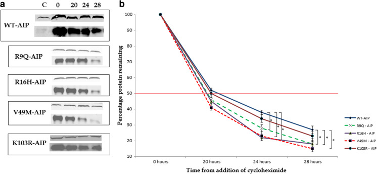
Since the N-terminus was proposed to regulate stability of the AIP [18], cycloheximide chase analysis was carried out to verify whether variations in the N-terminus of the protein would render it more unstable and therefore increases its rate of degradation. A time period of between 20 and 28 h was selected after trials at different time points ranging from 12 to 28 h (data not shown). The 20 to 28-h window was selected since it is the window within which the variation in degradation rate became most apparent. As observed in Fig. 6a, western blot analysis of AIP and β- actin revealed subtle but detectable differences between WT and N-terminus variant proteins after treatment with cycloheximide. At 20-h post-cycloheximide treatment, no significant differences were observed while at 24-h post-treatment, significant differences between the WT and V49M variant could be observed. At 28-h post-treatment, the differences were more pronounced, with significantly higher degradation observed for the R9Q, R16H and V49M N-terminus variants as compared to the WT AIP (Fig. 6b). The half-lives of the wild-type (T ½ = 21 h) and N-terminus mutants (T ½: R9Q = 18.5 h, R16H = 18 h, V49M = 16 h and K103R = 20 h) were also different although not significantly. Significant differences were observed at time points beyond the protein half-lives.
Fig. 6.
Cycloheximide chase analysis of WT and N-terminus variants of the AIP proteins. GH3 cells were transfected with WT or N-terminus variants of the AIP protein (R9Q, R16H, V49M and K103R), and cycloheximide was added 48 h after transfection. Cells were lysed at 0, 20, 24 and 28 h after addition of cycloheximide. a Western blot analysis of GH3 cell lysates with AIP (lower panel) and β-actin (upper panel) antibodies for the detection of endogenous and over-expressed AIP and its degradation at different time points from the cessation of translation by cycloheximide. b Degradation rate of WT and N-terminus variants of the AIP at different time points indicates a variation in half-lives. Each point represents the mean of five separate experiments. Error bars indicate standard error. (*p = <0.05) (C control; GH3 cells transfected with empty vector after 48 h)
Therefore, significant and consistent differences between the WT and N-terminus variants were observed, in particular, the R9Q, R16H and V49 M mutants. The K103R variant appeared not to alter protein activity significantly when compared to the WT. In fact, bioinformatics analysis of the impact of these mutations using the PolyPhen2 online tool identified the R16H and V49M mutations as being possibly damaging with a score of 0.969 and 0.975, respectively, while the R9Q and K103R variants were labelled as benign with a respective score of 0.099 and 0.185.
Discussion
The scope of this study was to functionally verify whether particular point mutations leading to amino acid substitutions in the N-terminus of the AIP could have a deleterious effect on the functional activity of this protein in the setting of pituitary adenomas. Although the AIP has been clearly identified as an important tumour suppressor in pituitary adenomas and its mutations have been strongly linked to familial cases of functional pituitary tumours, significant heterogeneity and uncertainty still exist as to how these mutations predispose to disease [2]. The AIP is a co-chaperone that interacts with a number of nuclear receptors, viral proteins, cytoplasmic enzymes and novel interacting proteins that are being discovered. Any genetic changes leading to disruptions of such interactions could have a pathologic effect [3]. We reported a role for AIP in decreasing cAMP signalling and GH secretion in forskolin-stimulated GH3 cells transfected with WT AIP, an effect which was not abolished with partial or total PDE inhibition. This led to us to hypothesise that the cAMP–mediated role of AIP must occur through other interactions such as the interactions with the G-proteins [14]. This was subsequently proven by Tuominen and colleagues [15] who demonstrated that AIP knock-down disrupted Gαi-2 and Gαi-3 protein function resulting in increased cAMP levels. A direct interaction between the regulatory subunit R1α of the protein kinase A (PKA) and AIP was also identified, and a knock-down of AIP again resulted in an increase in cAMP levels and an increase in downstream signalling [24]. Given the importance of AIP and its effect on cAMP, it was hypothesised that the functional effect of point mutations might involve dysregulation of the cAMP signalling pathway.
In pituitary adenoma patients, AIP alterations have been found throughout the gene located on chromosome 11q13, and loss of heterozygosity of this locus has been frequently observed in patient tumour DNA [25]. The mutations that have attracted most attention were those causing major disruption to the C-terminus of the AIP since this was thought to be mainly responsible for AIP’s ability to functionally interact with partner proteins. However, recent studies have identified the N-terminus of AIP as being essential for interactions with other proteins [16–18]. Equally, the N-terminus has been shown to be important for the overall stability of the protein and to be essential for the correct localization of the aryl hydrocarbon receptor (AHR) [18]. Particular N-terminus missense mutations occur very frequently in both familial and sporadic settings of pituitary adenomas. Three common N-terminus mutations were therefore selected for further analysis while another mutation discovered locally, the R9Q, was also analysed in order to determine the extent of the functional de-regulation suffered by the AIP protein. Three of these variants, the R9Q, R16H and V49M, were shown to lose the tumour suppressing ability displayed by the wild-type AIP at 96 h from transfection in the GH3 pituitary adenoma cell line (Fig. 1), indicating that a functional role may indeed be attributed to these variants.
The c.26G > A leading to the R9Q missense mutation was first discovered in Malta in 2009 [26]. Recent studies have also localized the same mutation in two other populations. Puig–Domingo and co-workers [27] identified this variant in a female patient diagnosed at age 20 with a GH-secreting macroadenoma, while screening of a large French cohort identified the R9Q alteration in two patients, a Cushing’s disease patient diagnosed at the age of 39, while the other patient was a young girl with a macroprolactinoma [28] Therefore, significant clinical heterogeneity is associated with this genetic mutation. The arginine residue at position 9 was found to be conserved in all mammals, supporting a significant role for this amino acid. Furthermore, the population frequency of the c.26G > A variant was assessed by screening 285 random cord blood DNA samples collected locally (data not shown). None of the samples screened tested positive for this variant. Recent updates in the SNP database (dbSNP) label the R9Q mutation as an extremely rare polymorphism with a frequency of about 1 in 10,000.
Data obtained from this study indicate that the R9Q variation alters the behaviour of the AIP and reduces its ability to inhibit cAMP signalling and GH secretion significantly under forskolin-stimulated conditions. Addition of IBMX, a general phosphodiesterase inhibitor, had little effect on the difference observed between the WT and R9Q proteins except in the GH secretion assay where the addition of IBMX was required to achieve a significant difference. Given this data alone, the amino acid substitution at position 9 of the protein appears to have a small but significant impact on the cAMP-inhibitory ability of the WT protein. Cycloheximide chase analyses of the degradation rate of the protein also reveal significant differences since at 24 and 28 h (Fig. 5b) the R9Q variant protein degraded significantly more than the WT. Therefore, the functional impact of this mutant variant on the protein function may occur through its decreased half-life, thereby reducing the concentration of the protein in the cells. Although the bioinformatics prediction using PolyPhen2 program indicated a benign change, this result has to be taken into consideration that bioinformatics analyses are purely in silico and do not always reflect the proper cellular physiology and biology. Additionally, preliminary data from protein interaction studies being undertaken indicate that the R9Q variant also influences the ability of AIP to fully interact with common interacting partners (personal communication).
The c.47G > A substitution resulting in the R16H polymorphism has also attracted wide attention since this variant of the AIP has been found in numerous studies of PA patients, particularly in familial cases. It was first described in two cousins with GH-secreting adenomas [29], later in two patients with non-functioning adenomas [30] and later replicated in other studies in both familial and sporadic settings [31–33]. Recently, this gene variant was also found in a patient with a corticotroph macroadenoma [34], again displaying varied clinical heterogeneity with this mutant genotype. The arginine residue at position 16 is conserved in all vertebrates and the G > A substitution is very rare occurring in less than 1 in 1000 of the population.
Functional studies carried out on this variant reveal conflicting data. One study revealed that the R16H alteration led to a reduction in the interaction between AIP and PDE4A5 proteins when tested using a β-galactosidase assay [35], while the interaction with RET was not affected [36]. Results from this study indicate that the R16H alteration is sufficient to change the behaviour of the wild type AIP in relation to its effect on cAMP signalling and GH secretion. Similarly, stability studies indicate a significant difference in protein stability after 28 h for a number of variants, although a previous study failed to show a significant difference in protein half-life between wild type and R16H AIP [37]. Our data also fail to uncover significant differences in protein half-lives of both WT and variants since significant differences were only observed at later time points. It was concluded that this change probably represents a rare polymorphism rather than a pathogenic variant [38] since this change was also observed in unaffected relatives of patients. However, modest functional effects can still have a significant effect over a longer period of time, and given the incomplete penetrance of the AIP mutation, it is likely that the R16H mutation may itself not be enough to elicit a pathological effect but it may be sufficient to allow a neoplastic transformation once the cell obtains “the second hit” required for tumour formation.
The c.145 G > A substitution leading to the V49M mutation was originally identified in a Japanese man with childhood-onset gigantism. However, no LOH was observed for the AIP in the tumour of the patient [39]. Once again, the G allele is very well conserved in most vertebrates, and the allele frequency of the A allele is less than 1 in 10,000. However, functional work for this variant has shown little effect on the protein. The binding affinity for PDE4A5 was not altered in a β-galactosidase assay [35], and the V49M variant did not show a change in proliferation in GH4C1 cells [40]. Our data strongly supports a functional role for this substitution since in all assays the V49M alteration caused a significant loss of function as compared to the wild type AIP. Similarly, a significant difference in protein stability was observed with this amino acid substitution. Therefore, even though no change in binding affinity was observed in the other study [35], and no LOH was observed in the patient, this protein may still lose functionality enough to alter physiology of the pituitary and lead to higher proliferative rates or a susceptibility to increased proliferation.
The last missense mutation analysed was the K103R (c.308 A > G) variant which was first identified in a paediatric patient with Cushing’s disease having a corticotroph adenoma [41]. This position is again very well conserved although no allele frequency data is available for the G allele (ensemble.org). The only functional work carried out on this AIP variant indicated a much reduced PDE4A5 binding ability when compared to the wild-type (8% of WT) [35]. Data presented here on the influence of the K103R variant on cAMP signalling and GH secretion in GH3 cells indicated very little difference in behaviour when compared to the WT protein. Similarly, stability analysis using cycloheximide chase showed little difference between the WT and K103R AIP proteins and the Polyphen2 algorithm classifies this alteration as unlikely to be pathogenic. Therefore, the functional significance of the K103R variant remains unknown.
In summary, the data obtained in this study indicate a possible functional role for the R9Q, R16H and V49M N-terminus missense alterations leading to changes in the cAMP and GH-reducing-ability of the wild-type AIP. Although the variations observed in cAMP concentrations and GH secretion appear small, such minute but consistent changes in important signalling pathways such as cAMP secondary messenger and its relative direct effect on GH which can act is an immediate growth factor in a autocrine/paracrine fashion, can have a very significant impact on pituitary cell hyperplasia. Given the cascading effect of cAMP as a secondary messenger, it is quite likely that sustained higher thresholds of cAMP in a tissue such as the pituitary which relies on this signalling pathway for constant pulsatile secretion, could have deleterious effects over prolonged periods. Therefore, although minor changes in these effectors and mediators of pituitary cell proliferation were observed, these changes were significant and constant enough to merit attention and overtime. Consistently higher threshold of cAMP signalling and GH secretion could prove the decisive difference between normal cell physiology and pituitary hyperplasia leading to tumour formation.
The changes observed in protein function with respect to cAMP signalling and GH secretion were similarly evident in the protein stability studies which indicated that these variants have increased degradation rates when compared to the wild type protein. This is in agreement with the previous observations by other groups with regards to the role of the N-terminus in regulating stability of the AIP [18]. However, various studies also indicate a direct role for the N-terminal domain in interacting with other proteins. A study by Schimmack and colleagues [16] revealed that AIP functionally interacts with the scaffolding protein CARMA1 (CARD-containing MAGUK protein 1) to bring about the formation of the CARMA1-BCL10-MALT1 (CBM) complex in T cells. This interaction occurs solely through the N-terminus of the AIP with the PDZ-SH3 domain of the CARMA1 with the TRP repeats of the AIP being dispensable for the interaction. Additionally, the N-terminal domain of the AIP was shown to lack any PPIase (peptidyl-propyl cis/trans isomerase) activity but was able to interact with HSP90 protein in pull-down assays using both the N-terminal domain alone and the complete protein [17]. Therefore, various mechanisms have been proposed through which alterations in the N-terminus of the AIP may lead to increased susceptibility toward pituitary adenoma formation.
Given the evidence provided by this group [14] and others [15, 24] regarding the role of AIP in limiting cAMP formation and signalling, the effect of these N-terminus missense variants on the wild-type inhibition of these important signalling pathways is not to be disregarded. A recent study has identified a novel N-terminus missense mutation that appears very interesting in the clinical setting. The I13N mutation was identified in a 19-year-old patient with a large somatotropinoma with LOH of the AIP locus, from which, the authors concluded that the mutation must be disease-causing [42].
The incomplete penetrance displayed by the variants of the AIP protein adds further complexity. Mutations within the AIP gene have been found in both cases and controls, and no particular mutation has been found to segregate solely with the disease, and not clear phenotype is associated with any particular genetic alteration. In fact, a general penetrance of 30% is assigned towards AIP gene alterations [35, 43], although some mutations appear to have higher penetrance than others, such as the R304X mutant with a reported 41% penetrance [44]. Therefore, when assessing AIP alterations in a clinical setting, it is important to remember that further genetic and/or environmental triggers are required for the complete phenotype of pituitary adenoma to become apparent. AIP alterations by themselves do not display the complete spectrum of biological dysfunction required for tumorigenesis of any pituitary cell type, which also explains the range of phenotypic characteristics obtained with the same AIP genetic defect, hence, requiring further genetic/biological defects to lead to tumour formation. In conclusion, this study identified a functional role for three N-terminus missense mutations in comparison to the wild-type AIP in relation to its inhibitory effect on cAMP signalling and subsequent GH secretion. Additionally, these missense variations also altered the protein degradation rate of the wild-type protein indicating an alternative mechanism by which dysregulation of the protein could occur.
Acknowledgments
The authors would like to acknowledge Dr. Anthony Fenech from the Department of Pharmacology and Dr. Godfrey Grech for the use of their laboratories. The research was funded by the University of Malta Research funds (MEDRP02-05) and the Faculty of Medicine and Surgery (MDSIN08-22).
Compliance with Ethical Standards
Conflict of Interest
The authors declare that they have no conflict of interest.
Contributor Information
Robert Formosa, Email: robert.formosa@um.edu.mt.
Josanne Vassallo, Phone: 00356 2340 1881, Email: josanne.vassallo@um.edu.mt.
References
- 1.Vierimaa O, Goergitsi M, Lehtonen R, Vahteristo P, Kokko A, Raitila A, Tuppurainen K, Ebeling TML, Salmela PI, Paschke R, et al. Pituitary adenoma predisposition caused by germline mutations in the AIP Gene. Science. 2006;312:1128–1230. doi: 10.1126/science.1126100. [DOI] [PubMed] [Google Scholar]
- 2.Leontiou CA, Gueorguiev M, van der Spuy J, Quinton R, Lolli F, Hassan S, Chahal HS, Igreja SC, Jordan S, Rowe J, et al. The role of the aip gene in familial and sporadic pituitary adenomas. J Clin Endocrinol Metab. 2008;93(6):2390–2401. doi: 10.1210/jc.2007-2611. [DOI] [PubMed] [Google Scholar]
- 3.Trivellin G, Korbonitz M. AIP and its interacting partners. J Endocrinol. 2011;210:137–155. doi: 10.1530/JOE-11-0054. [DOI] [PubMed] [Google Scholar]
- 4.Daly A, Tichomirowa M, Petrossians P, Heliovaara E, Jefrrain-Rea M, Barlier A, Naves LA, Ebeling T, Harhu A, Raappana A, et al. Clinical characteristics and therapeutic responses in patients with germ-line AIP mutations and pituitary adenomas: an international collaborative study. J Clin Endocrinol Metab. 2010;95:E373–E383. doi: 10.1210/jc.2009-2556. [DOI] [PubMed] [Google Scholar]
- 5.Bolger GB, Peden AH, Steele MR, MacKenzie C, McEwan DG, Wallace DA, Huston E, Baillie GS, Houslay MD. Attenuation of the activity of the cAMP-specific phosphodiesterase PDE4A5 by interaction with the immunophilin XAP2. J Biol Chem. 2003;278:33351–33363. doi: 10.1074/jbc.M303269200. [DOI] [PubMed] [Google Scholar]
- 6.de Oliviera SK, Hoffmeister M, Gambaryan S, Muller-Esterl W, Guimaraes JA, Smolenski AP. Phosphodiesterase 2A forms a complex with the Co-chaperone XAP2 and regulates nuclear translocation of the aryl hydrocarbon receptor. J Biol Chem. 2007;282(18):13656–13663. doi: 10.1074/jbc.M610942200. [DOI] [PubMed] [Google Scholar]
- 7.Beischlag TV, Morales JL, Hollingshead BD, Perdew GH. The aryl hydrocarbon receptor complex and the control of Gene expression. Crit Rev Eukaryot Gene Expr. 2008;18(3):207–250. doi: 10.1615/CritRevEukarGeneExpr.v18.i3.20. [DOI] [PMC free article] [PubMed] [Google Scholar]
- 8.Nakata A, Urano D, Fujii-Kuriyama Y, Mizuno N, Tago K, Itoh H. G-protein signalling negatively regulates the stability of aryl hydrocarbon receptor. EMBO Rep. 2009;10:622–628. doi: 10.1038/embor.2009.35. [DOI] [PMC free article] [PubMed] [Google Scholar]
- 9.Suzuki S, Yamamoto I, Arita J. Mitogen-activated protein kinase-Dependant stimulation of proliferation of rat Lactotrophs in culture by 3′,5′-cyclic adenosine monophosphate. Endocrinology. 1999;140(6):2850–2858. doi: 10.1210/endo.140.6.6775. [DOI] [PubMed] [Google Scholar]
- 10.Lania A, Mantovani G, Spada A. G protein mutations in endocrine diseases. Eur J Endocrinol. 2001;145:543–559. doi: 10.1530/eje.0.1450543. [DOI] [PubMed] [Google Scholar]
- 11.Lania AG, Mantovani G, Ferrero S, Pellegrini C, Bondioni S, Peverelli E, Braidotti P, Locatelli M, Zavanone ML, Ferrante E, et al. Proliferation of transformed somatotroph cells related to low or absent expression of protein kinase a regulatory subunit 1A protein. Cancer Res. 2004;64:9193–9198. doi: 10.1158/0008-5472.CAN-04-1847. [DOI] [PubMed] [Google Scholar]
- 12.Pertuit M, Barlier A, Enjalbert A, Gerard A. Singalling pathway alterations in pituitary adenomas: involvement of Gsα, cAMP and mitogen-activated protein kinases. J Endocrinol. 2009;21:269–877. doi: 10.1111/j.1365-2826.2009.01910.x. [DOI] [PubMed] [Google Scholar]
- 13.Ramirez JL, Castano JP, Torronteras R, Martinez-Fuentes AJ, Frawley LS, Garcia-Navarro S, Gracia-Navarro F. Growth hormone (GH) releasing factor differentially activates cyclic adenosine 3′,5′-monophosphate- and inositol phosphate-dependent pathways to stimulate GH release in two porcine somatotroph subpopulations. Endocrinology. 1999;140:1752–1759. doi: 10.1210/endo.140.4.6613. [DOI] [PubMed] [Google Scholar]
- 14.Formosa R, Xuereb-Anastasi A, Korbonits M, Vassallo J. Aip regulates cAMP signalling and GH secretion in GH3 cells. Endocr Relat Cancer. 2013;20:495–505. doi: 10.1530/ERC-13-0043. [DOI] [PubMed] [Google Scholar]
- 15.Tuominen I, Heliovaara E, Raitila A, Rautiainen MR, Mehine M, Katainen R et al (2014) AIP inactivation leads to pituitary tumorigenesis through defective Gαi-cAMP signalling. Oncogene:1–11 [DOI] [PubMed]
- 16.Schimmack G, Eitelhuber AC, Vincendeau M, Demski K, Shonohara H, Kurosaki T, Krappmann D (2014) AIP augments CARMA1-BCL10-MALT1 complex formation to facilitate NF-κB signalling upon T cell activation. Cell communication and signalling 12(49). doi:10.1186/s12964-014-0049-7 [DOI] [PMC free article] [PubMed]
- 17.Linnert M, Lin YJ, Manns A, Haupt K, Paschke AK, Fischer G, Weiwad M, Lucke C. The FKBP-type domain of the human aryl hydrocarbon receptor- interacting protein reveals an unusual Hsp90 interaction. Biochemistry. 2013;S2:2097–2107. doi: 10.1021/bi301649m. [DOI] [PubMed] [Google Scholar]
- 18.Kazlauskas A, Poellinger L, Pongratz I. Two distinct regions of the immunophilin-like protein XAP2 regulate dioxin receptor function and interaction with hsp90. Journal of Biological hemistry. 2002;277:11795–11801. doi: 10.1074/jbc.M200053200. [DOI] [PubMed] [Google Scholar]
- 19.Chepurny OG, Holz GG. A novel cyclic adenosine monophosphate-responsive luciferase reporter incorporating a nonpalindromic cyclic adenosine monophosphate response element provides optimal performance for use in G protein-coupled receptor drug discovery efforts. J Biomol Screen. 2007;12(5):740–746. doi: 10.1177/1087057107301856. [DOI] [PMC free article] [PubMed] [Google Scholar]
- 20.Ohta Y, Nakagawa K, Imai Y, Katagiri T, Kioke T, Takaoka K. Cycli cAMP enhances Smad—mediated BMP signaling through PKA-CREB pathway. J Bone Miner Metab. 2008;26(5):478–484. doi: 10.1007/s00774-008-0850-8. [DOI] [PubMed] [Google Scholar]
- 21.Yang J, Li X, Al-Lamki RS, Southwood M, Zhao J, Lever AM, Grimminger F, Schermuly RT, Morrell NW. Smad-dependent and Smad-independent induction of id1 by prostacyclin analogues inhibits proliferation of pulmonary artery smooth muscle cells in vivo. Circ Res. 2010;107(2):252–262. doi: 10.1161/CIRCRESAHA.109.209940. [DOI] [PubMed] [Google Scholar]
- 22.Zhang X, Odom DT, Koo SH, Conkright MD, Canettieri G, Best J, Chen H, Jenner R, Herbolsheimer E, Jacobsen E, et al. Genome-wise analysis of cAMP-response element binding protein occupancy, phosphorylation, and target gene activation in human tissues. PNAS. 2005;102(12):4459–4464. doi: 10.1073/pnas.0501076102. [DOI] [PMC free article] [PubMed] [Google Scholar]
- 23.San-Marina S, Han Y, Suarez Saiz F, Trus MR, Minden MD. Lyl1 interacts with CREB1 and alters expression of CREB1 target genes. Biochemica et Biophsica Acta. 2008;1783:503–517. doi: 10.1016/j.bbamcr.2007.11.015. [DOI] [PubMed] [Google Scholar]
- 24.Schernthaner-Reiter MH, Trivellin G, Nerterova M, Hernandez Ramirez LC, Aflorei ED, De La Luz Sierra M, Korbonits M, Stratakis CA (2014) Interaction of AIP and the cAMP-dependent protein kinase (PKA) pathway and its role in pituitary tumor formation. Endocrine Abstracts, PP09–4
- 25.Beckers A, Aaltonen LA, Daly AF, Karhu A. Familial isolated pituitary adenomas (FIPA) and the pituitary adenoma predisposition due to mutations in the aryl hydrocarbon receptor interacting protein (AIP) gene. Endocr Rev. 2013;34(2):239–277. doi: 10.1210/er.2012-1013. [DOI] [PMC free article] [PubMed] [Google Scholar]
- 26.Formosa R, Farrugia C, Xuereb-Anastasi A, Korbonits M, Vassallo J. Aryl hydrocarbon receptor-interacting protein: mutational analysis and functional validation in primary pituitary cell cultures. Endocr Abstr. 2010;22:P436. [Google Scholar]
- 27.Puig-Domingo M, Oriola J, Halperin I, Mora M, Diaz-Soto G, Perales MJ, Alvares-Escola C, Lucas-Morante T, Bernabeu I, Marazuela M. Prevalence of germline mutations of AIP gene in sporadic aggressive somatotropinomas. Endocr Abstr. 2011;13:258. [Google Scholar]
- 28.Cazabat L, Bouligand J, Salenave S, Bernier M, Gaillard S, Parker F, Young J, Giouchon-Mantel A, Chanson P. Germ-line AIP mutation in apparently sporadic pituitary adenomas: prevalence in a prospective single-Centre cohort of 443 patients. J Clin Endocrinol Metab. 2012;97(4):E663–E670. doi: 10.1210/jc.2011-2291. [DOI] [PubMed] [Google Scholar]
- 29.Daly AF, Vanbellinghen JF, Khoo SK, Jaffrain-Rea ML, Naves LA, Guitelman MA, Murat A, Emy P, Gimenez-Roqueplo AP, Tamburrano G, Raverot G, et al. Aryl hydrocarbon receptor-interacting protein Gene mutation in familial isolated pituitary adenomas: analysis in 73 families. J Clin Endocrinol Metab. 2007;92(5):1891–1896. doi: 10.1210/jc.2006-2513. [DOI] [PubMed] [Google Scholar]
- 30.Buchbinder S, Bierhaud A, Zorn M, Nowroth PP, Humpert P, Schilling T. Aryl hydrocarbon receptor interacting protein gene (AIP) mutations are rare in patients with hormone secreting or non-secreting pituitary adenomas. Exp Clin Endocrinol Diabtetes. 2008;116(10):625–628. doi: 10.1055/s-2008-1065366. [DOI] [PubMed] [Google Scholar]
- 31.Cazabat L, Libè R, Perlemoine K, René-Corail F, Burnichon N, Gimenez-Roqueplo AP, Dupasquier-Fediaevsky L, Bertagna X, Clauser E, Chanson P, Bertherat J, et al. Germ-line inactivating mutations of the aryl hydrocarbon receptor-interacting protein gene in a large cohort of sporadic acromegaly: mutations are found in a subset of young patients with macroadenomas. Eur J Endocrinol. 2007;157(1):1–8. doi: 10.1530/EJE-07-0181. [DOI] [PubMed] [Google Scholar]
- 32.Georgitsi M, Railita A, Karhu A, Tuppurainen K, Makinen MJ, Vierimaa O, et al. Molecular diagnosis of pituitary adenoma predisposition caused by aryl hydrocarbon receptor—interacting protein gene mutations. Proc Natl Acad Sci U S A. 2007;104(10):4101–4105. doi: 10.1073/pnas.0700004104. [DOI] [PMC free article] [PubMed] [Google Scholar]
- 33.Yaneva M, Daly AF, Tichomirowa MA, Vanbellinghen JF, Hagelstein MT, Bours V, Zacharieva S, Beckers A (2008) Aryl hydrocarbon receptor interacting protein gene mutations in bulgarian FIPA and young sporadic pituitary adenoma patients. Proc of the 90th Annual Meet of the Endocrine Soc 3–520
- 34.Dinesen PT, Dal J, Gabrovska P, Gaustadnes M, Gravholt CH, Stals K, et al. An unusual case of an ACTH-secreting macroadenoma with a germline variant in the aryl hydrocarbon receptor—interacting protein (AIP) gene. Endocrinol Diabetes Metab Case Rep. 2015 doi: 10.1530/EDM-14-0105. [DOI] [PMC free article] [PubMed] [Google Scholar]
- 35.Igreja S, Chahal HS, King P, Bolger GB, Srirangalingam U, Guasti L, Chapple JP, Trivellin G, Gueorguiev M, Guegan K, et al. Characterization of aryl hydrocarbon receptor interacting protein (AIP) mutations in familial isolated pituitary adenoma families. Hum Mutat. 2010;31:950–960. doi: 10.1002/humu.21292. [DOI] [PMC free article] [PubMed] [Google Scholar]
- 36.Vargiolu M, Fusco D, Kurelac I, Dirnberger D, Baumeister R, Morra I, Melcarne A, Rimondini R, Romeo G, Bonora E. The tyrosine kinase receptor RET interacts in vivo with aryl hydrocarbon receptor-interacting protein to alter surviving availability. J Clin Endocrinol Metab. 2009;94(7):2571–2578. doi: 10.1210/jc.2008-1980. [DOI] [PubMed] [Google Scholar]
- 37.Martucci F, Trivellin G, Garcia E, Dalantaeva N, Chapple P, Pecori Giraldi F, Grossman A, Korbonits M. Can the stability of variant aryl hydrocarbon receptor interacting protein (AIP) be a marker for pathogenicity in FIPA? Endocr Abstr. 2012;28:P241. [Google Scholar]
- 38.Raitila A, Georgitsi M, Karhu A, Tuppurainen K, Makinen MJ, Birkenkamp-Demtroder K, et al. No evidence of somatic aryl hydrocarbon receptor interacting protein mutations in sporadic endocrine neoplasia. Endorc Relat Cancer. 2007;14(3):901–906. doi: 10.1677/ERC-07-0025. [DOI] [PubMed] [Google Scholar]
- 39.Iwata T, Yamada S, Mizusawa N, Golam H, Sano T, Yoshimoto K. The aryl hydrocarbon receptor—interacting protein gene is rarely mutated in sporadic GH-secreting adenomas. Clin Endocrinol. 2007;66:499–502. doi: 10.1111/j.1365-2265.2007.02758.x. [DOI] [PubMed] [Google Scholar]
- 40.Garcia-Rendueles E, Diaz-Rodriguez G, Trivellin G, Garcia-Lavandeira M, Vila Vila T, Dieguez C, Korbonits M, Alvarez C. Characterisation of mutant AIPV49M in somatotroph cells. Endocr Abstr. 2012;29:P1354. [Google Scholar]
- 41.Beckers A, Vanbellinghen JF, Boikos S, Martari M, Verma S, Daly AF, et al. (2008) Germline AIP, MEN1, PRKAR1A, CDKN1B (p27Kip1) and CDKN2C(p18INK4c) gene mutations in a large cohort of pediatric patients with pituitary adenomas occurring in isolation or with associated syndromic features. Proc of the 90th Annual Meet of the Endocrine Soc; OR38–1
- 42.Salvatori R, Daly AF, Quinones-Hinojosa A, Thiry A, Beckers A. A clinically novel AIP mutation in a patient with a very large, apparently sporadic somatotroph adenoma. Endocrinol Diabetes Metabol Case Rep. 2014;2014:140048. doi: 10.1530/EDM-14-0048. [DOI] [PMC free article] [PubMed] [Google Scholar]
- 43.Stratakis CA, Tichomirowa MA, Boikos S, Azevedo MF, Lodish M, Martari M, et al. The role of germline AIP, MEN1, PRKAR1A, CDKN1B and CDKN2C mutations in causing pituitary adenomas in a large cohort of children, adolescents, and patients with genetic syndromes. Clin Genet. 2010;78(5):457–463. doi: 10.1111/j.1399-0004.2010.01406.x. [DOI] [PMC free article] [PubMed] [Google Scholar]
- 44.Occhi G, Jaffrain-Rae ML, Trivellin G, Albiger N, Caccato F, De Mensi E, Angelini M, Ferasin S, Beckers A, Mantero F, Scaroni C. The R304X mutation of the aryl hydrocarbon receptor—interacting protein gene in familial isolated pituitary adenomas: mutational hotspot or founder effect? J Endocrinol Investig. 2010;33(11):800–805. doi: 10.1007/BF03350345. [DOI] [PubMed] [Google Scholar]


