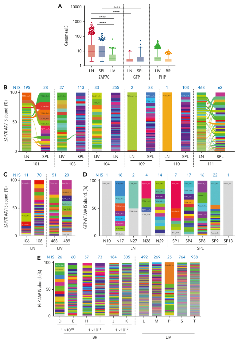
Figure 6.
Absolute and relative abundance of AAV integration sites. (A) Cellular genomes observed for each IS (Genomes/IS) in the different IS data sets are presented as indicated. (B-E) Stacked bar plots showing the relative abundance of AAV IS retrieved in ZAP70-treated mice (B,C), in WT mice intrathymically injected with a GFP-expressing AAV (D), and in Mecp2-deficient mice (E). In each column, every integration is represented by a different color whose height is proportional to the number of genomes retrieved for that specific IS (percentage of IS abundance, y-axis). The number of unique IS identified in each mouse is indicated in blue above each column. Ribbons connect AAV ISs tracked between different tissues of the same mouse. In A and B, statistical analyses were performed by a two-tailed Mann-Whitney test. ∗∗∗∗P < .0001.

