Abstract
Aurora kinase A (Aurora-A), a proto-oncogenic mitosis-regulating serine/threonine kinase, is frequently overexpressed in human breast cancer. While the kinase has been shown to cause functional inactivation of tumor suppressor protein p53, which binds estrogen receptor α (ERα) and downregulates its transcriptional activation function, significance of Aurora-A overexpression on p53 regulatory interactions in breast cancer cells has not been investigated. We describe in this report functional consequences of Aurora-A phosphorylation of p53 tumor suppressor protein on its subcellular distribution and binding with ERα that may be important in downregulating the transcription of p53-responsive growth inhibitory genes and development of resistance to DNA damage-induced apoptosis in Aurora-A overexpressing human breast cancer cells. Our results demonstrate that while estrogen activates Aurora-A expression in ERα-positive cells through ERα–GATA-3 signaling cascade, Aurora-A forms a ternary complex with p53 and ERα. Phosphorylation of p53 by Aurora-A sequesters the protein in the cytoplasm and enhances its interaction with ERα, thus repressing the transactivation functions of both p53 and ERα. These findings have significant clinical implications and suggest that prolonged estrogen exposure-mediated Aurora-A overexpression may be directly contributing to deregulated proliferation and resistance to DNA damage-induced apoptosis in breast cancer cells.
Keywords: Aurora-A, Estrogen receptor α, Breast cancer cells, p53, GATA-3, DNA damage-induced apoptosis
Introduction
Aurora-A is an evolutionarily conserved member of the Aurora protein serine/threonine kinase family that plays essential roles in mitotic entry, centrosome maturation, mitotic spindle assembly, and chromosome segregation processes [1]. The kinase is naturally overexpressed in many human epithelial malignancies, including those of breast and ovary [2], with some studies reporting that elevated expression of Aurora-A occurs more frequently in the in situ carcinomas compared with invasive cancers [3, 4]. Functionally, Aurora-A overexpression has been associated with centrosome anomalies and chromosomal instability as well as abrogation of DNA damage-induced apoptotic response and spindle assembly checkpoint override in tumor cells. Identification of Aurora-A gene as a tumor susceptibility locus and experimental evidence demonstrating tumorigenic potential of ectopically overexpressing kinase in human and rodent cells in vitro and in vivo [2, 5] suggest that overexpression of Aurora-A is involved in the genetic mechanism(s) underlying the development of aneuploidy, centrosome aberrations, and resistance to radiation and spindle-damaging chemotherapeutic drugs in human cancer cells.
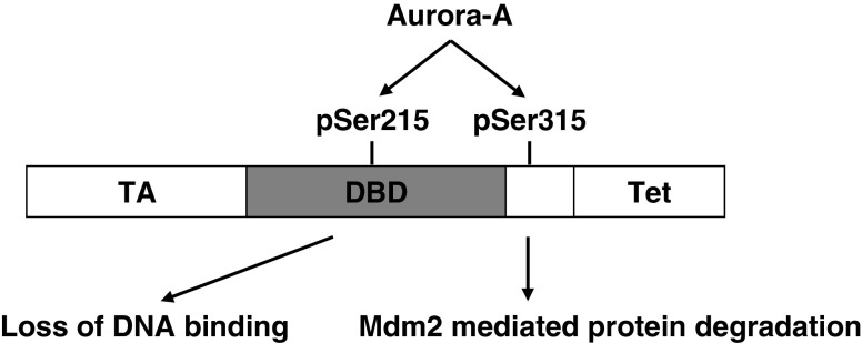
We and others have earlier shown that p53 tumor suppressor protein is a natural substrate of Aurora-A, which phosphorylates the residues serine 315 and serine 215 of the tumor suppressor protein [6, 7]. Phosphorylation of p53 at serine 315 facilitates its MDM2-mediated degradation, while serine 215 phosphorylation results in its loss of DNA binding activity. Therefore, these studies revealed that deregulated high levels of Aurora-A phosphorylation lead to functional inactivation of p53 tumor suppressor protein with consequential development of resistance to DNA damage-induced apoptosis response in Aurora-A overexpressing human cancer cells [6, 7]. The incidence of p53 mutations in breast cancers is rather infrequent, with about 80% of the cases expressing wild-type yet functionally impaired proteins that are also estrogen receptor (ER)-positive. Interestingly, most ER-negative breast tumors express mutant p53 protein [8–10], indicating that inactivation of the p53 tumor suppressor pathway is a critical event in the development of both ER-positive and ER-negative breast cancers.
In view of the central nodal positioning of p53 functions among the complex signaling networks required for the maintenance of normal cellular homeostasis and induction of damage response in cells, it is important that regulatory interactions influencing p53 functions be comprehensibly elucidated in order to understand their roles in the development of malignant transformation-associated phenotypes in breast cancer cells. It is relevant, in this context, that estrogen receptor α (ERα) has recently been reported to directly bind p53 and inhibit its transcription regulatory function [11–13], resulting in abrogation of p53-mediated cell cycle arrest and apoptosis response. Since Aurora-A elevated expression, reported to cause functional inactivation in p53, is detected among a similar proportion of functionally impaired wild-type p53 expressing breast tumor cases, we decided to investigate if Aurora-A also regulates p53–ERα interaction and if this regulation exacerbates the loss of function effects of the p53–ERα interaction in breast cancer cells. The studies were also important in view of our recent findings that estrogen signaling plays a direct role in regulating Aurora-A expression through activation of the ERα–GATA-3 signaling cascade [14], suggesting that abnormal expression of ERα–GATA-3–Aurora-A functional interactions may be critical in the development of estrogen-induced sporadic breast cancer.
We describe in this report that Aurora-A phosphorylation of p53 sequesters the protein in the cytoplasm and enhances its interaction with ERα in a ternary complex comprising of the three proteins. As a consequence, cells overexpressing Aurora-A also sequester ERα in the cytoplasm, indicating that prolonged estrogen exposure-induced Aurora-A overexpression is involved in the abrogation of ERα signaling in breast epithelial cells. Furthermore, increased interaction of Aurora-A phosphorylated p53 with ERα contributes to exacerbate ERα antagonism of p53 function, resulting in deregulated growth and development of resistance to DNA damage-induced apoptosis response in breast cancer cells.
Results
Estrogen Stimulates Proliferation of ERα-Positive Breast Cancer Cells Through ERα–GATA-3–Aurora-A Signaling Cascade
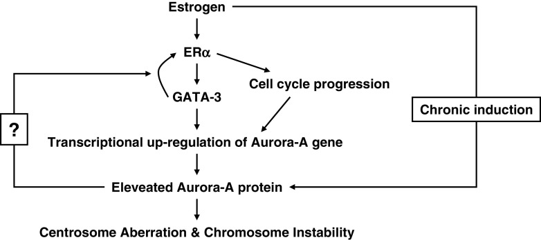
Estrogen (E2) stimulation of ERα-positive breast cancer cell growth has been reported to be mediated by the transcription factor GATA-3, that plays a role in maintaining ERα expression and sensitivity to the growth stimulatory effect of E2. This involves induction of pioneer factors, such as FOXA1, required to keep ERα-sensitive loci in a transcriptionally active conformation [15]. By doing functional analysis of a series of deletion constructs of the Aurora-A promoter in ERα-positive and -negative breast cancer cells, we observed that E2 mediates recruitment of GATA-3 to the Aurora-A promoter to activate its expression in ERα-positive breast cancer cells [14]. These findings established that E2-induced growth of breast cancer cells is regulated through activation of Aurora-A transcription, and prolonged E2 exposure is likely to result in deregulated proliferation with associated chromosomal instability and centrosomal anomalies [16], commonly detected in malignant breast epithelial cells (Fig. 1).
Fig. 1.
Hypothetical functional interaction of Aurora-A with estrogen induced ERα–GATA-3 transcriptional network
Aurora-A Phosphorylation Induces Functional Inactivation of p53
Tumor suppressor protein p53 is an important regulator of cell proliferation and maintenance of genomic integrity. Wild-type form of the protein plays a central role in the induction of cell cycle arrest and cell death in response to various stimuli causing DNA and spindle damage in cells. Loss of function mutations in p53, commonly occurring in a significant proportion of cancers, allow malignant cells to proliferate with altered genomic content and acquire resistance to various DNA- and microtubule-damaging therapeutic agents. We and others have demonstrated that p53 tumor suppressor protein is a natural substrate of Aurora-A kinase [6, 7]. Aurora-A phosphorylates the residues Serine 315 and Serine 215 of p53. Phosphorylation of Serine 315 leads to degradation of the protein due to proteolysis following ubiquitination by MDM2, while Serine 215 phosphorylation by Aurora-A abrogates p53 DNA binding and transcription activation function. Silencing of Aurora-A, on the other hand, results in reduced phosphorylation of p53 with restoration of in vivo transactivation function leading to expression of downstream target genes, making the cells sensitive to various damage-inducing stimuli. The results taken together revealed that Aurora-A overexpression in cancer cells causes loss of function of p53 even in the absence of inactivating mutations and is responsible for override of damage checkpoint response mechanisms consequentially developing resistance to DNA- and microtubule-targeting therapeutic agents (Fig. 2).
Fig. 2.
Functional consequences of Aurora-A-mediated phosphorylation of p53. Aurora-A inactivates p53 tumor suppressor pathway through phosphorylation of serine 215 in the DNA binding domain (DBD) and serine 315 in the C-terminus of p53 tumor suppressor protein. TA transactivation domain, Tet tetramerization domain
Phosphorylation of p53 at Serine 215 by Aurora-A Alters Subcellular Localization of p53
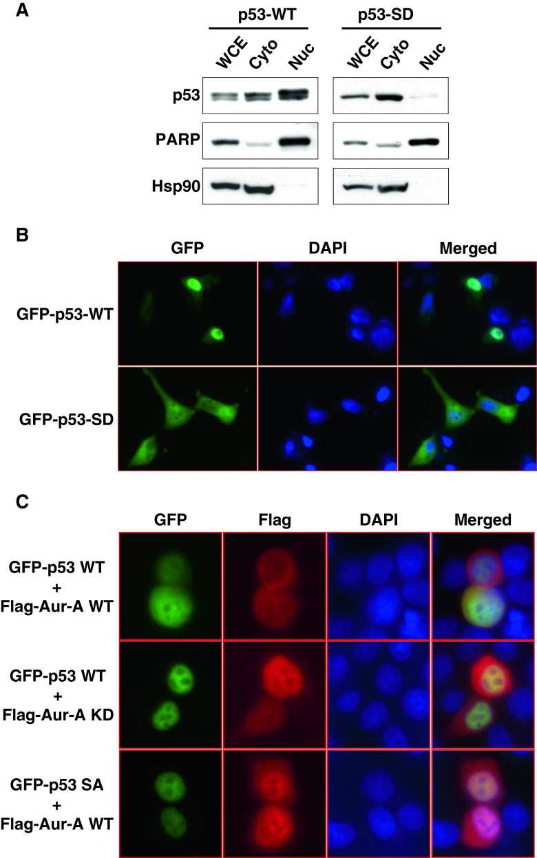
In order to investigate if loss of function changes in Aurora-A phosphorylated p53 is accompanied with altered subcellular distribution of the protein, we performed cell fractionation studies of p53 null lung cancer cell line H1299 transfected with wild-type or phosphor-mimetic mutants of p53. Results revealed that in the wild-type 53 expressing cells, p53 is distributed predominantly in the nuclear fraction, with the cytoplasmic fraction showing relatively less abundant amount of the protein. In contrast, the cells expressing the phosphor-mimetic S215D mutant of p53 demonstrated almost exclusive distribution of the protein in the cytoplasm (Fig. 3a). The distribution pattern of the S315A mutant of p53 was similar to that of the wild-type protein (data not shown). The purity of the nuclear and cytoplasmic fractions was verified with the amount of poly (ADP-ribose) polymerase (PARP) and the Hsp90 proteins detected in these respective extracts. To validate these findings further, we performed immunofluorescence microscopy analysis of the wild-type and phosphor-mimetic or phosphor-deficient mutant p53-expressing cells. In the first set of experiments, green fluorescent protein (GFP)-labeled wild-type protein was detected primarily in the nucleus and the S215D mutant in the cytoplasm, corroborating the cell fractionation results described above (Fig. 3b). In the second set of experiments, we confirmed the role of Aurora-A in the varying distribution patterns of p53 in the nucleus and in the cytoplasm. GFP-labeled p53 wild-type protein when co-expressed with Flag-tagged wild-type Aurora-A protein showed localization mostly in the cytoplasm, but when co-expressed with Flag-tagged kinase, inactive Aurora-A protein localized predominantly in the nucleus. GFP-labeled phosphor-deficient mutant was detected primarily in the nucleus, as seen in case of the wild-type protein (Fig. 3c). Preferential localization of the wild-type p53 protein in the cytoplasm only in the presence of wild-type Aurora-A and not in the presence of the kinase inactive mutant as well as nuclear distribution of the phosphor-deficient p53 mutant clearly document that Aurora-A phosphorylation facilitates cytoplasmic localization of the p53 protein.
Fig. 3.
Serine 215 phosphorylation of p53 by Aurora-A controls subcellular localization of p53. a H1299 cells transfected with GFP tagged wild-type (WT) or phosphor-mimetic mutant (SD) of p53 for 24 h were fractionated into cytosolic (Cyto) and nuclear (Nuc) fractions and analyzed by immunoblotting with anti-GFP antibody. The purity of cytosolic and nuclear fractions was confirmed by immunoblotting for anti-Hsp90 and anti-PARP antibodies, respectively. WCE whole cell lysates. b H1299 cells transfected with GFP-p53 WT or SD mutant were counterstained for DNA with DAPI (blue), and subcellular localization of GFP-p53 was analyzed by immunofluorescent microscopy. c WT or phosphor-deficient mutant (SA) of GFP-p53 was co-transfected with Flag-tagged WT or kinase dead mutant (KD) of Aurora-A into H1299 cells. Twenty-four hours after transfection, cells were immunostained with anti-flag antibody (red) and counterstained for DNA with DAPI (blue). Subcellular distribution of GFP-p53 was analyzed by by immunofluorescent microscopy
Aurora-A Complex with ERα–p53 Enhances the ERα–p53 Interaction and Localization in the Cytoplasm
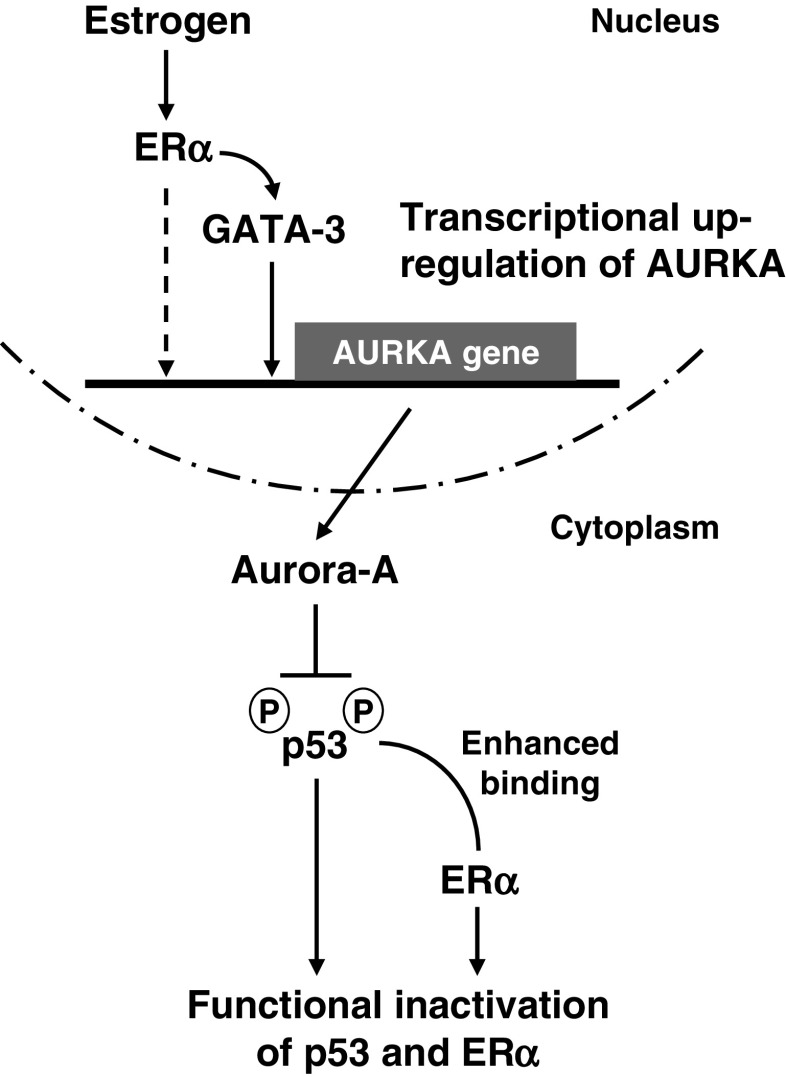
We next investigated if endogenous Aurora-A exists in the reported ERα–p53 complex in human breast cancer cells in vivo. Toward this end, we performed co-immunoprecipitation of cell lysates from ERα-positive MCF7 cells to detect interactions of the endogenous proteins in a possible ternary complex. Immunoprecipitation with antibodies against Aurora-A, ERα, and p53 detected the presence of the three proteins in a complex that were not seen with normal IgG (NIG) in these experiments, verifying the specificity of the interactions resolved in each case (Fig. 4a). Having observed the existence of a complex constituted of Aurora-A, ERα, and p53, we were interested to know if phosphorylation of p53 by Aurora-A regulates its interaction with ERα and also results in cytoplasmic sequestration of the complex. For the purpose, T7-tagged ERα was co-expressed in MCF7 cells with either Flag-tagged wild-type or phosphor-deficient S215A or phosphor-mimetic S215D mutants of p53. The cell lysates were then subjected to immunoprecipitation with anti-Flag antibody to pull down the three different p53 allele-encoded products and immunoblotted with anti-T7 and Flag antibodies to detect their interactions with ERα in each immune complex. Signal intensities of the immunoprecipitated proteins revealed that the amount of ERα present in the phosphor-mimetic mutant p53 complex was significantly higher than those detected in the wild-type p53 and the phosphor-deficient mutant p53 complexes. The results clearly reflected that Aurora-A-phosphorylated p53 enhances binding of ERα to the Aurora-A–ERα–p53 ternary complex (Fig. 4b). Since Aurora-A phosphorylation at serine 215 residue leads to cytoplasmic sequestration of p53, we decided to examine if ERα-positive human breast cancer cells overexpressing Aurora-A show enrichment of both ERα and p53 in the separated cytoplasmic fraction compared with the nuclear fraction of total cell extracts. These experiments were performed with MCF7 cells stably or transiently overexpressing Aurora-A. In both instances, there was clear enrichment of ERα and p53 in the cytoplasm compared with their presence in the nuclear fractions. The experiments allowed us to conclude that in Aurora-A overexpressing ERα-positive human breast cancer cells, ERα bound to Aurora-A-phosphorylated p53 along with Aurora-A remains in a complex that is predominantly localized in the cytoplasm. Based on these results and the previous findings on the role of ERα in positively regulating p53 [17] and Aurora-A [14] transcription, we propose that ERα, p53, and Aurora-A are involved in biphasic regulatory interactions. In the first phase, ERα is involved in activating both p53 and Aurora-A expression in a feed forward regulatory manner, while in the second phase, Aurora-A negatively feedback regulates p53 and ERα functions. This negative feedback regulation is achieved through cytoplasmic sequestration and proteolysis of p53 and also exacerbation of ERα antagonism of p53 function through enhanced binding in breast cancer cells (Fig. 5). As a consequence, it is logical to suggest that continued exposure of breast epithelial cells to estrogen triggers aberrant overexpression of Aurora-A, leading to deregulated proliferation accompanying cellular and molecular changes associated with oncogenic transformation, which involves functional inactivation of p53 and ERα as well as development of estrogen insensitivity.
Fig. 4.
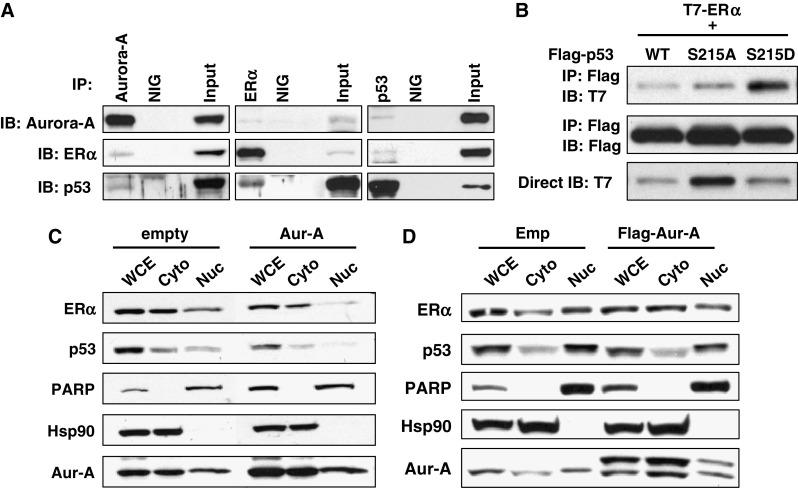
Aurora-A forms ternary complex with ERα and p53, and Aurora-A overexpression causes cytoplasmic sequesteration of them. a MCF7 cells were immunoprecipitated with indicated antibodies or normal IgG (NIG) followed by immunoblotting with indicated antibodies. Aliquots of the same whole cell lysates were directly immunoblotted with anti-Aurora-A antibody (bottom). b T7-tagged ERα was co-transfected with Flag-tagged p53 WT or SA or SD into MCF7 cells. Twenty-four hours after transfection, cells were subjected to immunoprecipitation with anti-Flag antibody followed by immunoblotting with indicated antibodies (top and middle). Whole cell lysates were directly immunoblotted with anti-T7 antibody (bottom). c Stable clone expressing empty vector and Aurora-A was fractionated into cytosolic (Cyto) and nuclear (Nuc) fractions and analyzed by immunoblotting with anti-ERα and p53 antibodies. The purity of cytosolic and nuclear fractions was confirmed by immunoblotting for anti-Hsp90 and anti-PARP antibodies, respectively. WCE whole cell lysates. d MCF7 cells transiently transfected with empty vector or flag-tagged Aurora-A for 24 h were fractionated as in c
Fig. 5.
Proposed model of Aurora-A-mediated functional inactivation of p53 and ERα explaining the signaling pathway, leading to functional inactivation of both ERα and p53 by Aurora-A
Discussion
Aurora-A has been reported to be highly expressed in a significant proportion of in situ and invasive breast cancers [4, 18, 19]. Elevated expression of the AURKA gene was recently identified as the most informative genetic marker in a panel of predictive genes selected from a 3D culture model that was associated with poor prognosis of ER-positive tumors analyzed from multiple independent micro-array datasets [20]. Polymorphisms in the Aurora-A gene have been associated with increased risk of breast cancer [21] that is further elevated when prolonged exposure to estrogen is taken into account [22]. Prognostic significance of Aurora-A gene status in association with chronic estrogen exposure in human breast cancers is recapitulated in the ACI female rat model of mammary tumors induced following long-term exposure to estrogen that was shown to be mediated by elevated expression of Aurora-A [16]. Our recent observation that estrogen positively regulates Aurora-A transcription through activation of the ERα–GATA-3 signaling cascade in ERα-positive breast cancer cells helps explain the plausible molecular mechanisms underlying the above findings. Thus, it appears that aberrant expression of ERα-regulated pathway is critically involved in the initiation and progression of Aurora-A overexpressing breast tumors. Since cross talk between Aurora-A and p53 as well as between ERα and p53 have been implicated to play important roles in breast tumorigenesis, it is imperative that the functional regulatory loop involving the three genes be properly elucidated to unravel the molecular mechanisms responsible for the development of cellular phenotypes associated with mammary tumors.
Regulatory cross talk between p53 and ERα, both at the gene expression level and at the level of protein–protein functional interactions, has been implicated to play roles in diverse cellular processes critical to development, differentiation, and tumorigenesis [23–25]. E2 has been shown to upregulate p53 promoter activity and expression in breast cancer cell lines [17] as well as expression and nuclear localization of p53 in mammary gland of female mice when administered in combination with progesterone in vivo [24]. The E2-mediated transactivation of the p53 promoter, which lacks a consensus estrogen response element (ERE), was proposed to involve ERα in an indirect role as a member of the multi-protein complex that requires CCAAT binding transcription factor-1 and NF-κB as essential components [17]. On the other hand, p53 was shown to regulate ER transcription by binding the promoter through a complex with CARM1, CBP, c-Jun, Sp1, and RNA polymerase II [26] that plausibly is maintained in vivo since in mouse models of breast tumor, a direct correlation of p53 genotype and gene dosage with mRNA and protein expression levels of ER was detected. Tumorigenesis in this mouse model of breast cancer was inhibited more effectively in animals with wild-type p53 genotype expressing normal levels of ER compared with those having p53 loss of function genotype and reduced ER expression [27]. These observations have clinical implications on the role of p53 genotype in determining the effect of hormones on tumors and their therapeutic response to anti-estrogens, such as tamoxifen. Interestingly, active p53-mediated repression of tumor cell proliferation has also recently been reported to occur as a consequence of antiestrogen inhibiting ERα binding to p53, thereby decreasing recruitment of ERα to p53-responsive promoters and allowing transcription of the p53 responsive genes, such as the cyclin-dependent kinase inhibitor p21waf1/cip1 [28]. This study demonstrated that ERα represses p53-mediated transcriptional activation in breast cancer by recruiting nuclear receptor corepressors (NCoR and SMRT) and histone deacetylase 1, thus implicating a dual role for ERα in promoting abnormal cell proliferation through enhancing the transcription of ERE containing pro-proliferative genes and repressing the transcription of p53-responsive anti-proliferative genes. Our findings of ERα activating mitosis—promoting Aurora-A gene transcription through GATA-3 signaling cascade—and Aurora-A phosphorylaton of p53—leading to functional inactivation of the tumor suppressor protein, add yet another layer of complexity to the p53–ERα regulatory network, expected to enhance the aberrant proliferation of breast cancer cells.
An interesting result in our study revealed that ERα forms a ternary complex with Aurora-A and p53, and that phosphorylation of p53 by Aurora-A enhances the level of bound ERα in this complex, which is predominantly sequestered in the cytoplasm. Since cytoplasmic sequestration of ERα is expected to abrogate its transcriptional activation function, it is logical to suggest that Aurora-A overexpressing cells with inactivated ERα may proliferate in an uncontrolled manner in the absence of normally expressing E2-induced ER signaling pathway. Although the mechanisms leading to ER-negative breast tumors are still not clearly known, an interesting hypothesis suggests that ER-negative carcinomas result from transformation of stem or progenitor cells [29]. It is relevant in this context that a potential role of ERα–p53 interaction inhibiting transcriptional activation by p53, detected in stem/progenitor cell containing murine mammospheres, has been suggested to be a critical event in mammary tissue homeostasis and cancer [28]. In view of the finding that ERα binding to p53 phosphorylated by Aurora-A is elevated in breast cancer cells, a possible role for Aurora-A in facilitating ERα-mediated p53 inactivation in breast cancer progenitor cells remains a possibility. Furthermore, since disruption of ERα–p53 interaction has recently been proposed as a novel therapeutic strategy for breast cancer [13], inactivating Aurora-A, likely to promote dissociation of the complex, in such a strategy deserves to be investigated. This can be pursued with the small molecule Aurora-A inhibitors currently in clinical trial for anti-tumor therapy.
In summary, the findings being reported in this paper on the role of Aurora-A in promoting p53 binding to ERα and induction of functional inactivation of both p53 and ERα in breast cancer cells have far-reaching implications in terms of the biology underlying mammary tumorigenesis as well as towards designing of novel therapeutic approaches against this malignancy. Future studies should provide more detailed insight into these possibilities.
Experimental Procedures
Plasmids and Cell Culture
pEGFP- and pcDNA3–Flag–p53 vectors were kindly provided by Dr. Zhi-min Yuan. pcDNA3.1–T7–ERα vector was kindly provided by Dr. Rakesh Kumar [30]. S215D mutation was generated by using Quik mutagenesis kit (Stratagene), and mutation was confirmed by sequencing. pcDNA3–Flag–Aurora-A WT and KD were described [31]. H1299 and MCF7 cell lines were obtained from ATCC and were maintained in DMEM with 10% FBS and antibiotics. MCF7 cells stably overexpressing Aurora-A or control vector were described [6].
Antibodies
Antibodies used in this study were: rabbit polyclonal anti-Aurora-A [2], mouse monoclonal anti-Aurora-A (BD Biosciences), anti-ERα (F-10), anti-p53 (FL-393), anti-Hsp90, control IgG (Santa Cruz Biotechnology), anti-PARP (Cell Signaling Biotechnology), anti-Flag M2 (Sigma), and anti-T7 (Bethyl Laboratories).
Transfection, Cell Fractionation, Immunofluorescence Labeling, and Immunoprecipitation
For transfection, Fugene 6 transfection reagent (Roche) was used according to manufacturer’s instruction. Twenty-four hours after transfection, cell fractionation of transfected cells was performed according to the manufacturer’s protocol (PIERCE).Whole cell extracts were prepared by using RIPA buffer. The subcellular localization of proteins was also determined by indirect immunofluorescence. Briefly, the cells were grown on glass coverslips, fixed in 4% paraformaldehyde for 20 min, permeabilized for 5 min in PBS with 0.5% Triton X-100, and blocked for 1 h in 3% BSA in PBS with 0.1% Triton X-100. Cells were incubated with anti-Flag M2 antibody for 12 h at 4°C, washed three times in PBS, and then incubated with secondary antibody conjugated with Alexa-568 (Invitrogen). DNA was counterstained with 4′,6-diamidino-2-phenylindole (Invitrogen). Microscopic analyses were performed using a Nikon Eclipse 80i fluorescence microscope. All experimental procedures and conditions for immunoprecipitation were described elsewhere [32].
Acknowledgements
This study was supported by grants from National Institutes of Health (NIH; RO1-CA89716) and The University of Texas M.D. Anderson Cancer Center (University Cancer Foundation). The M.D. Anderson Cancer Center DNA sequencing facility is supported by NIH Cancer Center Support Grant (CA16672).
Conflict of Interest
The authors declare that they have no conflict of interest.
References
- 1.Marumoto T, Zhang D, Saya H. Aurora-A—a guardian of poles. Nat Rev Cancer. 2005;5:42–50. doi: 10.1038/nrc1526. [DOI] [PubMed] [Google Scholar]
- 2.Zhou H, Kuang J, Zhong L, Kuo WL, Gray JW, Sahin A, Brinkley BR, Sen S. Tumour amplified kinase STK15/BTAK induces centrosome amplification, aneuploidy and transformation. Nat Genet. 1998;20:189–193. doi: 10.1038/2496. [DOI] [PubMed] [Google Scholar]
- 3.Gritsko TM, Coppola D, Paciga JE, Yang L, Sun M, Shelley SA, Fiorica JV, Nicosia SV, Cheng JQ. Activation and overexpression of centrosome kinase BTAK/Aurora-A in human ovarian cancer. Clin Cancer Res. 2003;9:1420–1426. [PubMed] [Google Scholar]
- 4.Hoque A, Carter J, Xia W, Hung MC, Sahin AA, Sen S, Lippman SM. Loss of aurora A/STK15/BTAK overexpression correlates with transition of in situ to invasive ductal carcinoma of the breast. Cancer Epidemiol Biomark Prev. 2003;12:1518–1522. [PubMed] [Google Scholar]
- 5.Bischoff JR, Anderson L, Zhu Y, Mossie K, Ng L, Souza B, Schryver B, Flanagan P, Clairvoyant F, Ginther C, et al. A homologue of Drosophila aurora kinase is oncogenic and amplified in human colorectal cancers. EMBO J. 1998;17:3052–3065. doi: 10.1093/emboj/17.11.3052. [DOI] [PMC free article] [PubMed] [Google Scholar]
- 6.Katayama H, Sasai K, Kawai H, Yuan ZM, Bondaruk J, Suzuki F, Fujii S, Arlinghaus RB, Czerniak BA, Sen S. Phosphorylation by aurora kinase A induces Mdm2-mediated destabilization and inhibition of p53. Nat Genet. 2004;36:55–62. doi: 10.1038/ng1279. [DOI] [PubMed] [Google Scholar]
- 7.Liu Q, Kaneko S, Yang L, Feldman RI, Nicosia SV, Chen J, Cheng JQ. Aurora-A abrogation of p53 DNA binding and transactivation activity by phosphorylation of serine 215. J Biol Chem. 2004;279:52175–52182. doi: 10.1074/jbc.M406802200. [DOI] [PubMed] [Google Scholar]
- 8.Cattoretti G, Rilke F, Andreola S, D’Amato L, Delia D. P53 expression in breast cancer. Int J Cancer. 1988;41:178–183. doi: 10.1002/ijc.2910410204. [DOI] [PubMed] [Google Scholar]
- 9.Miller LD, Smeds J, George J, Vega VB, Vergara L, Ploner A, Pawitan Y, Hall P, Klaar S, Liu ET, et al. An expression signature for p53 status in human breast cancer predicts mutation status, transcriptional effects, and patient survival. Proc Natl Acad Sci USA. 2005;102:13550–13555. doi: 10.1073/pnas.0506230102. [DOI] [PMC free article] [PubMed] [Google Scholar]
- 10.Olivier M, Langerød A, Carrieri P, Bergh J, Klaar S, Eyfjord J, Theillet C, Rodriguez C, Lidereau R, Bièche I, et al. The clinical value of somatic TP53 gene mutations in 1,794 patients with breast cancer. Clin Cancer Res. 2006;12:1157–1167. doi: 10.1158/1078-0432.CCR-05-1029. [DOI] [PubMed] [Google Scholar]
- 11.Liu W, Konduri SD, Bansal S, Nayak BK, Rajasekaran SA, Karuppayil SM, Rajasekaran AK, Das GM. Estrogen receptor-alpha binds p53 tumor suppressor protein directly and represses its function. J Biol Chem. 2006;281:9837–9840. doi: 10.1074/jbc.C600001200. [DOI] [PubMed] [Google Scholar]
- 12.Sayeed A, Konduri SD, Liu W, Bansal S, Li F, Das GM. Estrogen receptor alpha inhibits p53-mediated transcriptional repression: implications for the regulation of apoptosis. Cancer Res. 2007;67:7746–7755. doi: 10.1158/0008-5472.CAN-06-3724. [DOI] [PubMed] [Google Scholar]
- 13.Liu W, Ip MM, Podgorsak MB, Das GM. Disruption of estrogen receptor alpha–p53 interaction in breast tumors: a novel mechanism underlying the anti-tumor effect of radiation therapy. Breast Cancer Res Treat. 2009;115:43–50. doi: 10.1007/s10549-008-0044-z. [DOI] [PMC free article] [PubMed] [Google Scholar]
- 14.Jiang S, Katayama H, Wang J, Li SA, Hong Y, Radvanyi L, Li JJ, Sen S. Estrogen-induced Aurora Kinase-A (AURKA) gene expression is activated by GATA-3 in estrogen receptor-positive breast cancer cells. Horm Cancer. 2010;1:11–20. doi: 10.1007/s12672-010-0006-x. [DOI] [PMC free article] [PubMed] [Google Scholar]
- 15.Eeckhoute J, Keeton EK, Lupien M, Krum SA, Carroll JS, Brown M. Positive cross-regulatory loop ties GATA-3 to estrogen receptor alpha expression in breast cancer. Cancer Res. 2007;67:6477–6483. doi: 10.1158/0008-5472.CAN-07-0746. [DOI] [PubMed] [Google Scholar]
- 16.Li JJ, Weroha SJ, Lingle WL, Papa D, Salisbury JL, Li SA. Estrogen mediates Aurora-A overexpression, centrosome amplification, chromosomal instability, and breast cancer in female ACI rats. Proc Natl Acad Sci US. 2004;A101:18123–18128. doi: 10.1073/pnas.0408273101. [DOI] [PMC free article] [PubMed] [Google Scholar]
- 17.Qin C, Nguyen T, Stewart J, Samudio I, Burghardt R, Safe S. Estrogen up-regulation of p53 gene expression in MCF-7 breast cancer cells is mediated by calmodulin kinase IV-dependent activation of a nuclear factor kappaB/CCAAT-binding transcription factor-1 complex. Mol Endocrinol. 2002;16:1793–1809. doi: 10.1210/me.2002-0006. [DOI] [PubMed] [Google Scholar]
- 18.Tanaka T, Kimura M, Matsunaga K, Fukada D, Mori H, Okano Y. Centrosomal kinase AIK1 is overexpressed in invasive ductal carcinoma of the breast. Cancer Res. 1999;59:2041–2044. [PubMed] [Google Scholar]
- 19.Miyoshi Y, Iwao K, Egawa C, Noguchi S. Association of centrosomal kinase STK15/BTAK mRNA expression with chromosomal instability in human breast cancers. Int J Cancer. 2001;92:370–373. doi: 10.1002/ijc.1200. [DOI] [PubMed] [Google Scholar]
- 20.Martin KJ, Patrick DR, Bissell MJ, Fournier MV. Prognostic breast cancer signature identified from 3D culture model accurately predicts clinical outcome across independent datasets. PLoS ONE. 2008;3:e2994. doi: 10.1371/journal.pone.0002994. [DOI] [PMC free article] [PubMed] [Google Scholar]
- 21.Cox DG, Hankinson SE, Hunter DJ. Polymorphisms of the AURKA (STK15/Aurora Kinase) gene and breast cancer risk (United States) Cancer Causes Control. 2006;17:81–83. doi: 10.1007/s10552-005-0429-9. [DOI] [PubMed] [Google Scholar]
- 22.Dai Q, Cai QY, Shu XO, Ewart-Toland A, Wen WQ, Balmain A, Gao YT, Zheng W. Synergistic effects of STK15 gene polymorphisms and endogenous estrogen exposure in the risk of breast cancer. Cancer Epidemiol Biomark Prev. 2004;13:2065–2070. [PubMed] [Google Scholar]
- 23.Hurd C, Dinda S, Khattree N, Moudgil VK. Estrogen-dependent and independent activation of the P1 promoter of the p53 gene in transiently transfected breast cancer cells. Oncogene. 1999;18:1067–1072. doi: 10.1038/sj.onc.1202398. [DOI] [PubMed] [Google Scholar]
- 24.Sivaraman L, Conneely OM, Medina D, O’Malley BW. p53 is a potential mediator of pregnancy and hormone-induced resistance to mammary carcinogenesis. Proc Natl Acad Sci USA. 2001;98:12379–12384. doi: 10.1073/pnas.221459098. [DOI] [PMC free article] [PubMed] [Google Scholar]
- 25.Riley T, Sontag E, Chen P, Levine A. Transcriptional control of human p53-regulated genes. Nat Rev Mol Cell Biol. 2008;9:402–412. doi: 10.1038/nrm2395. [DOI] [PubMed] [Google Scholar]
- 26.Shirley SH, Rundhaug JE, Tian J, Cullinan-Ammann N, Lambertz I, Conti CJ, Fuchs-Young R. Transcriptional regulation of estrogen receptor-alpha by p53 in human breast cancer cells. Cancer Res. 2009;69:3405–3414. doi: 10.1158/0008-5472.CAN-08-3628. [DOI] [PMC free article] [PubMed] [Google Scholar]
- 27.Fuchs-Young R, Shirley SH, Lambertz I, Colby JK, Tian J, Johnston D, Gimenez-Conti IB, Donehower LA, Conti CJ, Hursting SD. P53 genotype as a determinant of ER expression and tamoxifen response in the MMTV-Wnt-1 model of mammary carcinogenesis. Breast Cancer Res Treat. 2010 doi: 10.1007/s10549-010-1308-y. [DOI] [PMC free article] [PubMed] [Google Scholar]
- 28.Konduri SD, Medisetty R, Liu W, Kaipparettu BA, Srivastava P, Brauch H, Fritz P, Swetzig WM, Gardner AE, Khan SA, et al. Mechanisms of estrogen receptor antagonism toward p53 and its implications in breast cancer therapeutic response and stem cell regulation. Proc Natl Acad Sci USA. 2010;107:15081–15086. doi: 10.1073/pnas.1009575107. [DOI] [PMC free article] [PubMed] [Google Scholar]
- 29.Varmus HE, Godley LA, Roy S, Taylor IC, Yuschenkoff L, Shi YP, Pinkel D, Gray J, Pyle R, Aldaz CM, et al. Defining the steps in a multistep mouse model for mammary carcinogenesis. Cold Spring Harb Symp Quant Biol. 1994;59:491–499. doi: 10.1101/sqb.1994.059.01.054. [DOI] [PubMed] [Google Scholar]
- 30.Wang RA, Mazumdar A, Vadlamudi RK, Kumar R. P21-activated kinase-1 phosphorylates and transactivates estrogen receptor-α and promotes hyperplasia in mammary epithelium. EMBO J. 2002;21:5437–5447. doi: 10.1093/emboj/cdf543. [DOI] [PMC free article] [PubMed] [Google Scholar]
- 31.Katayama H, Zhou H, Li Q, Tatsuka M, Sen S. Interaction and feedback regulation between STK15/BTAK/Aurora-A kinase and protein phosphatase 1 through mitotic cell division cycle. J Biol Chem. 2001;276:46219–46224. doi: 10.1074/jbc.M107540200. [DOI] [PubMed] [Google Scholar]
- 32.Katayama H, Sasai K, Kloc M, Brinkley BR, Sen S. Aurora kinase-A regulates kinetochore/chromatin associated microtubule assembly in human cells. Cell Cycle. 2008;7:2691–2704. doi: 10.4161/cc.7.17.6460. [DOI] [PubMed] [Google Scholar]


