Abstract
Progestins are reported to increase the risk of more aggressive estrogen receptor positive, progesterone receptor positive (ER+ PR+) breast cancers in postmenopausal women. Using an in vivo rat model of ER+ PR + mammary cancer, we show that tumors arising in the presence of estrogen and progesterone exhibit increased proliferation and decreased nuclear expression of the cell cycle inhibitor p27 compared with tumors growing in the presence of estrogen alone. In human T47D breast cancer cells, progestin increased proliferation and decreased nuclear p27 expression. The decrease of nuclear p27 protein was dependent on activation of Src and PI3K by progesterone receptor isoforms PRA or PRB. Importantly, increased proliferation and decreased nuclear p27 expression were observed in invasive breast carcinoma compared with carcinoma in situ. These results suggest that progesterone specifically regulates intracellular localization of p27 protein and proliferation. Therefore, progesterone-activated pathways can provide useful therapeutic targets for treatment of more aggressive ER+ PR+ breast cancers.
Electronic supplementary material
The online version of this article (doi:10.1007/s12672-013-0159-5) contains supplementary material, which is available to authorized users.
Keywords: Proliferate Cell Nuclear Antigen, T47D Cell, T47D Breast Cancer Cell, Progesterone Receptor Isoforms, Mammary Cancer Development
Introduction
There is compelling evidence that progestins (PS), synthetic molecules with activities similar to the natural hormone progesterone (P) [1], increase breast cancer risk in postmenopausal women receiving estrogen (E) plus progestin (E + PS) hormone therapy (HT) compared to women receiving therapy with E alone [2]. In human breast cancer cell lines in vitro, PS influence cell proliferation, survival, responsiveness to growth factors, and cell morphology [3–6]. However, how PS and the endogenous P contribute to growth of ER+ PR+ breast cancers in vivo remains poorly understood.
P action in the breast is conveyed by two progesterone receptor isoforms, PRA and PRB. PRB regulates expression of genes required for cell proliferation [7], while PRA controls expression of genes important for cell adherence, cell morphology, and resistance to apoptosis [6, 8, 9]. In the mouse mammary gland, PRB is crucial for lobuloalveolar mammary development during pregnancy, whereas PRA deficiency results in uterine and ovarian abnormalities and infertility [10, 11]. PRA overexpression leads to alterations in normal epithelial organization of the mouse mammary gland that are commonly observed in precancerous lesions, suggesting that P signaling via PRA may contribute to the development of mammary cancer [12].
Recent studies have shown that the cell cycle inhibitor p27 (Kip1) may play opposing roles in cancer cells depending on its intracellular localization. In the nucleus, p27 acts as a potent inhibitor of proliferation in the normal breast and breast cancer [13, 14]. In the cytosol of breast cancer cells, p27 promotes cytoskeletal remodeling and cell motility [15, 16]. Decreased nuclear and increased cytoplasmic expression of p27 is frequently observed in primary breast cancers and associated with poor clinical outcome [17, 18]. While it is established that the cytoplasmic localization of p27 is induced by the PI3/PKB [17–19] or Src phosphorylation [20] in vitro, the physiological regulators of subcellular p27 localization in vivo are not known.
Mice are widely used as a model for breast cancer research. Nevertheless, important differences exist between murine and human breast tissue; the mouse mammary gland has a different histoarchitecture of the adult gland compared to the human breast [21]. Furthermore, the developmental pattern of PR expression and PR isoform colocalization differ between the mouse and the human [22], and mouse mammary cancers are predominantly steroid receptor negative (ER− PR−), whereas human breast cancers are predominantly ER+ PR+. In contrast, the adult rat mammary glands have prominent ductal–lobular organization similar to the human breast, and the developmental profile of PRA and PRB expression and PR isoform colocalization in the rat closely resemble that in the human breast [13, 23]. Importantly, the majority of rat mammary cancers are ER+ PR+ and hormone dependent [24].
Using an in vivo rat model of mammary cancers, we have identified novel molecular mechanisms by which P or PS may enhance proliferation of ER+ PR+ human breast cancers. We show here that P and PS promote cytoplasmic localization of p27 acting via cytoplasmic signaling pathways. This mechanism may explain increased aggressiveness of breast cancers that develop in premenopausal women who produce endogenous E and P or postmenopausal women receiving E + PS HRT. These data raise the possibility that blocking progesterone-activated pathways in patients with ER+ PR+ breast cancer may produce additional therapeutic benefits that are not achievable by conventional antiestrogen treatments.
Methods
Animals
Fifty-day-old female Sprague–Dawley rats (Charles-River Laboratory, Raleigh, NC) were ovariectomized, implanted with silastic pellets containing E (2.5 mg/1 cm) with or without four pellets containing P (50 mg/4 cm) and treated with a single intragastric dose (50 mg/kg) of 7,12-dimethylbenz[α]anthracene (DMBA) (N = 16 E-treated rats, N = 17 E + P-treated rats). Sham-operated rats (N = 19 animals) received pellets containing cholesterol and served as ovary-intact (OI) control. Hormone-releasing pellets were replaced every 10 weeks. These replacement regimens produced systemic hormone levels within the physiological range (Online Resources 6). Plasma hormone levels were measured as previously described [41]. For proliferation studies, animals received i.p. injection with 5-bromo-2-deoxyuridine (BrdU) (Sigma, St. Louis, MO) (70 mg/kg) 2 h prior to being euthanized. Normal mammary tissues and tumors were fixed in 10 % buffered formalin and paraffin-embedded for immunohistochemical analysis or flash-frozen in liquid nitrogen and stored at −80 °C for mRNA or protein analysis. Rat tumors were evaluated by a board-certified veterinary pathologist (I.M.L) following the criteria from the Annapolis meeting on mammary pathology of genetically engineered mice [42]. The total number of palpable tumors was similar among all experimental groups. However, many palpable tumors in OI or E-treated rats were benign fibroadenomas with or without atypical hyperplasia.
Cell Lines
Human breast cancer T47D cells co-expressing PRA and PRB were purchased from ATCC (Manassas, VA). T47D cell lines, Y (lacking PR), YA (expressing only PRA), and YB (expressing only PRB) were generously provided by Dr. KB Horwitz (University of Colorado). Cells were grown in minimal essential media and 5 % fetal bovine serum as described [28]. Cells were cultured in phenol red and serum-free media for 48 h prior to treatments with synthetic PS promegestone R5020 (20 nM), PR inhibitor RU486 (100 nM) (both from PerkinElmer Life Sciences, Boston, MA), estradiol (E, 10 nM) (Sigma, St. Louis, MO), Src inhibitor PP2 (20 μM) (Calbiochem, La Jolla, CA), PI3 kinase inhibitor LY294002 (10 μM), and MEKK1/2 inhibitor U0126 (10 μM) (both from Cell Signaling, Beverly, MA) for 24 h. The aliquots of these kinase inhibitors were tested for their ability to block proliferation (LY and U0126) in primary culture of normal mouse mammary epithelial organoids in 3-D cultures and for their ability to block organogenesis and decrease phosphorylated Src levels (PP2) in 3-D cultures of normal mouse mammary epithelial organoids [43]. R5020 was used for in vitro studies because P is rapidly metabolized in vitro [31].
Human Archival Breast Cancer Samples
Human breast cancers were collected in 2000 at Sparrow Hospital, Lansing, MI. All samples were positive for ERα and PR. According to corresponding pathology reports, breast cancer specimens consisted of invasive ductal carcinoma with variable proportions of intraductal carcinoma in situ. Archival specimens were obtained with a protocol approved by the institutional review boards of Michigan State University and Sparrow Hospital.
Immunoblot, Cell Cycle Analysis, and RT-PCR-Based Microarray Analysis
Analyses (Rat Cancer Pathway Finder PCR Array, Qiagen, Frederick, MD) were performed as previously described [41, 44]. Protein loading in immunoblots was normalized by intensity of β-actin bands. Rat keratin 5 (K5) and keratin 18 primers for RT-PCR were from Qiagen, Frederick, MD.
Immunofluorescent Staining
Cells were fixed in 10 % buffered formalin for 30 min, washed with PBS, permeabilized with 0.05 % Triton-X100, blocked in 1 % BSA, incubated overnight with primary antibody, and developed with AlexaFluor488-labeled goat anti-rabbit antibody (Invitrogen, Carlsbad, CA, 1:400). Immunohistochemistry was performed as previously described [13]. The specificity of anti-PRA antibody has been shown by Mote et al. and Aupperlee et al. [22, 45]. The specificity of anti-rodent PRB antibody has been demonstrated by Kariagina et al. [13]. Antibodies used in the study are described in Online Resource (7). Nuclei were counterstained with 4′,6-diamidino-2-phenylindole (DAPI). Since the cytoplasmic p27 is poorly detected by immunocytochemistry, only quantitative analysis of the immunofluorescent nuclear p27 staining was performed using MetaMorph software (Molecular Devices, Sunnyvale, CA).
Quantitation and Statistical Analysis
The number of BrdU, proliferating cell nuclear antigen (PCNA), Ki-67, and p27 positive cells was quantitated as previously described [13] and was expressed as the percent of total epithelial cells with at least 1,000 tumor cells counted. Results were presented as mean ± SEM and differences were considered significant at P < 0.05 by ANOVA/MANOVA with Duncan’s post hoc test where appropriate.
Results
E + P Treatment Increases Incidence of ER+ PR+ Mammary Carcinoma in Carcinogen-Treated Rats
Control OI and ovariectomized rats treated with E alone or E + P were given carcinogen DMBA to induce mammary cancers. According to histopathological analysis, OI rats frequently developed benign fibroadenomas (about 50 % of palpable tumors). The most common malignant mammary neoplasm in OI rats was the papillary type of carcinoma (total number of carcinomas N = 11 per 19 rats). In contrast, the predominant types of lesions in E-treated rats were atypical ductal hyperplasia and cribriform ductal carcinoma in situ (DCIS), with some tumors progressing to cribriform adenocarcinoma (N = 12 carcinomas per 16 rats). E + P treatment significantly enhanced the development of adenocarcinomas compared to treatment with E alone (N = 29 carcinomas per 17 rats, P = 0.0016, χ2 test = 12.839, dF = 2). These invasive tumors varied from cribriform to tubular and papillary type and frequently exhibited features commonly found in more aggressive breast cancers, such as areas of necrosis and infiltration of inflammatory cells.
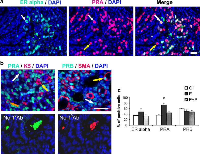
Mammary epithelium in both human and rodents consists of luminal and basal/myoepithelial cells. Luminal cells express steroid receptors ERα, PRA, and PRB [25]. Basal/myoepithelial cells that form the outer layer in the normal mammary gland express α-smooth muscle actin (SMA), transcription factor p63, K5, and keratin 14 (K14) [26]. Mammary tumors in all three treatment groups expressed ERα (Fig. 1a). ERα expression frequently colocalized with PRA expression; however, many PRA-expressing tumor cells did not express ERα, at least at levels detectable by immunofluorescent staining (Fig. 1a). Both PRA and PRB were expressed in luminal tumor cells, whereas PRB was also frequently detected in basal/myoepithelial tumor cells expressing SMA (Fig. 1b, c). Experimental treatments did not influence the percentage of ERα or PRB-expressing cells with the exception of E treatment, which significantly increased the percentage of PRA-expressing cells (Fig. 1d).
Fig. 1.
Carcinogen-induced rat mammary tumors express steroid receptors in luminal and basal/myoepithelial tumor cells. Tumors were from carcinogen-treated ovary-intact (OI) or ovariectomized rats treated with estradiol alone (E) or estradiol plus progesterone (E + P). a Representative images of staining with anti-ERα (teal, white arrows) and anti-PRA antibodies (magenta, white and yellow arrows). White arrows indicate a cell co-expressing ERα and PRA, and yellow arrow indicates a cell expressing only PRA. b On the left, representative images of staining with anti-PRA (teal, a white arrow) and anti-keratin 5 antibodies (K5, red, cytoplasmic stain, yellow arrows) and, on the right, staining with anti-PRB (teal, white arrow) and anti-smooth muscle actin (SMA) antibodies (red, cytoplasmic stain, yellow arrow). An orange arrow indicates a cell co-expressing PRA and basal/myoepithelial marker K5. A yellow arrow indicates a cell co-expressing PRB and basal/myoepithelial marker SMA. Bottom left panel shows staining with anti-rabbit secondary antibody labeled with Alexa 488, bottom right panel shows staining with anti-mouse secondary antibody labeled with Alexa546. a, b Nuclei are stained with DAPI (blue). Scale bars, 50 μm. c Quantitation of cells expressing steroid receptors (N = 5–8 samples/group). *P < 0.05, the percent of PRA positive cells is significantly greater in tumors from E-treated rats compared with that in OI or rats treated with E + P
P/PS Increases Proliferation in ER+ PR+ Mammary Cancers In Vivo
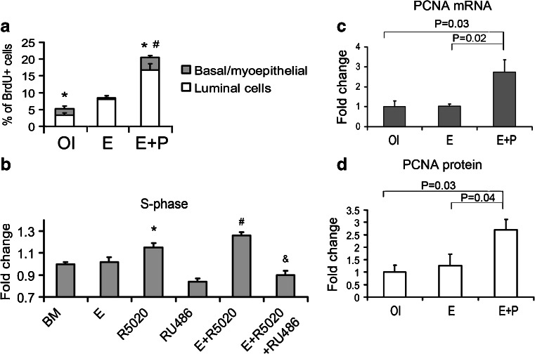
Cell type-specific proliferation was measured by BrdU incorporation. The baseline proliferation of luminal tumor cells was ∼3 % in tumors from OI rats. E + P treatment significantly increased proliferation of luminal tumor cells compared with E-alone treatment (Fig. 2a). Proliferation of basal/myoepithelial tumor cells was significantly greater in tumors from OI or E + P-treatment groups compared with tumors from E-treated rats (Fig. 2a), suggesting that P specifically induced proliferation of basal/myoepithelial tumor cells.
Fig. 2.
Combined treatment with estrogen plus progesterone increases proliferation in rat mammary tumors in vivo and human T47D breast cancer cells in vitro. a, c, d Tumors were from carcinogen-treated ovary-intact (OI) or ovariectomized rats treated with estradiol alone (E) or estradiol plus progesterone (E + P). a Quantitation of BrdU + SMA-luminal cells and BrdU+ SMA+ basal/myoepithelial cells in tumors (N = 8–17 samples/group). *P < 0.05, the percent of basal/myoepithelial proliferating cells is greater in tumors from OI or E + P-treated rats compared with tumors from E-treated rats. #P < 0.05, the percent of luminal proliferating cells is greater in tumors from E + P-treated rats compared with tumors from OI or E-treated rats. b T47D cells were treated for 24 h with estradiol (E), synthetic progestin R5020, PR inhibitor RU486, or their combinations. Bars represent mean ± SEM fold change relative to BM (N = 5–9 samples/treatment). *, #, P < 0.05, the percent of cells in S-phase is increased by R5020 or E + R5020, respectively, compared with basal media (BM). & P < 0.05, the percent of cells in S-phase is decreased by E + R5020 + RU486 compared to E + R5020. c, d Relative expression of PCNA mRNA (c) and densitometry of PCNA immunoblot (d) in tumors (N = 5 tumors/group). Bars represent mean ± SEM fold change compared with levels in OI tumors
Next, we investigated the roles of E and P in human ERα, PRA, and PRB-positive T47D breast cancer cells, which are widely used as model of luminal breast cancer. Consistent with the results obtained in vivo in rat mammary tumors, combined treatment with E plus synthetic PS R5020 (E + R5020) increased proliferation of T47D cells (as measured by the number of cells in S-phase) compared with either E or R5020 alone (Fig. 2b). The PR antagonist RU486 efficiently blocked the proliferation of cells grown without any treatment and cells stimulated to proliferate by E + R5020.
Cell proliferation is controlled by a number of positive and negative cell cycle regulatory proteins. The expression of PCNA mRNA and protein was significantly increased in E + P-treated tumors (Fig. 2c, d). Levels of other positive cell cycle regulators (e.g., cyclin D1, E1, and B1) varied among individual tumors, but averaged levels were not significantly different among the treatment groups (Online Resource 1). Levels of phosphorylated Rb protein, which is required for the cell cycle progression, were not significantly greater in tumors from E and E + P-treatment groups compared with tumors from OI rats (Online Resource 1). Additionally, gene expression of cell cycle inhibitors p21 and p16 was decreased in tumors from E and E + P groups compared with OI group (Online Resource 2). The levels of p21 and p16 proteins in tumors from all treatment groups were below the level of detection by immunoblot (data not shown).
P/PS Decreases Nuclear Localization of p27 via Cytoplasmic, Non-Genomic Signaling
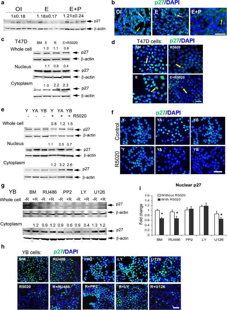
Whole cell protein levels of the cell cycle inhibitor p27 were variable among individual tumors, but mean levels were similar among the treatment groups (Fig. 3a). Importantly, in tumors from E + P-treated rats, p27 expression was localized mainly to the cytoplasm compared with predominant nuclear localization in tumors from OI and E-treated rats (Fig. 3b), suggesting inactivation of the inhibitory function of p27 on cell cycle progression [27]. Consistent with the findings in vivo, R5020 and E + R5020 treatments increased p27 protein in the cytoplasm and decreased p27 protein in the nucleus without altering total p27 levels in T47D cells (Fig. 3c, d). To investigate which PR isoform mediated the effect of P on subcellular localization of p27, we used T47D cells that lack PR (Y cells), express only PRA (YA cells), or express only PRB (YB cells) [28]. Treatment with R5020 did not alter the total cellular p27, but markedly increased p27 protein levels in the cytoplasm (Fig. 3e) and decreased nuclear p27 protein levels and nuclear p27 staining in both YA and YB cells (Fig. 3e, f). These results indicate that PS acting through either PRA or PRB may cause cytoplasmic localization of p27. The effect of R5020 to reduce nuclear localization of p27 was not blocked by PR antagonist RU486, but was abrogated by inhibitors of Src (PP2) and PI3 kinase (LY) signaling in both YB (Fig. 3g–i) and T47D cells (Online Resource 3). When YB cells were treated with a combination of R5020 and Src or PI3K inhibitors, we observed a noticeable decrease of cytoplasmic p27 and strong nuclear p27 staining, while the total levels of p27 was not changed (Fig. 3g–i). These findings suggest that the regulation of p27 localization by PS occurs primarily in the cytoplasm via interaction of PS-bound PR and cytoplasmic kinases. Collectively, these results demonstrate a strong positive association between a P/PS-mediated decrease of nuclear p27 protein and increased proliferation in both rat mammary tumors in vivo and human breast cancer cells in vitro.
Fig. 3.
Combined treatment with estrogen and progesterone causes cytoplasmic localization of p27 in rat mammary tumors in vivo and T47D cells in vitro. a, b Tumors were from carcinogen-treated ovary-intact (OI) or ovariectomized rats treated with estradiol alone (E) or estradiol plus progesterone (E + P). a Immunoblot of p27 in tumor whole cell extracts (N = 5 tumors/group). Numbers above p27 bands indicate mean ± SEM levels of p27 compared with OI tumors. b Representative images of p27 immunostaining in tumors. Cytoplasmic p27 (green, white arrows), nuclear p27 (teal, yellow arrows), and nuclei (blue). Scale bar 100 μm. c–f T47D cells co-expressing PRA and PRB were treated with estradiol (E), R5020 (R), or E + R for 24 h. Y, YA, and YB cells were treated with or without R5020 for 24 h. Representative images of nuclear p27 staining (teal, yellow arrows). c, e Immunoblot of p27 in whole cell, nuclear, and cytoplasmic extracts. Numbers above bands indicate p27 levels in treated cells compared to basal media control (BM). d, f Representative images of p27 immunostaining in T47D cells (d) or Y, YA, and YB cells (f). h, i YB cells were treated with or without R5020 and with or without PR inhibitor RU486, PI3 kinase inhibitor LY294002 (LY), Src inhibitor PP2, and MEKK1/2 inhibitor (U126). g Immunoblot of p27 in whole cell and cytoplasmic extracts. The levels of p27 in whole cell extract were not changed by any treatment. Numbers above bands indicate p27 levels in the cytosol of treated cells compared to basal media control (BM). h Representative images of cells stained with anti-p27 antibody (teal) and i quantitative analysis of nuclear p27 staining in treated cell relative to BM. * P < 0.05 compared to BM. b, d, f, h Nuclei are stained with DAPI (blue). Scale bars 50 μm
Transition of ER+ PR+ Human Breast Cancers from In Situ to Invasive Carcinoma
Next, we investigated proliferation and p27 expression in archival samples of ER+ PR+ primary human breast cancers that contained both a preinvasive DCIS component and an invasive ductal carcinoma (IDC) component (N = 21 patients). DCIS were composed of luminal cells circumscribed by a layer of basal/myoepithelial cells expressing K14 and p63 (Fig. 4a–c). The percentage of proliferating cells expressing PCNA and Ki-67 increased, while the percentage of cells expressing nuclear p27 decreased in IDC compared with DCIS (Fig. 4d, Online Resource 4). These data indicate that the transition from DCIS to invasive tumor is associated with increased proliferation and decreased nuclear p27 expression.
Fig. 4.
Invasiveness of human ER+ PR+ breast cancers is associated with increased proliferation and decreased nuclear p27 expression. Human breast cancer samples that contained ductal carcinoma in situ and invasive ductal carcinoma were immunostained with a proliferating cell nuclear antigen (PCNA) and p63, b Ki-67 and basal/myoepithelial keratin K14, and c p27 and K14. A–c Carcinoma in situ (top panels) and invasive tumors (bottom panels). White arrows indicate basal/myoepithelial cells, yellow arrows indicate PCNA or K-67 positive cells (a, b), or cells expressing nuclear p27 (c). White dotted lines indicate the margin of carcinoma in situ. d Quantitation of the percent of PCNA+, Ki-67+, and nuclear p27+ cells in human tumors. *P < 0.05 for comparisons indicated by brackets. Nuclei are stained with DAPI (blue). Scale bars 50 μm
Discussion
Although a stimulatory effect of P/PS on cell proliferation in breast cancer cell lines and in mouse cancer models has been described previously [3, 5, 12], this study provides the first demonstration that P/PS regulates the intracellular localization of the cell cycle inhibitor p27 in vivo and in vitro that may enhance cell proliferation. The treatment with E + P in ovariectomized rats markedly increased incidence of mammary carcinoma and resulted in tumors with increased proliferation and decreased expression of nuclear p27 compared with E treatment. We further showed that PS specifically regulates intracellular localization of p27 in human breast cancer cells. Importantly, greater proliferation and decrease of nuclear p27 localization were observed in IDC compared with DCIS, suggesting that P signaling may accelerate transition to IDC. Since cytoplasmic expression of p27 is strongly associated with poor prognosis in breast cancer patients, treatments that prevent p27 localization to the cytoplasm may provide clinical benefits. Specific contribution of P/PS in breast cancer development in vivo and in vitro implies that therapies targeted to P/PS-activated pathways may complement conventional antiestrogen treatments. This may be particularly important for treatment of antiestrogen-resistant ER+ PR+ breast cancers.
While it is recognized that synthetic PS increases the risk of more aggressive breast cancers in postmenopausal women receiving combined E + PS HT [2, 29], the current results indicate that the natural hormone P is also highly effective in promoting proliferation of mammary tumors in rats and is likely to act similarly in humans. Furthermore, the continuous administration of E + P resulted in the development of faster growing tumors compared with tumors from OI animals, where the hormonal milieu fluctuates during the estrus cycle. Indeed, the continuous use of E + PS HRT has been shown to be associated with a greater breast cancer risk than E + PS HRT taken in a cyclic manner [30]. It was also shown that the cyclic intake of E + PS increases the breast cancer risk compared with the cyclic intake of E + P [2]. Because P is metabolized significantly faster and has a shorter half-life than synthetic PS [31], the lack of P effects in this study may have resulted from potentially reduced P levels due to metabolic inactivation.
The predominant papillary tumor type that develops in OI rats is not very common in humans. In contrast, mammary cancers in hormone-treated ovariectomized E or E + P-treated rats are more similar to human breast cancers in regards to tumor histopathology. Because ovariectomy completely blocks development of carcinogen-induced tumors and P alone does not support mammary cancer development in ovariectomized rats [32], we conclude that E is a critical factor for tumor initiation and development. In general, the presence of E is permissive for P action in vivo because E signaling stimulates PR expression and, thus, allows robust P action [33]. However, the majority of lesions in E-treated rats were DCIS or noninvasive carcinomas. In contrast, E + P treatment drastically accelerated tumor progression and increased incidence of carcinoma more than twofold. The faster development of carcinomas in E + P-treated rats is likely due to a dual action of P on tumor cells; the direct stimulation of genes required for proliferation and the relief from cell cycle inhibition by nuclear p27.
In the rat mammary tumors, P indeed increased proliferation of luminal cells that expressed both PRA and PRB and basal/myoepithelial cells that expressed PRB only. It has been reported by others that P acting specifically through PRB stimulates expression of PCNA in YB cells [7]. Therefore, it is possible that P acting through PRB homodimers (in basal/myoepithelial cells) or PRA/PRB heterodimers (in luminal cells) directly stimulates proliferation by inducing the PCNA gene. Besides direct induction of proliferation in cells expressing PRB, P may stimulate proliferation of PR-negative cells by inducing paracrine factors. For example, the P-dependent paracrine factor Wnt-4 has been reported to be important for proliferation in the normal mouse mammary gland [34]. Another P-regulated paracrine factor, receptor activator of nuclear factor kappa B ligand (RANKL), has been demonstrated to increase proliferation and mammary cancer development in the mouse and mediates P-induced proliferation in the normal human breast tissue [35–37]. However, whether or not Wnt-4 and RANKL contribute to breast cancer development or proliferation of breast cancer cells in vivo remains unclear.
High nuclear p27 expression is associated with a state of proliferative quiescence in the rat mammary gland during development [13]. In contrast, decreased nuclear p27 expression is observed during pregnancy when mammary glands actively proliferate [13]. In agreement with studies in the rat, it has been shown that decreased nuclear p27 levels may facilitate proliferation in human breast cancer cell lines [18]. According to previous reports, prolonged treatment with PS (for more than 36 h) blocks proliferation in T47D cells due to accumulation of p27 protein [38]. In contrast, present observations in the rat mammary tumors in vivo showed that total cellular p27 protein was not increased by chronic E + P treatment. It is possible that the long-term regulation of p27 protein in the rat tumors in vivo and regulation of p27 protein in the T47D breast cancer cells in vitro differ.
Both progesterone receptor isoforms, PRA and PRB, mediated the effect of PS on p27 localization to the cytoplasm in T47D cells. Similar effect of PS was noted in MCF7 breast cancer cells expressing both PRA and PRB (Online Resource 5). Importantly, the activation of the cytoplasmic kinases Src and PI3K was required for cytoplasmic localization of p27. Indeed, both PRA and PRB can produce rapid activation of cytoplasmic kinases via direct interaction between the SH2 domain of progestin-bound PRA or PRB and Src [39]. Because PRA predominantly localizes to the nucleus, it is more likely that PRB, which localizes to both the nucleus and cytoplasm, mediates the effect of P/PS on p27 localization in vivo. In addition to Src activation, PS treatment rapidly activates the PI3 kinase pathway in T47D cells [40]). It has been shown that the PI3 kinase is required for p27 phosphorylation on Thr157 and that phosphorylated p27 is sequestered in the cytosol [17–19]. Our findings that activation of both Src and PI3 kinase are necessary for cytoplasmic localization of p27 are in agreement with previous reports by other groups. Importantly, the PR antagonist RU486 did not influence intracellular localization of p27 but completely blocked PS-induced proliferation (Fig. 2b). This suggests that the major mechanism by which P induces proliferation involves genomic signaling though activated PR. The genomic induction of genes driving proliferation such as PCNA is, possibly, a primary mechanism responsible for proliferation induced by R5020; however, the non-genomic signaling of PRA or PRB that leads to activation of Src and PI3 kinases and cytoplasmic localization of p27 protein may facilitate and enhance tumor proliferation by releasing inhibition of cyclin-dependent kinases in the nucleus.
In conclusion, the data obtained in vivo in the rat indicate that physiological levels of the natural hormone P can significantly contribute to mammary cancer development. ER+ PR+ mammary cancers in ovariectomized E + P-treated rats may be a useful preclinical model of human IDC for testing novel therapeutic interventions. Our results obtained in T47D cells and similarities between ER+ PR+ human and rat mammary cancers strongly suggest that P or PS specifically contribute to the aggressiveness of human breast cancers by regulating proliferation and intracellular localization of the cell cycle inhibitor p27. Thus, assessment of the systemic P levels in breast cancer patients, analysis of PRA and PRB expression, and evaluation of biomarkers indicative of P signaling may provide novel prognostic tools as well as targets for interventions in patients with more aggressive ER+ PR+ breast cancers.
Declaration
All animal handling and experimental procedures were approved by Michigan State University committee on animal use and care. Analysis of human breast cancer specimens was approved by the institutional review boards of Michigan State University and Sparrow Hospital.
Electronic Supplementary Material
Below is the link to the electronic supplementary material.
Effects of hormone treatments on cell cycle regulatory proteins in rat mammary tumors. a, b Ovary-intact (OI) or ovariectomized rats treated with estradiol alone (E) or estradiol plus progesterone (E+P) were treated with DMBA to induce mammary tumors. Immunoblot analysis of cell cycle regulatory proteins cyclin D1, cyclin E1, cyclin B1, and phosphorylated Rb protein (phospho-Rb) in whole cell tumor extracts (N = 4–6 tumors/group). a Numbers above bands indicate mean ± SEM fold change compared with OI tumors. b Densitometry of phospho-Rb protein showing a trend for increased phospho-Rb levels in tumors from rats treated with E+P. (PDF 172 kb)
Effects of hormone treatments on mRNA expression of cell cycle regulatory proteins, p21 and p16, in rat mammary tumors. Ovary-intact (OI) or ovariectomized rats treated with estradiol alone (E) or estradiol plus progesterone (E + P) were treated with DMBA to induce mammary tumors. Real-time RT-PCR analysis of p16 and p21 mRNA expression was performed (N = 3–5 tumors/group). The bars represent the mean ± SEM fold difference. The level of p16 mRNA expression in OI tumors was arbitrarily taken as one. The levels of p21 mRNA is presented as fold change compared to the levels of p16 mRNA expression in OI tumors. *P < 0.05, p21 mRNA is decreased by E and E + P treatment compared with OI control. #, P < 0.05, p16 mRNA is decreased by E and E + P treatment compared with OI control. (PDF 222 kb)
Effects of kinase inhibitors on intracellular localization of p27 protein in T47D cells treated with progestin. a Representative images of cells cultured without serum for 48 h and treated for 24 h with or without R5020 (R) with or without PR antagonist RU486, Src inhibitor PP2, PI3 kinase inhibitor LY294002 (LY), and MEKK1/2 inhibitor (U126) and stained with anti-p27 antibody (teal) and b quantitative analysis of nuclear p27 staining in treated cells relative to basal media (BM) control. * - P < 0.05 compared to BM. Nuclei are stained with DAPI (blue). Scale bar, 50 μm. (PDF 951 kb)
(PDF 24 kb)
Intracellular localization of p27 protein in MCF7 cells treated with progestin. a Representative images of cells cultured without serum for 48 h and treated for 24 h with basal media (BM), estrogen (E), synthetic progestin R5020 (R), and their combination (E+R5020) and stained with anti-p27 antibody (teal). Nuclei are stained with DAPI (blue). Scale bar, 50 μm. b Immunoblot of p27 in whole cell extracts. Numbers above bands indicate p27 levels in treated cells compared to basal media control (PDF 1184 kb)
Effect of hormone treatments on serum estradiol and progesterone levels ovary-intact (OI) or ovariectomized rats treated with estradiol alone (E) or estradiol plus progesterone (E + P) were treated with DMBA to induce mammary tumors. Hormone levels were measured in rats that developed mammary tumors. Serum 17-β-estradiol levels (a) (N = 5–6 animals/group) and progesterone levels (b) (N = 6-8 animals/group). *P < 0.05, progesterone levels in E-treated rats are lower than in OI or E + P-treated rats. (PDF 488 kb)
(PDF 27 kb)
Acknowledgments
The authors thank Kristina Miller, Lyndsi Davenport, Kyle Pohl, Alicia Kramer, Kristen Bullard, Anthony Yuhas III, Sharmila Kulkarni, Bennett Cho, Howard Her, Jang Park, and Michael DeVisser for the excellent technical assistance, Dr. Valentina Factor for critical reading of the manuscript, and Dr. Horowitz for providing Y, YA, and YB cells. This work was supported by the US Army Medical Research and Materiel Command under W81XWH-07-1-0502 (to S.Z.H), W81XWH-11-1-0108 (to A.K) and by the Breast Cancer and the Environment Research Centers grant U01 ES/CA 012800 (to S.Z.H.) from the National Institute of Environment Health Science (NIEHS) and the National Cancer Institute, National Institutes of Health, Department of Health and Human Services. The contents of the study are solely the responsibility of the authors and do not necessarily represent the official views of the NIEHS or NCI, NIH.
References
- 1.Bray JD, Jelinsky S, Ghatge R, Bray JA, Tunkey C, Saraf K, Jacobsen BM, Richer JK, Brown EL, Winneker RC, Horwitz KB, Lyttle CR. Quantitative analysis of gene regulation by seven clinically relevant progestins suggests a highly similar mechanism of action through progesterone receptors in T47D breast cancer cells. J Steroid Biochem Mol Biol. 2005;97:328–341. doi: 10.1016/j.jsbmb.2005.06.032. [DOI] [PubMed] [Google Scholar]
- 2.Campagnoli C, Clavel-Chapelon F, Kaaks R, Peris C, Berrino F. Progestins and progesterone in hormone replacement therapy and the risk of breast cancer. J Steroid Biochem Mol Biol. 2005;96:95–108. doi: 10.1016/j.jsbmb.2005.02.014. [DOI] [PMC free article] [PubMed] [Google Scholar]
- 3.Skildum A, Faivre E, Lange CA. Progesterone receptors induce cell cycle progression via activation of mitogen-activated protein kinases. Mol Endocrinol. 2005;19:327–339. doi: 10.1210/me.2004-0306. [DOI] [PubMed] [Google Scholar]
- 4.Moore MR, Spence JB, Kiningham KK, Dillon JL. Progestin inhibition of cell death in human breast cancer cell lines. J Steroid Biochem Mol Biol. 2006;98:218–227. doi: 10.1016/j.jsbmb.2005.09.008. [DOI] [PubMed] [Google Scholar]
- 5.Carvajal A, Espinoza N, Kato S, Pinto M, Sadarangani A, Monso C, Aranda E, Villalon M, Richer JK, Horwitz KB, Brosens JJ, Owen GI. Progesterone pretreatment potentiates EGF pathway signaling in the breast cancer cell line ZR-75*. Breast Cancer Res Treat. 2005;94:171–183. doi: 10.1007/s10549-005-7726-6. [DOI] [PubMed] [Google Scholar]
- 6.McGowan EM, Weinberger RP, Graham JD, Hill HD, Hughes JA, O’Neill GM, Clarke CL. Cytoskeletal responsiveness to progestins is dependent on progesterone receptor A levels. J Mol Endocrinol. 2003;31:241–253. doi: 10.1677/jme.0.0310241. [DOI] [PubMed] [Google Scholar]
- 7.Richer JK, Jacobsen BM, Manning NG, Abel MG, Wolf DM, Horwitz KB. Differential gene regulation by the two progesterone receptor isoforms in human breast cancer cells. J Biol Chem. 2002;277:5209–5218. doi: 10.1074/jbc.M110090200. [DOI] [PubMed] [Google Scholar]
- 8.McGowan EM, Clarke CL. Effect of overexpression of progesterone receptor A on endogenous progestin-sensitive endpoints in breast cancer cells. Mol Endocrinol. 1999;13:1657–1671. doi: 10.1210/me.13.10.1657. [DOI] [PubMed] [Google Scholar]
- 9.Graham JD, Yager ML, Hill HD, Byth K, O’Neill GM, Clarke CL. Altered progesterone receptor isoform expression remodels progestin responsiveness of breast cancer cells. Mol Endocrinol. 2005;19:2713–2735. doi: 10.1210/me.2005-0126. [DOI] [PubMed] [Google Scholar]
- 10.Mulac-Jericevic B, Lydon JP, DeMayo FJ, Conneely OM. Defective mammary gland morphogenesis in mice lacking the progesterone receptor B isoform. Proc Natl Acad Sci U S A. 2003;100:9744–9749. doi: 10.1073/pnas.1732707100. [DOI] [PMC free article] [PubMed] [Google Scholar]
- 11.Mulac-Jericevic B, Conneely OM. Reproductive tissue selective actions of progesterone receptors. Reproduction. 2004;128:139–146. doi: 10.1530/rep.1.00189. [DOI] [PubMed] [Google Scholar]
- 12.Shyamala G, Yang X, Cardiff RD, Dale E. Impact of progesterone receptor on cell-fate decisions during mammary gland development. Proc Natl Acad Sci U S A. 2000;97:3044–3049. doi: 10.1073/pnas.97.7.3044. [DOI] [PMC free article] [PubMed] [Google Scholar]
- 13.Kariagina A, Aupperlee MD, Haslam SZ. Progesterone receptor isoforms and proliferation in the rat mammary gland during development. Endocrinology. 2007;148:2723–2736. doi: 10.1210/en.2006-1493. [DOI] [PubMed] [Google Scholar]
- 14.Han S, Park K, Kim HY, Lee MS, Kim HJ, Kim YD. Reduced expression of p27Kip1 protein is associated with poor clinical outcome of breast cancer patients treated with systemic chemotherapy and is linked to cell proliferation and differentiation. Breast Cancer Res Treat. 1999;55:161–167. doi: 10.1023/A:1006258222233. [DOI] [PubMed] [Google Scholar]
- 15.Larrea MD, Hong F, Wander SA, da Silva TG, Helfman D, Lannigan D, Smith JA, Slingerland JM. RSK1 drives p27Kip1 phosphorylation at T198 to promote RhoA inhibition and increase cell motility. Proc Natl Acad Sci U S A. 2009;106:9268–9273. doi: 10.1073/pnas.0805057106. [DOI] [PMC free article] [PubMed] [Google Scholar]
- 16.Wu FY, Wang SE, Sanders ME, Shin I, Rojo F, Baselga J, Arteaga CL. Reduction of cytosolic p27(Kip1) inhibits cancer cell motility, survival, and tumorigenicity. Cancer Res. 2006;66:2162–2172. doi: 10.1158/0008-5472.CAN-05-3304. [DOI] [PubMed] [Google Scholar]
- 17.Liang J, Zubovitz J, Petrocelli T, Kotchetkov R, Connor MK, Han K, Lee JH, Ciarallo S, Catzavelos C, Beniston R, Franssen E, Slingerland JM. PKB/Akt phosphorylates p27, impairs nuclear import of p27, and opposes p27-mediated G1 arrest. Nat Med. 2002;8:1153–1160. doi: 10.1038/nm761. [DOI] [PubMed] [Google Scholar]
- 18.Viglietto G, Motti ML, Bruni P, Melillo RM, D’Alessio A, Califano D, Vinci F, Chiappetta G, Tsichlis P, Bellacosa A, Fusco A, Santoro M. Cytoplasmic relocalization and inhibition of the cyclin-dependent kinase inhibitor p27(Kip1) by PKB/Akt-mediated phosphorylation in breast cancer. Nat Med. 2002;8:1136–1144. doi: 10.1038/nm762. [DOI] [PubMed] [Google Scholar]
- 19.Shin I, Yakes FM, Rojo F, Shin NY, Bakin AV, Baselga J, Arteaga CL. PKB/Akt mediates cell-cycle progression by phosphorylation of p27(Kip1) at threonine 157 and modulation of its cellular localization. Nat Med. 2002;8:1145–1152. doi: 10.1038/nm759. [DOI] [PubMed] [Google Scholar]
- 20.Chu I, Sun J, Arnaout A, Kahn H, Hanna W, Narod S, Sun P, Tan CK, Hengst L, Slingerland J. p27 phosphorylation by Src regulates inhibition of cyclin E-Cdk2. Cell. 2007;128:281–294. doi: 10.1016/j.cell.2006.11.049. [DOI] [PMC free article] [PubMed] [Google Scholar]
- 21.Ball SM. The development of the terminal end bud in the prepubertal–pubertal mouse mammary gland. Anat Rec. 1998;250:459–464. doi: 10.1002/(SICI)1097-0185(199804)250:4<459::AID-AR9>3.0.CO;2-S. [DOI] [PubMed] [Google Scholar]
- 22.Aupperlee MD, Smith KT, Kariagina A, Haslam SZ. Progesterone receptor isoforms A and B: temporal and spatial differences in expression during murine mammary gland development. Endocrinology. 2005;146:3577–3588. doi: 10.1210/en.2005-0346. [DOI] [PubMed] [Google Scholar]
- 23.Russo J, Russo IH. Experimentally induced mammary tumors in rats. Breast Cancer Res Treat. 1996;39:7–20. doi: 10.1007/BF01806074. [DOI] [PubMed] [Google Scholar]
- 24.Russo J, Gusterson BA, Rogers AE, Russo IH, Wellings SR, van Zwieten MJ. Comparative study of human and rat mammary tumorigenesis. Lab Invest. 1990;62:244–278. [PubMed] [Google Scholar]
- 25.Clarke RB, Howell A, Potten CS, Anderson E. Dissociation between steroid receptor expression and cell proliferation in the human breast. Cancer Res. 1997;57:4987–4991. [PubMed] [Google Scholar]
- 26.Heatley M, Maxwell P, Whiteside C, Toner P. Cytokeratin intermediate filament expression in benign and malignant breast disease. J Clin Pathol. 1995;48:26–32. doi: 10.1136/jcp.48.1.26. [DOI] [PMC free article] [PubMed] [Google Scholar]
- 27.Coqueret O. New roles for p21 and p27 cell-cycle inhibitors: a function for each cell compartment? Trends Cell Biol. 2003;13:65–70. doi: 10.1016/S0962-8924(02)00043-0. [DOI] [PubMed] [Google Scholar]
- 28.Sartorius CA, Groshong SD, Miller LA, Powell RL, Tung L, Takimoto GS, Horwitz KB. New T47D breast cancer cell lines for the independent study of progesterone B- and A-receptors: only antiprogestin-occupied B-receptors are switched to transcriptional agonists by cAMP. Cancer Res. 1994;54:3868–3877. [PubMed] [Google Scholar]
- 29.Chlebowski RT, Anderson GL, Gass M, Lane DS, Aragaki AK, Kuller LH, Manson JE, Stefanick ML, Ockene J, Sarto GE, Johnson KC, Wactawski-Wende J, Ravdin PM, Schenken R, Hendrix SL, Rajkovic A, Rohan TE, Yasmeen S, Prentice RL. Estrogen plus progestin and breast cancer incidence and mortality in postmenopausal women. JAMA. 2010;304:1684–1692. doi: 10.1001/jama.2010.1500. [DOI] [PMC free article] [PubMed] [Google Scholar]
- 30.Porch JV, Lee IM, Cook NR, Rexrode KM, Burin JE. Estrogen–progestin replacement therapy and breast cancer risk: the Women’s Health Study (United States) Cancer Causes Control. 2002;13:847–854. doi: 10.1023/A:1020617415381. [DOI] [PubMed] [Google Scholar]
- 31.Horwitz KB, Pike AW, Gonzalez-Aller C, Fennessey PV. Progesterone metabolism in T47Dco human breast cancer cells–II. Intracellular metabolic path of progesterone and synthetic progestins. J Steroid Biochem. 1986;25:911–916. doi: 10.1016/0022-4731(86)90323-7. [DOI] [PubMed] [Google Scholar]
- 32.McCormick DL, Mehta RG, Thompson CA, Dinger N, Caldwell JA, Moon RC. Enhanced inhibition of mammary carcinogenesis by combined treatment with N-(4-hydroxyphenyl) retinamide and ovariectomy. Cancer Res. 1982;42:508–512. [PubMed] [Google Scholar]
- 33.Brisken C, O’Malley B. Hormone action in the mammary gland. Cold Spring Harb Perspect Biol. 2010;2:a003178. doi: 10.1101/cshperspect.a003178. [DOI] [PMC free article] [PubMed] [Google Scholar]
- 34.Brisken C, Heineman A, Chavarria T, Elenbaas B, Tan J, Dey SK, McMahon JA, McMahon AP, Weinberg RA. Essential function of Wnt-4 in mammary gland development downstream of progesterone signaling. Genes Dev. 2000;14:650–654. [PMC free article] [PubMed] [Google Scholar]
- 35.Schramek D, Leibbrandt A, Sigl V, Kenner L, Pospisilik JA, Lee HJ, Hanada R, Joshi PA, Aliprantis A, Glimcher L, Pasparakis M, Khokha R, Ormandy CJ, Widschwendter M, Schett G, Penninger JM. Osteoclast differentiation factor RANKL controls development of progestin-driven mammary cancer. Nature. 2010;468:98–102. doi: 10.1038/nature09387. [DOI] [PMC free article] [PubMed] [Google Scholar]
- 36.Gonzalez-Suarez E, Jacob AP, Jones J, Miller R, Roudier-Meyer MP, Erwert R, Pinkas J, Branstetter D, Dougall WC. RANK ligand mediates progestin-induced mammary epithelial proliferation and carcinogenesis. Nature. 2010;468:103–107. doi: 10.1038/nature09495. [DOI] [PubMed] [Google Scholar]
- 37.Tanos T, Sflomos G, Echeverria PC, Ayyanan A, Gutierrez M, Delaloye JF, Raffoul W, Fiche M, Dougall W, Schneider P, Yalcin-Ozuysal O, Brisken C. Progesterone/RANKL is a major regulatory axis in the human breast. Sci Transl Med. 2013;5:182. doi: 10.1126/scitranslmed.3005654. [DOI] [PubMed] [Google Scholar]
- 38.Groshong SD, Owen GI, Grimison B, Schauer IE, Todd MC, Langan TA, Sclafani RA, Lange CA, Horwitz KB. Biphasic regulation of breast cancer cell growth by progesterone: role of the cyclin-dependent kinase inhibitors, p21 and p27(Kip1) Mol Endocrinol. 1997;11:1593–1607. doi: 10.1210/me.11.11.1593. [DOI] [PubMed] [Google Scholar]
- 39.Faivre EJ, Lange CA. Progesterone receptors upregulate Wnt-1 to induce epidermal growth factor receptor transactivation and c-Src-dependent sustained activation of Erk1/2 mitogen-activated protein kinase in breast cancer cells. Mol Cell Biol. 2007;27:466–480. doi: 10.1128/MCB.01539-06. [DOI] [PMC free article] [PubMed] [Google Scholar]
- 40.Saitoh M, Ohmichi M, Takahashi K, Kawagoe J, Ohta T, Doshida M, Takahashi T, Igarashi H, Mori-Abe A, Du B, Tsutsumi S, Kurachi H. Medroxyprogesterone acetate induces cell proliferation through up-regulation of cyclin D1 expression via phosphatidylinositol 3-kinase/Akt/nuclear factor-kappaB cascade in human breast cancer cells. Endocrinology. 2005;146:4917–4925. doi: 10.1210/en.2004-1535. [DOI] [PubMed] [Google Scholar]
- 41.Zhao Y, Tan YS, Haslam SZ, Yang C. Perfluorooctanoic acid effects on steroid hormone and growth factor levels mediate stimulation of peripubertal mammary gland development in C57BL/6 mice. Toxicol Sci. 2010;115:214–224. doi: 10.1093/toxsci/kfq030. [DOI] [PMC free article] [PubMed] [Google Scholar]
- 42.Cardiff RD, Anver MR, Gusterson BA, Hennighausen L, Jensen RA, Merino MJ, Rehm S, Russo J, Tavassoli FA, Wakefield LM, Ward JM, Green JE. The mammary pathology of genetically engineered mice: the consensus report and recommendations from the Annapolis meeting. Oncogene. 2000;19:968–988. doi: 10.1038/sj.onc.1203277. [DOI] [PubMed] [Google Scholar]
- 43.Meyer G, Leipprandt J, Xie J, Aupperlee MD, Haslam SZ. A potential role of progestin-induced laminin-5/alpha6-integrin signaling in the formation of side branches in the mammary gland. Endocrinology. 2012;153:4990–5001. doi: 10.1210/en.2012-1518. [DOI] [PMC free article] [PubMed] [Google Scholar]
- 44.Kariagina A, Xie J, Leipprandt JR, Haslam SZ. Amphiregulin mediates estrogen, progesterone, and EGFR signaling in the normal rat mammary gland and in hormone-dependent rat mammary cancers. Hormones & Cancer. 2010;1:229–244. doi: 10.1007/s12672-010-0048-0. [DOI] [PMC free article] [PubMed] [Google Scholar]
- 45.Mote PA, Johnston JF, Manninen T, Tuohimaa P, Clarke CL. Detection of progesterone receptor forms A and B by immunohistochemical analysis. J Clin Pathol. 2001;54:624–630. doi: 10.1136/jcp.54.8.624. [DOI] [PMC free article] [PubMed] [Google Scholar]
Associated Data
This section collects any data citations, data availability statements, or supplementary materials included in this article.
Supplementary Materials
Effects of hormone treatments on cell cycle regulatory proteins in rat mammary tumors. a, b Ovary-intact (OI) or ovariectomized rats treated with estradiol alone (E) or estradiol plus progesterone (E+P) were treated with DMBA to induce mammary tumors. Immunoblot analysis of cell cycle regulatory proteins cyclin D1, cyclin E1, cyclin B1, and phosphorylated Rb protein (phospho-Rb) in whole cell tumor extracts (N = 4–6 tumors/group). a Numbers above bands indicate mean ± SEM fold change compared with OI tumors. b Densitometry of phospho-Rb protein showing a trend for increased phospho-Rb levels in tumors from rats treated with E+P. (PDF 172 kb)
Effects of hormone treatments on mRNA expression of cell cycle regulatory proteins, p21 and p16, in rat mammary tumors. Ovary-intact (OI) or ovariectomized rats treated with estradiol alone (E) or estradiol plus progesterone (E + P) were treated with DMBA to induce mammary tumors. Real-time RT-PCR analysis of p16 and p21 mRNA expression was performed (N = 3–5 tumors/group). The bars represent the mean ± SEM fold difference. The level of p16 mRNA expression in OI tumors was arbitrarily taken as one. The levels of p21 mRNA is presented as fold change compared to the levels of p16 mRNA expression in OI tumors. *P < 0.05, p21 mRNA is decreased by E and E + P treatment compared with OI control. #, P < 0.05, p16 mRNA is decreased by E and E + P treatment compared with OI control. (PDF 222 kb)
Effects of kinase inhibitors on intracellular localization of p27 protein in T47D cells treated with progestin. a Representative images of cells cultured without serum for 48 h and treated for 24 h with or without R5020 (R) with or without PR antagonist RU486, Src inhibitor PP2, PI3 kinase inhibitor LY294002 (LY), and MEKK1/2 inhibitor (U126) and stained with anti-p27 antibody (teal) and b quantitative analysis of nuclear p27 staining in treated cells relative to basal media (BM) control. * - P < 0.05 compared to BM. Nuclei are stained with DAPI (blue). Scale bar, 50 μm. (PDF 951 kb)
(PDF 24 kb)
Intracellular localization of p27 protein in MCF7 cells treated with progestin. a Representative images of cells cultured without serum for 48 h and treated for 24 h with basal media (BM), estrogen (E), synthetic progestin R5020 (R), and their combination (E+R5020) and stained with anti-p27 antibody (teal). Nuclei are stained with DAPI (blue). Scale bar, 50 μm. b Immunoblot of p27 in whole cell extracts. Numbers above bands indicate p27 levels in treated cells compared to basal media control (PDF 1184 kb)
Effect of hormone treatments on serum estradiol and progesterone levels ovary-intact (OI) or ovariectomized rats treated with estradiol alone (E) or estradiol plus progesterone (E + P) were treated with DMBA to induce mammary tumors. Hormone levels were measured in rats that developed mammary tumors. Serum 17-β-estradiol levels (a) (N = 5–6 animals/group) and progesterone levels (b) (N = 6-8 animals/group). *P < 0.05, progesterone levels in E-treated rats are lower than in OI or E + P-treated rats. (PDF 488 kb)
(PDF 27 kb)


