Abstract
Background
Informed decisions on myopia management require an understanding of financial impact. We describe methodology for estimating lifetime myopia costs, with comparison across management options, using exemplars in Australia and China.
Methods
We demonstrate a process for modelling lifetime costs of traditional myopia management (TMM=full, single-vision correction) and active myopia management (AMM) options with clinically meaningful treatment efficacy. Evidence-based, location-specific and ethnicity-specific progression data determined the likelihood of all possible refractive outcomes. Myopia care costs were collected from published sources and key informants. Refractive and ocular health decisions were based on standard clinical protocols that responded to the speed of progression, level of myopia, and associated risks of pathology and vision impairment. We used the progressions, costs, protocols and risks to estimate and compare lifetime cost of myopia under each scenario and tested the effect of 0%, 3% and 5% annual discounting, where discounting adjusts future costs to 2020 value.
Results
Low-dose atropine, antimyopia spectacles, antimyopia multifocal soft contact lenses and orthokeratology met our AMM inclusion criteria. Lifetime cost for TMM with 3% discounting was US$7437 (CI US$4953 to US$10 740) in Australia and US$8006 (CI US$3026 to US$13 707) in China. The lowest lifetime cost options with 3% discounting were antimyopia spectacles (US$7280, CI US$5246 to US$9888) in Australia and low-dose atropine (US$4453, CI US$2136 to US$9115) in China.
Conclusions
Financial investment in AMM during childhood may be balanced or exceeded across a lifetime by reduced refractive progression, simpler lenses, and reduced risk of pathology and vision loss. Our methodology can be applied to estimate cost in comparable scenarios.
Keywords: Optics and Refraction, Public health
Introduction
Myopia, high myopia and associated pathological complications are large and growing problems with significant costs.1–6 Traditional myopia management (TMM) provides single-vision optical correction of a person’s full refractive error throughout childhood and beyond, responding to progression and complication as they occur. There are now also a range of active myopia management (AMM) options, including atropine, novel spectacle designs and specialised contact lenses.7 AMM options have demonstrated around 50% reduction in myopia progression compared with TMM during childhood.7–13 Some efficacy questions remain, but the lack of broader value analysis creates perhaps greater uncertainty regarding public health, industry and clinical approaches to childhood myopia.7 14 15
AMM options have implementation costs in childhood, whereas savings from lower myopia are likely later in life.16 For instance, the frequency of myopia complications later in life increases markedly with the level of myopia.6 17 18 Individuals, and the health practitioners, systems and funders that support them, face an early choice between TMM and various AMM options. In addition to efficacy evidence, they deserve evidence on the value proposition of their choices.
We compared estimated lifetime societal costs between TMM and AMM options for an 8-year-old child who presents with −0.75D myopia in both eyes. Estimates were completed for ethnic majority, urban dwellers in Australia and China, providing contrasting myopia and health system situations. We present these as exemplars of a methodology that can be applied to myopia of any amount, at any age, in any place and ethnicity, and can be updated for new costs or new AMM options as they become available.
Methods
TMM served as our reference case.19 AMM options were identified via PubMed without restriction using terms “myopia”, “progression” AND “clinical trial”, together with “contact lens”, “spectacle” OR “atropine”. We excluded combination trials, studies not describing efficacy and studies less than 12 months in duration. We included only products that were available in both exemplar countries and supported by peer-reviewed randomised controlled trials. We used spherical equivalent results to indicate treatment efficacy except in orthokeratology, which can only report axial length data.
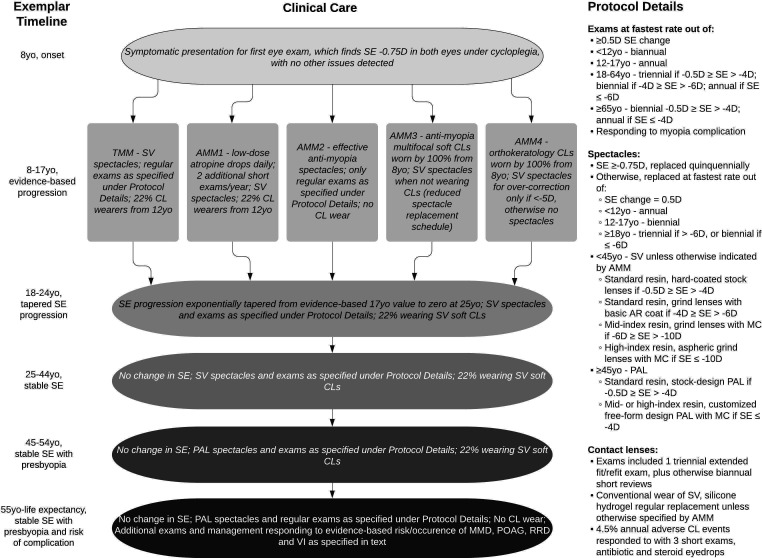
We chose specific starting scenarios to develop methodology that can be adapted to explore lifetime societal costs associated with TMM and AMM options in any situation. Our exemplars were an 8-year-old European Australian in an urban area of Australia and an 8-year-old Han Chinese in an urban area of China, each presenting to an eye examination reporting blurred distance vision and found to have −0.75 DS cycloplegic refraction in both eyes but otherwise normal. We assumed they began TMM and AMM options from this first symptomatic myopia presentation and tracked the consequences through life expectancy following the Consolidated Health Economic Evaluation Reporting Standards and figure 1.20
Figure 1.
Clinical care flow diagram. AMM, active myopia management; AMM1, low-dose atropine; AMM2, antimyopia spectacles; AMM3, antimyopia multifocal soft contact lenses; AMM4, orthokeratology; AR, antireflection; CL, contact lens; D, diopter; MC, multicoat; MMD, myopic macular degeneration; PAL, progressive addition lens; POAG, primary open-angle glaucoma; RRD, rhegmatogenous retinal detachment; SE, spherical equivalent; SV, single vision; TMM, traditional myopia management; VI, vision impairment; yo, year old.
Cost estimates
In Australia, costs associated with myopia-related clinical care were based on Medicare scheduled fees21; spectacle and contact lens costs were based on the Optical Distributors and Manufacturers Association price reference guide22; spectacle frame costs were based on the Victorian Eyecare Service23; and the majority of pharmaceutical costs were based on the Pharmaceutical Benefits Scheme (PBS).24 Prepackaged low-dose atropine is not available in Australia, not covered by the PBS and only available from compounding pharmacies.25 Key informants at the Zhongshan Ophthalmic Centre in Guangzhou, Guangdong, and the Aier Eye Hospital system with centres across China provided representative ophthalmic examination, vision correction, myopia control and myopia complication management costs for China.26 27 Given our reliance on key informants in China and the evolving nature of Chinese healthcare delivery systems, we checked for outliers by reviewing any costs that our standard methodology suggested were more than three times more expensive in China than Australia. Our checking process was to ask a wider array of key informants including a cost comparison with Hong Kong Special Autonomous Region, China.
In addition to the direct costs, we accounted for related and productivity costs associated with myopia care. Related costs include transport to relevant examinations, spectacle collection, collection of myopia control options and access to management of myopia complications. Productivity costs monetised time spent receiving eye care, both for adults attending their own myopia-related examinations and taking dependents to theirs. Disability weights were used as a proxy to estimate the potential productivity impact of each level of vision impairment.28 All related and productivity costs were based on estimates for travel, and average adult income adjusted for labour force participation rates and employment rates.26 27 29 30
We took a societal perspective in our cost approach, incorporating all myopia-related costs regardless of who paid for them.19 All costs were current in 2020, and converted from local currency to US dollars at the rate provided by the US Federal Reserve for 20 October 2020.31 We have not predicted future inflation nor price changes as technologies age and become mainstream. Similarly, we have not predicted new technologies or combinations of techniques that may improve myopia control over the single options that are currently validated. Our main outcome has future costs discounted at 3% per annum, and we tested the effects of 0% and 5% per annum discounting as a sensitivity analysis.32 33 Discounting adjusts future costs to present value (2020 value in this case). There is broad consensus in economics that discounting is important, but there is ongoing debate regarding the rate that should be applied in various circumstances, resulting in our sensitivity analysis range.
Likelihood of refractive outcomes
From the common starting point (−0.75D at 8 years of age), average and variance in myopia progression under TMM were modelled on data from Guangzhou (Guangdong, China) and Shanghai (China).34 35 This progression was used directly for the Han child in urban China and adjusted to an urban European Australian child using the method of Donovan et al through to 18 years of age.36 The spherical equivalent progression at 18 years of age was exponentially decayed towards an assumed zero progression at 25 years of age. The average and variance data were used to generate probability profiles for TMM refractive error outcomes at 25 years of age for each exemplar.
We analysed the treatment efficacy results of all AMM options that satisfied our inclusion criteria. Results from different studies of the same treatment type were weighted by sample size and averaged. After averaging, we additionally rejected treatment types with <25% treatment efficacy.
Clinical care protocols
Clinical care protocols and evidence-based risk calculations governed all decisions on myopia-related need—when and what type of ophthalmic examinations would be provided21 37–39; spectacle type and replacement schedule22 23 26 27 40 41; additional care required by AMM options8 37 42–44; contact lens wear, type, replacement schedule and complications21 45–49; risk of myopia complications and responses to them21 26 50–75; and risk of vision impairment and responses to it.17 18 21 76–78 Figure 1—our clinical care flow diagram—illustrates and describes the protocols in all age groups.
Adding up the costs
We modelled and compared TMM and each AMM option in Australia and China. Each scenario had its own unique refractive error outcome profile (see the Likelihood of refractive outcomes section) that defined the probability of an 8-year-old child with −0.75 D progressing to each different spherical equivalent level by adulthood. Costs were added up for each possible spherical equivalent level, within each scenario, across life expectancy and then weighted according to the refractive error outcome probability profile for each scenario. These weighted costs across the spectrum of spherical equivalent outcomes were added to obtain our overall cost estimate for each scenario.
Threshold analysis
We performed a threshold analysis for any AMM option in either country which exceeded the TMM cost of the same country under 3% annual discounting. The threshold analysis determined the critical, price-adjustable item of each identified AMM option, and the price point of that item that would equalise the AMM and TMM cost in that country.
Sensitivity analysis
Upper and lower limits of our cost estimates were derived from varying spherical equivalent outcomes and all costs to reasonable extremes. The upper and lower limits for spherical equivalent outcome represent 95% CIs. Cost limits depended on type and location. Examples of cost limits include Medicare’s ‘no gap’ option for lowest, and industry accepted private rates for highest, cost of services in Australia21 and ±SD across ophthalmic lens options in Australia.22 Tenth and seventy-fifth percentiles were used to define the lower and upper limits of adult income.30 Potential productivity lost to each level of vision impairment was bracketed using the WHO recommendations.28
As an additional sensitivity analysis, we also varied the discount rate from 3% (our estimate) to 0% and 5%.32
Results
Our online-only online supplemental result 1 provide all relevant unit costs in online supplemental table S1, estimated spherical equivalent progressions in online supplemental figure S1 and estimated myopia complications, related protocols and resulting vision impairment in online supplemental table S2) for our exemplar scenarios. Four AMM options were identified by our methodology to have adequate evidence supporting clinically meaningful effects on childhood myopia progression:
bjophthalmol-2021-320318supp001.pdf (727.6KB, pdf)
Low-dose (0.01%–0.05%) atropine was designated as AMM1, with a 46% reduction in spherical equivalent progression compared with TMM.8 9 79
Effective antimyopia spectacle designs were designated as AMM2, with a 49% reduction based on executive bifocal and defocus incorporated multiple segment designs.10 11
Antimyopia multifocal soft contact lenses were designated as AMM3, with a 42% reduction.12 80–89
Orthokeratology was designated as AMM4, with a 46% reduction.13 90–95
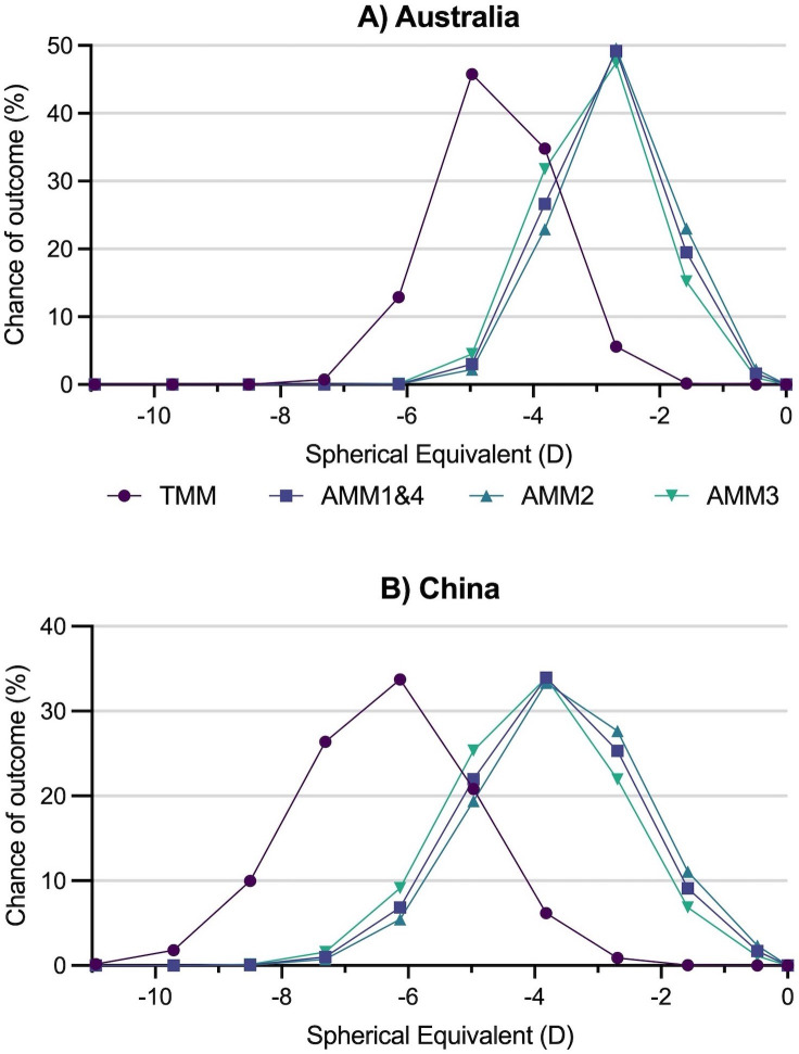
The effect of the AMM options on the likelihood of adult refractive error outcomes is shown in figure 2.
Figure 2.
Probability profiles for adult spherical equivalent outcomes from a starting point of an 8-year-old child presenting with −0.75 D. Profiles are for (A) European in Australia and (B) Han in China. AMM, active myopia management; AMM1, low-dose atropine; AMM2, antimyopia spectacles; AMM3, antimyopia multifocal soft contact lenses; AMM4, orthokeratology; D, diopter; TMM, traditional myopia management.
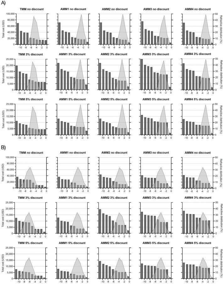
The lifetime cost estimates for all possible spherical equivalent outcomes under each scenario are shown in figure 3. The cost implications of high myopia are clearly demonstrated. They derive from more technical and expensive corrective lenses that are replaced more often, best-practice protocols that include more regular and specialised ophthalmic examinations, higher frequency of pathological complications and higher likelihood of vision impairment. The variations between management options are less than the changes between spherical equivalent powers in most cases. The intuitive implication is that early investment to reduce myopia progression may be worthwhile to an individual.
Figure 3.
Lifetime costs of myopia for each scenario (dark grey bars against left vertical axis), at all possible spherical equivalent outcomes (in dioptres on the horizontal axis), with likelihood of each spherical equivalent outcome (light grey area curves against the right vertical axis). Costs are shown with no discounting, 3% discounting and 5% discounting in Australia (A) and China (B). AMM, active myopia management; AMM1, low-dose atropine; AMM2, antimyopia spectacles; AMM3, antimyopia multifocal soft contact lenses; AMM4, orthokeratology; TMM, traditional myopia management.
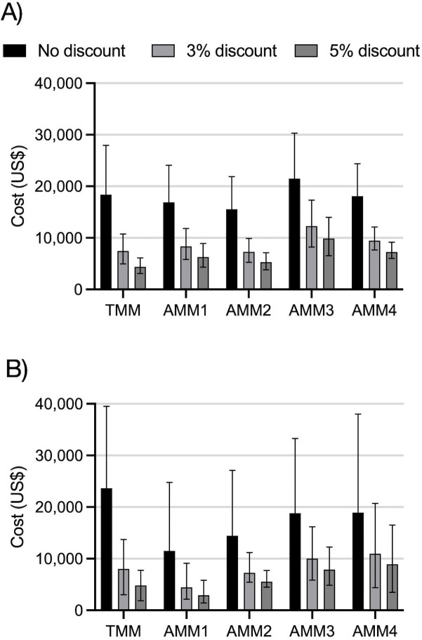
Our estimated lifetime societal costs of myopia under each exemplar scenario were derived by multiplying the two components of figure 3—that is, by weighting the cost estimate for each possible spherical equivalent (dark grey columns) by the likelihood of reaching that spherical equivalent (light grey area). The resultant cost estimates are shown in figure 4 and online supplemental table S3 in our online-only online supplemental results.
Figure 4.
Lifetime costs of myopia for five interventions in urban Australia (A) and China (B). Error bars show bounds of sensitivity analysis. AMM, active myopia management; AMM1, low-dose atropine; AMM2, antimyopia spectacles; AMM3, antimyopia multifocal soft contact lenses; AMM4, orthokeratology; TMM, traditional myopia management.
China shows greater variation in overall costs between interventions than Australia. One reason is that Han Chinese have higher risk of greater myopia progression, meaning the percentage effect of AMM interventions spreads the probability distributions further across the power spectrum. Another reason is that the cost increment from standard spectacles and contact lenses (used for correcting lower refractive powers) to premium spectacles and contact lenses (used for controlling myopia progression and correcting higher refractive powers) is steeper in China than in Australia.
Age at which costs are incurred
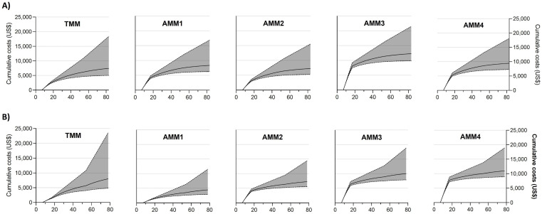
Figure 5 presents the costs associated with each scenario against age. AMM options show additional costs compared with TMM during childhood with one exception: low-dose atropine is cheaper than TMM at 18 years in China. TMM is more expensive than all AMM options later in life, with the relative cost by the end of life depending on place, ethnicity and the discount applied. The TMM curve is noticeably steeper in later life for Han Chinese than European Australians due to increased risk of higher myopia.
Figure 5.
Cumulative costs of myopia in US dollars against age in years, showing each intervention and discount level in Australia (A) and China (B). Solid line shows estimate with 3% annual discounting, upper and lower bounds are estimates with no and 5% annual discounting respectively.
Threshold analysis
In our exemplars with 3% annual discounting, only AMM2 is cheaper than TMM in Australia, whereas both AMM1 and AMM2 are cheaper than TMM in China across a lifetime. Threshold analysis was undertaken at the 3% discount level to determine the price point of critical items that would equalise the cost of other AMM options with TMM:
-
AMM1 critical item: 1-month supply of low-dose atropine.
In Australia: current price US$21.52, threshold price US$8.33.
-
AMM3 critical item: 3-month supply of antimyopia multifocal soft contact lenses.
In Australia: current price US$166.47, threshold price US$27.22.
In China: current price US$157.25, threshold price US$99.22.
-
AMM4 critical item: 2-year period of subsequent orthokeratology care and lenses.
In Australia: current price US$599.85, threshold price US$85.54.
In China: current price US$1623.87, threshold price US$865.93.
Discussion
In our exemplars, the additional early costs of AMM options are substantially balanced across a lifetime by reduced refractive progression, simpler corrective lenses, fewer lens replacements, reduced risk of eye disease and vision loss, and reduced management of myopia complications. The cheapest lifetime cost of all scenarios tested was low-dose atropine in China, regardless of discount level.
Pricing extremes—such as a bottle of low-dose atropine being 14 times more expensive in Australia than China—suggest likely fluidity over time. Low-dose atropine is currently diluted from 1% atropine by specialised compounding pharmacies in Australia; changing to direct manufacture would likely reduce AMM1 costs. Distribution and supply of antimyopia multifocal soft contact lenses are likely to improve in China and result in lower AMM3 costs. Our exemplars are based on static 2020 prices. New technologies, competition, generic versions, professional preferences, consumer trends and demand are likely to change prices over time.
Specialty contact lenses represent a significant step in commitment and cost for a family who otherwise intended to wear spectacles, but a relatively small step for a family who intended to pursue regular contact lenses regardless of any AMM option. We have compared the contact lens-based AMM options with an average TMM case, that is, someone with a 22% chance of wearing standard soft contact lenses to correct (not control) their myopia between 12 and 54 years of age. However, families most likely to consider AMM3 or AMM4 are likely to be interested in contact lenses regardless. As such, it is worth knowing the comparison between TMM, AMM3 and AMM4 for a child/family who want to correct myopia using contact lenses rather than spectacles between 8 and 18 years of age, that is, having a 100% chance of wearing standard soft contact lenses to correct myopia in TMM. Under this variation, lifetime TMM costs in Australia increase to US$22 809 without discounting, US$11 345 with 3% discounting or US$7851 with 5% discounting. Under the same variation in China, TMM costs increase to US$25 787 without discounting, US$9896 with 3% discounting, or US$6561 with 5% discounting. The lifetime cost of AMM4 under this variation is less than TMM in Australia at the 3% discount level. Combining 100% contact lens wear in TMM during childhood with threshold analysis for the remaining scenarios at the 3% discount level:
Threshold price of AMM3 in Australia would be US$139.55 per 3-month supply of antimyopia multifocal soft contact lenses.
Threshold price of AMM3 in China would be US$153.60 per 3-month supply of contact lenses.
Threshold price of AMM4 in China would be US$1352.04 for care and lenses per subsequent 2-year periods.
Our exemplars persist with each AMM intervention for 10 years regardless of myopia progress in the specific case. This would effectively mean persisting with a specific AMM in an individual even when the AMM appears ineffective in slowing myopia progression in that individual. It also effectively means persisting with an AMM when it appears to have been effective enough to consider ceasing treatment. In reality, clinicians make adaptive, iterative decisions based on evidence and observation. They are likely to change management as cases evolve, potentially to a different AMM if rapid myopia progression continues with the first option, or ceasing intervention if progression drops below an acceptable threshold.37 The rigidity of our assumptions is a sensibly conservative modelling approach, increasing our estimated cost of AMM options compared with TMM.
The strengths of our methodology include the use of evidence-based refractive outcomes, the application of evidence-based AMM effectivity, the probabilistic approach to refractive outcome in each scenario, and the broad consideration and sourcing of cost data. Our exemplars provide a comparison between an established health system dealing with relatively low myopia prevalence and progression (Australia) with an evolving health system dealing with very high myopia prevalence and progression (China).
There are also several limitations. First, we have excluded some broader indirect costs and benefits. We have not factored in potential utility reductions that could occur with AMM options, such as in response to daily atropine instillation, or cosmetic issues with antimyopia spectacles. Likewise, we have not factored in potential utility benefits of reduced myopia progression, such as thinner corrective lenses, less dependency on refractive correction, or less anxiety regarding complications or vision impairment. We have also not accounted for the potential productivity impact of uncorrected and undercorrected myopia, nor care costs for assisting a person with vision impairment.
Second, we have taken a simple average of product options for items within a scenario, for example, across the 12 options for single vision aspheric 1.67 index spectacle lenses listed in Australia’s EyeTalk,22 rather than weighting costs by market share. Weighted costs would potentially improve the accuracy of the estimates for policymakers but distort the choice for the end user.
Third, we have taken antimyopia treatment efficacy data from trials of 1–5 years and extended them to 10 years, unmodified for place, ethnicity or age. It is possible that efficacy will vary over time, and between places, ethnicities and ages, but there is no clear data to dictate modifications at this stage.
Fourth, we have done our best to model profession and industry norms, but management variations exist, and some may be cheaper and/or better. Changes in review schedules or lens replacement schedules would alter costs. Orthokeratology is perhaps the clearest example in which this might change the estimation significantly.
Fifth, we based risk of myopia complications on the best population-based evidence, which suggests the risk does not rise dramatically until 55 years of age. However, clinicians know that complications occur earlier and there is certainly evidence in support. It is possible that the population-based evidence is distorted by the tendency to survey those over 50 years of age far more frequently than younger groups.
Our estimates suggest the greatest impact on lifetime cost of myopia in Australia and China is the level of myopia reached. The financial benefits of AMM are likely to be greatest in communities experiencing larger myopia progression, that is, in this comparison, more in China than Australia. While AMM options have upfront cost impacts in childhood, these are mostly balanced or exceeded across a lifetime by savings associated with the reduced myopia achieved.
Specific supply and distribution issues are influencing relative costs in some newer or niche products— contact lens options in China and low-dose atropine in Australia are the most obvious examples from 2020. However, healthy threshold prices for each suggest supply should be commercially viable judged on lifetime cost alone. The methodology presented here could be used as a guide to test cost-effective prices for antimyopia products and services.
In addition to new products and services, there are multiple other existing strategies and options that we have not tested but could be explored using this methodology. Any antimyopia managements that have efficacy and cost data can be tested. The effect of refractive surgery at different stages of adulthood could be tested. Local costs or protocol variations can provide comparisons in or between any location/s.
In summary, the methodology established here can be applied to any myopia scenario to increase clinician confidence in the outcomes that are likely under each intervention. Costs can be updated and/or adjusted to local circumstance, and modelling can be adjusted for additional evidence on TMM progression or AMM option efficacy.
Acknowledgments
We acknowledge the assistance of Liqiong Xie (Zhongshan Ophthalmic Center), Rebecca Yan Shun Weng (Brien Holden Vision Institute), Qingyao Chen (BHVI China), Yinyi Song (BHVI China) and Carl Zeiss Vision (Germany) in gathering Chinese cost and protocol information.
Footnotes
Twitter: @ProfKevinFrick
Contributors: TRF: guarantor for the paper and involved in all aspects of its conception, design, data collection, analysis, manuscript creation and editing. PS: conception, project design, data collection, analysis and manuscript editing. TN: conception and data analysis. SR: design, analysis, review, manuscript creation and editing. NT: data analysis, review and editing. MH: analysis and review. KDF: involved in conception, project design, analysis and review.
Funding: This work was supported by a public health grant (no award/grant number) from the Brien Holden Vision Institute, Sydney, Australia.
Competing interests: TF and SR consult for BHVI, and PS, TN and NT are employed by Brien Holden Vision Institute (BHVI), which holds patents in myopia control.
Provenance and peer review: Not commissioned; externally peer reviewed.
Supplemental material: This content has been supplied by the author(s). It has not been vetted by BMJ Publishing Group Limited (BMJ) and may not have been peer-reviewed. Any opinions or recommendations discussed are solely those of the author(s) and are not endorsed by BMJ. BMJ disclaims all liability and responsibility arising from any reliance placed on the content. Where the content includes any translated material, BMJ does not warrant the accuracy and reliability of the translations (including but not limited to local regulations, clinical guidelines, terminology, drug names and drug dosages), and is not responsible for any error and/or omissions arising from translation and adaptation or otherwise.
Ethics statements
Patient consent for publication
Not applicable.
Ethics approval
This study does not involve human participants.
References
- 1. Holden BA, Fricke TR, Wilson DA, et al. Global prevalence of myopia and high myopia and temporal trends from 2000 through 2050. Ophthalmology 2016;123:1036–42. 10.1016/j.ophtha.2016.01.006 [DOI] [PubMed] [Google Scholar]
- 2. Naidoo KS, Fricke TR, Frick KD, et al. Potential lost productivity resulting from the global burden of myopia: systematic review, meta-analysis, and modeling. Ophthalmology 2019;126:338–46. 10.1016/j.ophtha.2018.10.029 [DOI] [PubMed] [Google Scholar]
- 3. Modjtahedi BS, Ferris FL, Hunter DG, et al. Public health burden and potential interventions for myopia. Ophthalmology 2018;125:628–30. 10.1016/j.ophtha.2018.01.033 [DOI] [PubMed] [Google Scholar]
- 4. Resnikoff S, Jonas JB, Friedman D, et al. Myopia – a 21st century public health issue. Invest. Ophthalmol. Vis. Sci. 2019;60:Mi-Mii:Mi. 10.1167/iovs.18-25983 [DOI] [PMC free article] [PubMed] [Google Scholar]
- 5. Morgan IG, French AN, Ashby RS, et al. The epidemics of myopia: aetiology and prevention. Prog Retin Eye Res 2018;62:134–49. 10.1016/j.preteyeres.2017.09.004 [DOI] [PubMed] [Google Scholar]
- 6. Haarman AEG, Enthoven CA, Tideman JWL, et al. The complications of myopia: a review and meta-analysis. Invest. Ophthalmol. Vis. Sci. 2020;61:49. 10.1167/iovs.61.4.49 [DOI] [PMC free article] [PubMed] [Google Scholar]
- 7. Wildsoet CF, Chia A, Cho P, et al. Imi – interventions for controlling myopia onset and progression report. Invest. Ophthalmol. Vis. Sci. 2019;60:M106–31. 10.1167/iovs.18-25958 [DOI] [PubMed] [Google Scholar]
- 8. Chia A, Lu Q-S, Tan D. Five-Year Clinical Trial on Atropine for the Treatment of Myopia 2: Myopia Control with Atropine 0.01% Eyedrops. Ophthalmology 2016;123:391–9. 10.1016/j.ophtha.2015.07.004 [DOI] [PubMed] [Google Scholar]
- 9. Yam JC, Jiang Y, Tang SM, et al. Low-concentration Atropine for Myopia Progression (LAMP) Study: a randomized, double-blinded, placebo-controlled trial of 0.05%, 0.025%, and 0.01% atropine eye drops in myopia control. Ophthalmology 2019;126:113–24. 10.1016/j.ophtha.2018.05.029 [DOI] [PubMed] [Google Scholar]
- 10. Cheng D, Woo GC, Drobe B, et al. Effect of bifocal and prismatic bifocal spectacles on myopia progression in children: three-year results of a randomized clinical trial. JAMA Ophthalmol 2014;132:258–64. 10.1001/jamaophthalmol.2013.7623 [DOI] [PubMed] [Google Scholar]
- 11. Lam CSY, Tang WC, Tse DY-Y, et al. Defocus incorporated multiple segments (DIMS) spectacle lenses slow myopia progression: a 2-year randomised clinical trial. Br J Ophthalmol 2020;104:363–8. 10.1136/bjophthalmol-2018-313739 [DOI] [PMC free article] [PubMed] [Google Scholar]
- 12. Chamberlain P, Peixoto-de-Matos SC, Logan NS, et al. A 3-year randomized clinical trial of MiSight lenses for myopia control. Optom Vis Sci 2019;96:556–67. 10.1097/OPX.0000000000001410 [DOI] [PubMed] [Google Scholar]
- 13. Hiraoka T, Kakita T, Okamoto F, et al. Long-Term effect of overnight orthokeratology on axial length elongation in childhood myopia: a 5-year follow-up study. Invest Ophthalmol Vis Sci 2012;53:3913–9. 10.1167/iovs.11-8453 [DOI] [PubMed] [Google Scholar]
- 14. Woodward MA, Prasov L, Newman-Casey PA. The debate surrounding the pathogenesis of myopia. JAMA Ophthalmol 2019;137:894–5. 10.1001/jamaophthalmol.2019.1487 [DOI] [PubMed] [Google Scholar]
- 15. Prousali E, Haidich A-B, Fontalis A, et al. Efficacy and safety of interventions to control myopia progression in children: an overview of systematic reviews and meta-analyses. BMC Ophthalmol 2019;19:106. 10.1186/s12886-019-1112-3 [DOI] [PMC free article] [PubMed] [Google Scholar]
- 16. Modjtahedi BS, Abbott RL, Fong DS, et al. Reducing the global burden of myopia by delaying the onset of myopia and reducing myopic progression in children: the Academy's task force on myopia. Ophthalmology 2021;128:816–26. 10.1016/j.ophtha.2020.10.040 [DOI] [PubMed] [Google Scholar]
- 17. Tideman JWL, Snabel MCC, Tedja MS, et al. Association of axial length with risk of uncorrectable visual impairment for Europeans with myopia. JAMA Ophthalmol 2016;134:1355–63. 10.1001/jamaophthalmol.2016.4009 [DOI] [PubMed] [Google Scholar]
- 18. Verhoeven VJM, Wong KT, Buitendijk GHS, et al. Visual consequences of refractive errors in the general population. Ophthalmology 2015;122:101–9. 10.1016/j.ophtha.2014.07.030 [DOI] [PubMed] [Google Scholar]
- 19. Russell LB, Gold MR, Siegel JE, et al. The role of cost-effectiveness analysis in health and medicine. panel on cost-effectiveness in health and medicine. JAMA 1996;276:1172–7. [PubMed] [Google Scholar]
- 20. Husereau D, Drummond M, Petrou S, et al. Consolidated Health Economic Evaluation Reporting Standards (CHEERS)--explanation and elaboration: a report of the ISPOR Health Economic Evaluation Publication Guidelines Good Reporting Practices Task Force. Value Health 2013;16:231–50. 10.1016/j.jval.2013.02.002 [DOI] [PubMed] [Google Scholar]
- 21. Australian Government, Department of Health . Medicare Benefits Schedule (MBS) Online (including Medicare Benefits Schedule Book, 2019. Available: http://www9.health.gov.au/mbs/fullDisplay.cfm?type=note&q=AN.0.10&qt=noteID&criteria=optometry [Accessed 13 Jun 2019].
- 22. Optical Distributors and Manufacturers Association . EyeTalk reference guide, April 2019 ED. Terrey Hills, NSW, Australia: ODMA, 2019. [Google Scholar]
- 23. Victorian State Government, Australian College of Optometry . Victorian Eyecare service, 2020. Available: https://www.aco.org.au/victorian-eyecare-service/
- 24. Australian Government,, Department of Health . PBS - The Pharmaceutical Benefits Scheme. Canberra, Australia: Australian Government, 2020. https://www.pbs.gov.au/browse/medicine-listing [Google Scholar]
- 25. Fricke T. Informal survey of low-dose atropine prices at major Australian compounding pharmacies, 2020. Available: https://customcarepharmacy.com.au/,%20https://sladepharmacy.com.au/,%20http://www.pharmacysmart.com.au/
- 26. Zhongshan Ophthalmic Center . Zhongshan ophthalmic center; sun Yat-Sen university. Guangzhou, Guangdong, China: ZOC, 2020. http://www.gzzoc.com/ [Google Scholar]
- 27. Aier Eye Hospital . About Aier eye Hospital group. Guangzhou, Guangdong, China: Aier Eye Hospital, 2020. http://en.aierchina.com/about [Google Scholar]
- 28. World Health Organization . Who methods and data sources for global burden of disease estimates 2000-2015 (global health estimates technical paper WHO/HIS/IER/GHE/2017.1). Geneva: who department of information evidence and research, 2017. Available: http://www.who.int/gho/mortality_burden_disease/en/index.html
- 29. Australian Taxation Office . Travel expenses. Canberra, ACT, Australia: Australian Government, 2019. https://www.ato.gov.au/Business/Income-and-deductions-for-business/Deductions/Deductions-for-motor-vehicle-expenses/Cents-per-kilometre-method/ [Google Scholar]
- 30. Australian Bureau of Statistics . ABS statistics on Australian earnings and work hours. Belconnen, act, Australia: Australian Bureau of statistics. accessed 23 October, 2020. Available: https://www.abs.gov.au/statistics/labour/earnings-and-work-hours
- 31. US Federal Reserve . Foreign exchange rates. US federal reserve, 2020. Available: https://www.federalreserve.gov/
- 32. Weinstein MC, Siegel JE, Gold MR, et al. Recommendations of the panel on cost-effectiveness in health and medicine. JAMA 1996;276:1253–8. 10.1001/jama.1996.03540150055031 [DOI] [PubMed] [Google Scholar]
- 33. Atik A, Barton K, Azuara-Blanco A, et al. Health economic evaluation in ophthalmology. Br J Ophthalmol 2021;105:602–7. 10.1136/bjophthalmol-2020-316880 [DOI] [PubMed] [Google Scholar]
- 34. Holden B, Sankaridurg P, Smith E, et al. Myopia, an underrated global challenge to vision: where the current data takes us on myopia control. Eye 2014;28:142–6. 10.1038/eye.2013.256 [DOI] [PMC free article] [PubMed] [Google Scholar]
- 35. Sankaridurg PR, Holden BA. Practical applications to modify and control the development of ametropia. Eye 2014;28:134–41. 10.1038/eye.2013.255 [DOI] [PMC free article] [PubMed] [Google Scholar]
- 36. Donovan L, Sankaridurg P, Ho A, et al. Myopia progression rates in urban children wearing single-vision spectacles. Optometry Vis Sci 2012;89:27–32. 10.1097/OPX.0b013e3182357f79 [DOI] [PMC free article] [PubMed] [Google Scholar]
- 37. Gifford KL, Richdale K, Kang P, et al. IMI - Clinical Management Guidelines Report. Invest Ophthalmol Vis Sci 2019;60:M184–203. 10.1167/iovs.18-25977 [DOI] [PubMed] [Google Scholar]
- 38. Celorio JM, Pruett RC. Prevalence of lattice degeneration and its relation to axial length in severe myopia. Am J Ophthalmol 1991;111:20–3. 10.1016/S0002-9394(14)76891-6 [DOI] [PubMed] [Google Scholar]
- 39. American Academy of ophthalmology . Posterior vitreous detachment, retinal breaks, and lattice degeneration preferred practice pattern. San Francisco, Ca, USA, 2019. [DOI] [PubMed] [Google Scholar]
- 40. Wilson DA, Daras S. Practical optical dispensing. 3 edn. Sydney: TAFE NSW - The Open Training and Education Network, 2014. [Google Scholar]
- 41. Fricke TR, Holden BA, Wilson DA, et al. Global cost of correcting vision impairment from uncorrected refractive error. Bull World Health Organ 2012;90:728–38. 10.2471/BLT.12.104034 [DOI] [PMC free article] [PubMed] [Google Scholar]
- 42. Wu P-C, Yang Y-H, Fang P-C. The long-term results of using low-concentration atropine eye drops for controlling myopia progression in schoolchildren. J Ocul Pharmacol Ther 2011;27:461–6. 10.1089/jop.2011.0027 [DOI] [PubMed] [Google Scholar]
- 43. Wu P-C, Chuang M-N, Choi J, et al. Update in myopia and treatment strategy of atropine use in myopia control. Eye 2019;33:3–13. 10.1038/s41433-018-0139-7 [DOI] [PMC free article] [PubMed] [Google Scholar]
- 44. Swarbrick HA. Orthokeratology review and update. Clin Exp Optom 2006;89:124–43. 10.1111/j.1444-0938.2006.00044.x [DOI] [PubMed] [Google Scholar]
- 45. Lee YC, Lim CW, Saw SM, et al. The prevalence and pattern of contact lens use in a Singapore community. Clao J 2000;26:21–5. [PubMed] [Google Scholar]
- 46. Morgan P, Woods CA, Tranoudis IG. International contact lens prescribing 2019. contact lens spectrum, 2020: 1–11. [Google Scholar]
- 47. Cheng X, Brennan NA, Toubouti Y, et al. Safety of soft contact lenses in children: retrospective review of six randomized controlled trials of myopia control. Acta Ophthalmol 2020;98:e346–51. 10.1111/aos.14283 [DOI] [PubMed] [Google Scholar]
- 48. Bullimore MA. The safety of soft contact lenses in children. Optom Vis Sci 2017;94:638–46. 10.1097/OPX.0000000000001078 [DOI] [PMC free article] [PubMed] [Google Scholar]
- 49. Lim CHL, Stapleton F, Mehta JS. Review of contact lens-related complications. Eye Contact Lens 2018;44 Suppl 2:S1–10. 10.1097/ICL.0000000000000481 [DOI] [PubMed] [Google Scholar]
- 50. Vongphanit J, Mitchell P, Wang JJ. Prevalence and progression of myopic retinopathy in an older population. Ophthalmology 2002;109:704–11. 10.1016/S0161-6420(01)01024-7 [DOI] [PubMed] [Google Scholar]
- 51. Wong YL, Zhu X, Tham YC, et al. Prevalence and predictors of myopic macular degeneration among Asian adults: pooled analysis from the Asian eye epidemiology Consortium. Br J Ophthalmol 2021;105:1140–8. 10.1136/bjophthalmol-2020-316648 [DOI] [PubMed] [Google Scholar]
- 52. Cheung CMG, Arnold JJ, Holz FG, et al. Myopic choroidal neovascularization: review, guidance, and consensus statement on management. Ophthalmology 2017;124:1690–711. 10.1016/j.ophtha.2017.04.028 [DOI] [PubMed] [Google Scholar]
- 53. Chan NS-W, Teo K, Cheung CMG. Epidemiology and diagnosis of myopic choroidal neovascularization in Asia. Eye Contact Lens 2016;42:48–55. 10.1097/ICL.0000000000000201 [DOI] [PubMed] [Google Scholar]
- 54. Lai TYY, Cheung CMG. Myopic choroidal neovascularization: diagnosis and treatment. Retina 2016;36:1614–21. 10.1097/IAE.0000000000001227 [DOI] [PubMed] [Google Scholar]
- 55. Leveziel N, Caillaux V, Bastuji-Garin S, et al. Angiographic and optical coherence tomography characteristics of recent myopic choroidal neovascularization. Am J Ophthalmol 2013;155:913–9. 10.1016/j.ajo.2012.11.021 [DOI] [PubMed] [Google Scholar]
- 56. Kasahara K, Moriyama M, Morohoshi K, et al. Six-Year outcomes of intravitreal bevacizumab for choroidal neovascularization in patients with pathologic myopia. Retina 2017;37:1055–64. 10.1097/IAE.0000000000001313 [DOI] [PubMed] [Google Scholar]
- 57. Flitcroft DI. The complex interactions of retinal, optical and environmental factors in myopia aetiology. Prog Retin Eye Res 2012;31:622–60. 10.1016/j.preteyeres.2012.06.004 [DOI] [PubMed] [Google Scholar]
- 58. Flitcroft DI, He M, Jonas JB, et al. Imi – defining and classifying myopia: a proposed set of standards for clinical and epidemiologic studies. Invest Ophthalmol Vis Sci 2019;60:M20–30. 10.1167/iovs.18-25957 [DOI] [PMC free article] [PubMed] [Google Scholar]
- 59. Mitchell P, Hourihan F, Sandbach J, et al. The relationship between glaucoma and myopia: the blue Mountains eye study. Ophthalmology 1999;106:2010–5. 10.1016/s0161-6420(99)90416-5 [DOI] [PubMed] [Google Scholar]
- 60. Australian Government National Health and Medical Research Council . NH&MRC Guidelines for the screening, prognosis, diagnosis, management and prevention of glaucoma. Canberra, ACT, Australia: Commonwealth of Australia, 2010. [Google Scholar]
- 61. Tang W, Zhang F, Liu K, et al. Efficacy and safety of prostaglandin analogues in primary open-angle glaucoma or ocular hypertension patients: a meta-analysis. Medicine 2019;98:e16597. 10.1097/MD.0000000000016597 [DOI] [PMC free article] [PubMed] [Google Scholar]
- 62. Mitry D, Charteris DG, Fleck BW, et al. The epidemiology of rhegmatogenous retinal detachment: geographical variation and clinical associations. Br J Ophthalmol 2010;94:678–84. 10.1136/bjo.2009.157727 [DOI] [PubMed] [Google Scholar]
- 63. Kim MS, Park SJ, Park KH, et al. Different mechanistic association of myopia with rhegmatogenous retinal detachment between young and elderly patients. Biomed Res Int 2019;2019:1–6. 10.1155/2019/5357241 [DOI] [PMC free article] [PubMed] [Google Scholar]
- 64. Wong TY, Tielsch JM, Schein OD. Racial difference in the incidence of retinal detachment in Singapore. Arch Ophthalmol 1999;117:379–83. 10.1001/archopht.117.3.379 [DOI] [PubMed] [Google Scholar]
- 65. Polkinghorne PJ, Craig JP. Northern New Zealand rhegmatogenous retinal detachment study: epidemiology and risk factors. Clin Exp Ophthalmol 2004;32:159–63. 10.1111/j.1442-9071.2004.00003.x [DOI] [PubMed] [Google Scholar]
- 66. Li X, Beijing Rhegmatogenous Retinal Detachment Study Group . Incidence and epidemiological characteristics of rhegmatogenous retinal detachment in Beijing, China. Ophthalmology 2003;110:2413–7. 10.1016/s0161-6420(03)00867-4 [DOI] [PubMed] [Google Scholar]
- 67. Wakabayashi T, Oshima Y, Fujimoto H, et al. Foveal microstructure and visual acuity after retinal detachment repair: imaging analysis by Fourier-domain optical coherence tomography. Ophthalmology 2009;116:519–28. 10.1016/j.ophtha.2008.10.001 [DOI] [PubMed] [Google Scholar]
- 68. Stirpe M, Heimann K. Vitreous changes and retinal detachment in highly myopic eyes. Eur J Ophthalmol 1996;6:50–8. 10.1177/112067219600600111 [DOI] [PubMed] [Google Scholar]
- 69. Heimann H, Bartz-Schmidt KU, Bornfeld N, et al. Scleral buckling versus primary vitrectomy in rhegmatogenous retinal detachment: a prospective randomized multicenter clinical study. Ophthalmology 2007;114:2142–54. 10.1016/j.ophtha.2007.09.013 [DOI] [PubMed] [Google Scholar]
- 70. Sun Q, Sun T, Xu Y, et al. Primary vitrectomy versus scleral buckling for the treatment of rhegmatogenous retinal detachment: a meta-analysis of randomized controlled clinical trials. Curr Eye Res 2012;37:492–9. 10.3109/02713683.2012.663854 [DOI] [PubMed] [Google Scholar]
- 71. Brucker AJ, Hopkins TB. Retinal detachment surgery: the latest in current management. Retina 2006;26:S28–33. 10.1097/01.iae.0000236462.31264.1d [DOI] [PubMed] [Google Scholar]
- 72. Jackson TL, Donachie PHJ, Sallam A, et al. United Kingdom national ophthalmology database study of vitreoretinal surgery: report 3, retinal detachment. Ophthalmology 2014;121:643–8. 10.1016/j.ophtha.2013.07.015 [DOI] [PubMed] [Google Scholar]
- 73. Jackson TL, Donachie PHJ, Sparrow JM, et al. United Kingdom national ophthalmology database study of vitreoretinal surgery: report 1; case mix, complications, and cataract. Eye 2013;27:644–51. 10.1038/eye.2013.12 [DOI] [PMC free article] [PubMed] [Google Scholar]
- 74. Liao L, Zhu X-H. Advances in the treatment of rhegmatogenous retinal detachment. Int J Ophthalmol 2019;12:660–7. 10.18240/ijo.2019.04.22 [DOI] [PMC free article] [PubMed] [Google Scholar]
- 75. Do DV, Gichuhi S, Vedula SS, et al. Surgery for post-vitrectomy cataract. Cochrane Database Syst Rev 2013;12:CD006366. 10.1002/14651858.CD006366.pub3 [DOI] [PMC free article] [PubMed] [Google Scholar]
- 76. Markowitz SN. Principles of modern low vision rehabilitation. Can J Ophthalmol 2006;41:289–312. 10.1139/I06-027 [DOI] [PubMed] [Google Scholar]
- 77. Bourne RRA, Flaxman SR, Braithwaite T, et al. Magnitude, temporal trends, and projections of the global prevalence of blindness and distance and near vision impairment: a systematic review and meta-analysis. Lancet Glob Health 2017;5:e888–97. 10.1016/S2214-109X(17)30293-0 [DOI] [PubMed] [Google Scholar]
- 78. Flaxman SR, Bourne RRA, Resnikoff S, et al. Global causes of blindness and distance vision impairment 1990–2020: a systematic review and meta-analysis. The Lancet Global Health 2017;5:e1221–34. 10.1016/S2214-109X(17)30393-5 [DOI] [PubMed] [Google Scholar]
- 79. Fu A, Stapleton F, Wei L, et al. Effect of low-dose atropine on myopia progression, pupil diameter and accommodative amplitude: low-dose atropine and myopia progression. Br J Ophthalmol 2020;104:1535–41. 10.1136/bjophthalmol-2019-315440 [DOI] [PubMed] [Google Scholar]
- 80. Lam CSY, Tang WC, Tse DY-Y, et al. Defocus incorporated soft contact (disc) lens slows myopia progression in Hong Kong Chinese schoolchildren: a 2-year randomised clinical trial. Br J Ophthalmol 2014;98:40–5. 10.1136/bjophthalmol-2013-303914 [DOI] [PMC free article] [PubMed] [Google Scholar]
- 81. Walline JJ, Greiner KL, McVey ME, et al. Multifocal contact lens myopia control. Optometry Vis Sci 2013;90:1207–14. 10.1097/OPX.0000000000000036 [DOI] [PubMed] [Google Scholar]
- 82. Sankaridurg P, Bakaraju RC, Naduvilath T, et al. Myopia control with novel central and peripheral plus contact lenses and extended depth of focus contact lenses: 2 year results from a randomised clinical trial. Ophthalmic Physiol Opt 2019;39:294–307. 10.1111/opo.12621 [DOI] [PMC free article] [PubMed] [Google Scholar]
- 83. Walline JJ, Walker MK, Mutti DO, et al. Effect of high add power, medium add power, or single-vision contact lenses on myopia progression in children: the blink randomized clinical trial. JAMA 2020;324:571–80. 10.1001/jama.2020.10834 [DOI] [PMC free article] [PubMed] [Google Scholar]
- 84. Cheng X, Xu J, Chehab K, et al. Soft contact lenses with positive spherical aberration for myopia control. Optom Vis Sci 2016;93:353–66. 10.1097/OPX.0000000000000773 [DOI] [PubMed] [Google Scholar]
- 85. Sankaridurg P, Holden B, Smith E, et al. Decrease in rate of myopia progression with a contact lens designed to reduce relative peripheral hyperopia: one-year results. Invest Ophthalmol Vis Sci 2011;52:9362–7. 10.1167/iovs.11-7260 [DOI] [PubMed] [Google Scholar]
- 86. Aller TA, Liu M, Wildsoet CF. Myopia control with bifocal contact lenses: a randomized clinical trial. Optom Vis Sci 2016;93:344–52. 10.1097/OPX.0000000000000808 [DOI] [PubMed] [Google Scholar]
- 87. Ruiz-Pomeda A, Pérez-Sánchez B, Valls I, et al. MiSight assessment study Spain (mass). A 2-year randomized clinical trial. Graefes Arch Clin Exp Ophthalmol 2018;256:1011–21. 10.1007/s00417-018-3906-z [DOI] [PubMed] [Google Scholar]
- 88. Pauné J, Morales H, Armengol J, et al. Myopia control with a novel peripheral gradient soft lens and orthokeratology: a 2-year clinical trial. Biomed Res Int 2015;2015:1–10. 10.1155/2015/507572 [DOI] [PMC free article] [PubMed] [Google Scholar]
- 89. Fujikado T, Ninomiya S, Kobayashi T, et al. Effect of low-addition soft contact lenses with decentered optical design on myopia progression in children: a pilot study. Clin Ophthalmol 2014;8:1947–56. 10.2147/OPTH.S66884 [DOI] [PMC free article] [PubMed] [Google Scholar]
- 90. Cho P, Cheung S-W. Retardation of myopia in Orthokeratology (ROMIO) study: a 2-year randomized clinical trial. Invest. Ophthalmol. Vis. Sci. 2012;53:7077–85. 10.1167/iovs.12-10565 [DOI] [PubMed] [Google Scholar]
- 91. Santodomingo-Rubido J, Villa-Collar C, Gilmartin B, et al. Myopia control with orthokeratology contact lenses in Spain: refractive and biometric changes. Invest Ophthalmol Vis Sci 2012;53:5060–5. 10.1167/iovs.11-8005 [DOI] [PubMed] [Google Scholar]
- 92. Walline JJ, Jones LA, Sinnott LT. Corneal reshaping and myopia progression. British Journal of Ophthalmology 2009;93:1181–5. 10.1136/bjo.2008.151365 [DOI] [PubMed] [Google Scholar]
- 93. Cho P, Cheung SW, Edwards M. The longitudinal orthokeratology research in children (LORIC) in Hong Kong: a pilot study on refractive changes and myopic control. Curr Eye Res 2005;30:71–80. 10.1080/02713680590907256 [DOI] [PubMed] [Google Scholar]
- 94. Chen C, Cheung SW, Cho P. Myopia control using toric orthokeratology (TO-SEE study). Invest Ophthalmol Vis Sci 2013;54:6510–7. 10.1167/iovs.13-12527 [DOI] [PubMed] [Google Scholar]
- 95. Charm J, Cho P. High myopia-partial reduction ortho-k: a 2-year randomized study. Optom Vis Sci 2013;90:530–9. 10.1097/OPX.0b013e318293657d [DOI] [PubMed] [Google Scholar]
Associated Data
This section collects any data citations, data availability statements, or supplementary materials included in this article.
Supplementary Materials
bjophthalmol-2021-320318supp001.pdf (727.6KB, pdf)


