Abstract
In this work, we present DrugSolver CavitomiX, a novel computational pipeline for drug repurposing and identifying ligands and inhibitors of target enzymes. The pipeline is based on cavity point clouds representing physico-chemical properties of the cavity induced solely by the protein. To test the pipeline’s ability to identify inhibitors, we chose enzymes essential for SARS-CoV-2 replication as a test system. The active-site cavities of the viral enzymes main protease (Mpro) and papain-like protease (Plpro), as well as of the human transmembrane serine protease 2 (TMPRSS2), were selected as target cavities. Using active-site point-cloud comparisons, it was possible to identify two compounds—flufenamic acid and fusidic acid—which show strong inhibition of viral replication. The complexes from which fusidic acid and flufenamic acid were derived would not have been identified using classical sequence- and structure-based methods as they show very little structural (TM-score: 0.1 and 0.09, respectively) and very low sequence (~ 5%) identity to Mpro and TMPRSS2, respectively. Furthermore, a cavity-based off-target screening was performed using acetylcholinesterase (AChE) as an example. Using cavity comparisons, the human carboxylesterase was successfully identified, which is a described off-target for AChE inhibitors.
Subject terms: Drug development, Viral proteins, Computational science
Introduction
The comparison of biomolecular active sites is an emerging method to predict enzymatic function and potential ligands1. Especially since AI-based structure predictions2–4 now provide access to billions of novel protein structures, the annotation, interpretation, and comparison of binding sites has become an essential part of everyday bioinformatics. In this work, we examine the potential of the Catalophore™ technology5,6 to predict possible inhibitors for selected enzymes using Catalophores. In short, Catalophores are 3D multivariate point clouds representing the spatial distribution of a set of physico-chemical properties including, among others, hydrophobicity and electrostatics.
Recently, Catalophores have been used in the prediction of the binding affinity of human angiotensin-converting enzyme 2 (hACE2) to the receptor-binding domain (RBD) of SARS-CoV-2 spike protein7, to track changes in the binding interface of SARS-CoV-2 spike variants8, and for protein engineering9.
Catalophores enable the study of enzymes beyond the structural level. They allow an analysis based on cavities of active sites represented by 3D point clouds. Representing cavities and their properties as 3D point clouds has a great advantage in the usage of machine-learning methods, where it corresponds to voxelization. This technology “makes biology machine-readable”, and we refer to it as CavitomiX.
Herein we report a CavitomiX-based pipeline for drug repurposing and inhibitor search. In this method, Catalophore point clouds are compared by an iterative-closest-point matching algorithm. It is important to understand that this comparison is independent of sequence- or structural identity.
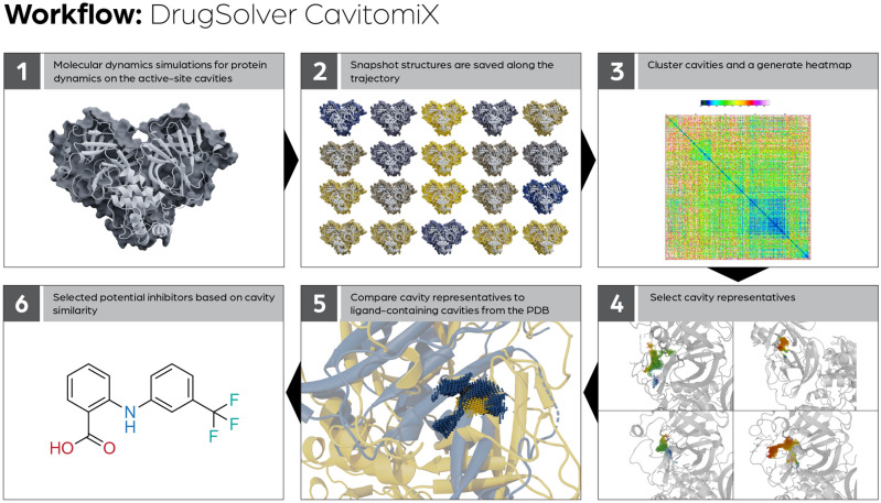
In Fig. 1, an overview of the workflow is shown: Starting from the models of the structures, molecular dynamics (MD) simulations are performed to address the influence of protein dynamics on the active-site cavities. Snapshot structures are saved along the trajectory. In these snapshot structures, the cavities are detected automatically, and the active-site cavities are selected. (A cavity is chosen as an active-site cavity if it aligns the active-site residues of the enzyme).
Figure 1.
General description of the DrugSolver CavitomiX workflow to identify inhibitors based on active-site cavity similarity.
To reduce the number of cavities used for comparisons against cavities from the Protein Data Bank (PDB), all active-site cavities calculated in the snapshot structures are compared to each other. Based on this comparison, the cavities are clustered, and a heatmap is calculated. For the larger clusters, cavity representatives are selected. These cavities represent a specific dynamic state of the cavity observed in the trajectory. The cavity representatives are then compared to a dataset of ligand-containing cavities (e.g., with cavities of ligand-containing proteins from the PDB). The ligands of the most similar cavities to the cavity of the target enzyme are selected as potential inhibitors.
We tested the ability of DrugSolver CavitomiX as a tool for drug repurposing for the case of SARS-CoV-2 to identify possible inhibitors of the SARS-CoV-2 Mpro, the SARS-CoV-2 Plpro, and the host TMPRSS2, all enzymes essential for viral replication. In the following, we briefly describe these three proteins and their role. Viral cell entry depends on the binding of the S (Spike) protein to the human ACE2 receptor10. The S1-domain of the spike protein binds to the receptor. In addition to that, a process called "S-priming" is necessary. Here the S protein is cleaved at the S1/S2 site, which enables membrane fusion—a process driven by the S2 subunit11. The human transmembrane protease serine 2 (TMPRSS2) is used for S protein priming12–14.
Consequently, inhibition of this protease could result in decreased virus uptake into the cell. Therefore, this enzyme is an interesting drug target. After cell entry, the non-structural proteins (NSPs) of SARS-CoV-2 are responsible for viral replication in human cells. One of the essential NSPs is the 3Cl-like protease or main protease (Mpro).
Mpro is a cysteine protease and employs a catalytic diad, which consists of a cysteine and a histidine. Cysteine proteases can also employ a catalytic triad containing glutamine, glutamate, asparagine, or aspartate as the third catalytic residues. In the case of the Mpro, a water molecule takes over the role of this third residue15–17. Mpro is an attractive drug target because it is essential to cleave the coronavirus polyprotein at 11 cleavage sites downstream of NSP418–20. This function makes it a crucial enzyme for processing non-structural proteins. Essential viral enzymes cannot be active without further proteolytic release21. Thus, Mpro is a key enzyme for viral replication18–20.
Furthermore, its substrate specificity—Mpro only cleaves polyprotein sequences after a glutamine residue—is interesting for a drug target since no human protease is known with this substrate specificity. This could lower the potential side effects of a drug targeting Mpro22–25.
The papain-like protease (Plpro) is the second viral protease. Like Mpro, Plpro is a cysteine protease. Its catalytic triad consists of Asp, His, and Cys. It plays a role in polyprotein processing and is essential for viral replication. It cleaves NSP1, NSP2, and NSP3 at the recognition site ”LXGG↓XX”26,27. This is essential for the maturation of the polyprotein and makes Plpro a promising drug target. The predicted inhibitors were tested for their ability to inhibit the target enzymes in wet-lab experiments using a Caco-2 cell-based viral-infection assay.
In the second validation experiment, we tested the ability of the Catalophore™ based DrugSolver CavitomiX platform to identify off-targets. For this application, the example of acetylcholinesterase (AChE) was chosen. AChE is an essential enzyme in the central nervous system of animals and the target for drugs against Alzheimer’s disease (AD)28 as well as the target of the highly potent organophosphorus nerve agents, like Sarin or Novichok29. AChE terminates neurotransmission at cholinergic synapses by hydrolyzing acetylcholine (ACh), consequently regulating the neurotransmitter’s concentration30. In AD treatment, that process is slowed down through inhibitors to enhance the low concentrations of ACh in the synaptic cleft. The Novichok agents, on the other hand, covalently bind to the catalytic triad of AChE and thus permanently inactivates AChE, leading to lethal levels of ACh accumulation29. The identification of off-targets is highly interesting since it helps to predict and potentially avoid possible side effects of AD drugs or harmful effects of nerve agents. Off-targets are identified by the similarity of their binding-site cavities to the active-site cavity of AChE. To this end, a search was performed in an uncurated dataset of cavities of PDB-proteins (i.e. all cavities from PDB proteins, calculated with standard parameters, without any manual selection or curation).
Results
Calculated active-site cavities and cavity representatives for different dynamic states
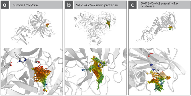
First all detectable cavities of the main protease, TMPRSS2, and papain-like protease were calculated; the Catalophore™ technology5,6 makes it possible to represent cavities as 3D-point clouds and attribute a set of physico-chemical properties to each cavity point. After that, the cavity aligning the active-site residues was selected as the active-site cavity. (as shown in Fig. 2). Then, MD simulations were carried out: 10 ns (TMPRSS2, PIPro) and 100 µs (Mpro). 1000 snapshot structures per protein were created along the trajectory and the active-site cavities were calculated in all these structures. Using hierarchical clustering methods all the resulting active-site cavities were grouped into clusters based on point cloud similarities. These clusters can be interpreted to represent different states of the cavities created by protein dynamics. For each larger cluster, a cavity representative was calculated and used in an additional set of cavity comparisons. This way, protein dynamics and their influence on protein binding pockets can be considered.
Figure 2.
Active-site cavities in (A) human TMPRSS2, (B) SARS-CoV-2 Mpro, and (C) SARS-CoV-2 Plpro.
Cavity-based identification of potential inhibitors
As mentioned above, the following strategy was used to identify potential inhibitors for Mpro, Plpro, and TMPRSS2: similar cavities to the calculated active-site cavity representatives with bound ligands were identified. Enzymes with a similar active site should show similar small-molecule-binding properties. The calculated cavity representatives were compared with a set of cavities from the PDB with the condition that the cavity must contain a co-crystallized ligand. This way, it was possible to gather a dataset of approximately 100,000 cavities with a known binding partner to which the cavity cluster representatives could be compared. The ligands of the most similar cavities to the target cavities were selected for testing their inhibitory effect on SARS-CoV-2 replication.
Potential inhibitors identified for TMPRSS2 and the SARS-CoV-2 protease
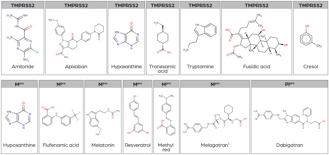
The following compounds (shown in Fig. 3) were identified as potential inhibitors for the SARS-CoV-2 Mpro, SARS-CoV-2 Plpro, and the human TMPRSS2 by the high similarity of the compound-binding cavities to the active-site cavities of Mpro/Plpro/TMPRSS2.
Figure 3.
Structures of potential inhibitors identified by the cavity-based workflow. (1At the time of this work, melagatran was not available to purchase at Sigma-Aldrich; therefore, the prodrug ximelagatran (the alkylated version of melagatran) was ordered and tested instead).
Cell-based virus replication assay test to determine the inhibitory effects of selected compounds on the replication of SARS-CoV-2
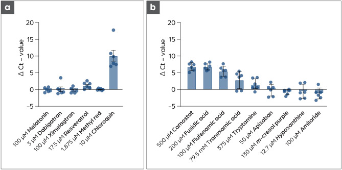
First compounds targeted against the Mpro were tested on Vero CCL81 cells at the highest concentration where no cytotoxic effect were observed (Fig. S1). None of the tested substances showed a strong antiviral effect, except chloroquine, which was used as a control substance as it is known to inhibit viral replication in Vero cells31 despite not showing therapeutic benefit in clinical trials33. Only low antiviral activity was found for resveratrol. This substance is known to bind unspecifically to many proteins and influences a broad variety of cellular factors. Binding to the Mpro cannot be excluded, but many other factors may also be influenced by resveratrol and result in the low antiviral effect observed34. In parallel, a cell-based virus replication assay suitable to test TMPRSS2 inhibitors was established. A Caco-2 cell-based assay was chosen since Caco-2 cells were shown to express TMPRSS2 (Fig. S4). This assay was used to test potential inhibitors for TMPRSS2 and also for testing potential inhibitors of Mpro and Plpro, which were not tested in the first round using the Vero cell-based assay. Caco-2 cells were infected at a high viral load (MOI: 0.825) to identify a strong antiviral effect of the compounds and to exclude possible false-positive results. Samples were taken after 48 h and analyzed with qRT-PCR for the quantification of viral replication35,36. Two compounds, fusidic acid and flufenamic acid, show a antiviral effect comparable with the known TMPRSS2 inhibitor Camostat Mesylate as shown in Fig. 4. Additionally, the antiviral effect was confirmed using immunohistochemistry staining of the infected cells. In this semiquantitative assay, infected cells were visualized utilizing a SARS-CoV-2 antibody directed against the nucleocapsid protein (N). As shown in Fig. S3, fewer cells got infected compared to an infected control when treated with fusidic acid or flufenamic acid during the assay and incubation period.
Figure 4.
Testing of putative inhibitors in cell-based based virus replication assays. (a) None of the tested substances show an antiviral effect against SARS-CoV-2 on Vero CCL81 cells. Substances were added for ½ h before infection of each well with 12 PFU SARS-CoV-2. The cell culture supernatant was harvested after 48 h, followed by RNA-extraction and qRT-PCR for the nucleocapsid target (N1 primer). Chloroquine was used as a control substance31,32. ∆Ct values were calculated for better comparability by subtracting the Ct-value of the infected samples without substance pre-treatment from the infected samples with substance pre-treatment n = 6. (b) Fusidic acid and flufenamic acid show inhibition of SARS-CoV-2 replication on Caco-2 cells. Cells were pretreated with the respective substance for 1/2 h, followed by SARS-CoV-2 infection (MOI: 0.825). The supernatant was harvested after 48 h followed by RNA extraction and qRT-PCR analysis (N2 primer).
Fusidic acid and flufenamic acid were identified as inhibitors of viral replication
Fusidic acid
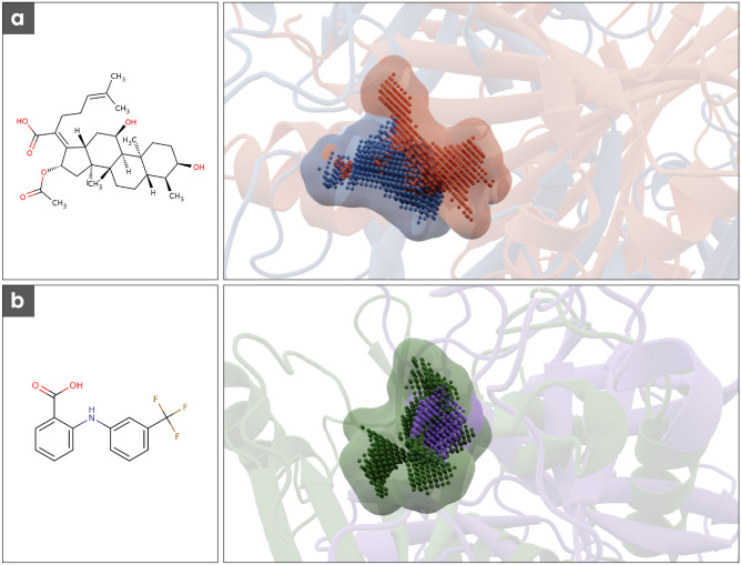
Active-site cavity representatives of TMPRSS2 show a high similarity to the ligand-binding cavity of the Escherichia coli enzyme chloramphenicol acetyltransferase 3 (PDB-ID: 1QCA). The corresponding cavity match can be seen in Fig. 5a. Chloramphenicol acetyltransferase and TMPRSS2 show no significant sequence identity (~ 5%) or structural similarity (Tm score: 0.09). Fusidic acid is a potent competitive inhibitor of chloramphenicol acetyltransferase 337 and is an approved drug that has been used as an antibiotic since the 1960s38, making it an attractive candidate as a potential inhibitor of TMPRSS2.
Figure 5.
(a) Chemical structure of fusidic acid and the corresponding cavity match. In blue structure and cavity of the Mpro in red transcription factor TEAD2 (b) Chemical structure of flufenamic acid and the corresponding cavity match. In purple structure and cavity of TMPRSS2 in purple Chloramphenicol acetyltransferase.
Flufenamic acid
The active-site cavity representatives of the Mpro show a high similarity to a cavity of transcription factor TEAD2 with bound flufenamic acid (Fig. 5b). The main protease and the transcription factor TEAD2 are sequentially and structurally diverse and therefore show a very low sequence identity of about 4% and a very low Tm score of 0.1. Flufenamic acid was invented and approved in the 1960s39 and is mainly used as an analgesic drug in treating the pain associated with rheumatoid diseases40.
Cavity-based identification of CSE1 as an off-target for AChE inhibitors
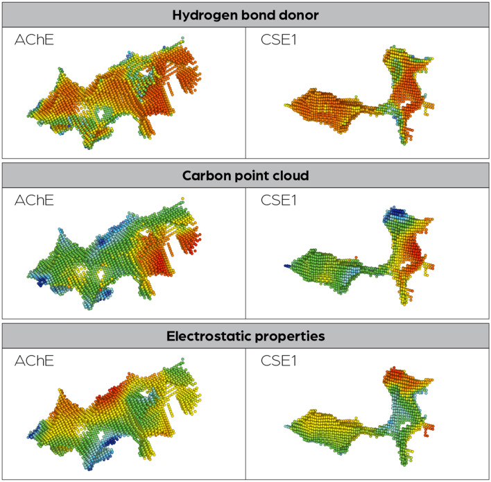
The cavity-comparison results were ranked by similarity to the acute-site cavity of AChE. Among the top hits, human carboxylesterase 1 (CES1) was identified (PDB-ID: 5a7h), the active-site cavities of AChE and CSE1 are shown in Fig. 6. CES1 is known to bind AChE inhibitors, making it an off-target for drugs targeting AChE. The fact that it can be identified using the Catalophore™ DrugSolver platform shows that also off-target identification is possible, despite the actual geometric shape of the cavities, which were identified to have similar properties, differ significantly.
Figure 6.
Side-by-side comparison of active-site cavities from AChE and CSE1.
Discussion
In this work, we established a workflow based on active site cavity comparisons, capable of identifying inhibitors for selected target enzymes. This technology enables a novel approach towards drug repurposing since it is independent of structure and sequence alignments. DrugSolver CavitomiX delivered two already approved drugs that show anti-viral activity against SARS-CoV-2 in infection assays: fusidic and flufenamic acid. These two compounds were found as proteins binding them had similar cavities as our target enzymes. Despite their high active-site cavity similarity, they show very little structural (TM-score41 of 0.09 and 0.1) and very low sequence (around 5%) identity to Mpro and TMPRSS2, respectively. Thus, these two proteins (and consequently their ligands) would never have been identified using sequence- and structure-based comparisons.
Now that the computational pipeline is established, it can quickly be modified and adapted to address new pathogens as needed. In future versions, a combination with technologies such as DrugSolver pathogen-seqscan42, which enables the automatic detection of druggable targets in new pathogens and is part of Innophore’s DrugSolver platform, makes it possible to address newly occurring pathogens even faster. Also, in the case of mutating drug targets, like SARS-CoV-2 Mpro43, the mutated targets can quickly be re-addressed if needed.
In the last years, more and more efforts have been devoted to the development of novel methods of protein structure prediction. Methods like AlphaFold22, RoseTTAFold4, ESM fold3, and the BioNeMo toolbox (https://www.nvidia.com/en-us/gpu-cloud/bionemo/) were made available to the public, and follow-up versions of these methods are currently in development. These new methods, among other drug repurposing approaches44, make DrugSolver CavitomiX even more potent. It enables its users to quickly predict the proteome of newly emerging pathogens. Using the Catalophore™ CavitomiX approach, the cavitome of new pathogens can be calculated, and the here-described workflow enables the rapid identification of potential inhibitors.
CES1 is already described to be a target of AChE inhibitors. The enzyme was even suggested to be applied as a bio-scavenger to protect against organophosphorus nerve agents, which are known to bind to AChE45. Finding the CES1 among the best hits in the Catalophore™ workflow is a promising result, showing the potential of the Catalophore™ DrugSolver CavitomiX to be used for off-target identification. A combination of drug identification and off-target identification in one workflow has great potential. Identifying off-targets is of great interest for predicting and preventing side effects. The given example here serves as a proof-of-principle, and the off-target identification using 3D cavity property point cloud information will be further improved in future work.
Methods
Homology model
For TMPRSS2 and Plpro, SWISS-models46–49 were built using default settings. While Plpro had an 83% sequence identity with its crystal template (PDB-ID: 5y3e), the best template for TMPRSS2 revealed only 38% identity with serine protease Hepsin (PDB-ID: 5ce1) automatically selected as a template. For the main protease, we used the model as well as MD-simulation data of the D.E. Shaw research center, which had been published in March 202050.
Molecular dynamics simulations
Own MD simulations have been carried out using Yasara version 20.4.2451 with the Amber03 force field. For each system, 1000 structural snapshots with a constant time interval of 10 ps in between had been sampled, covering a total time range of 10 ns. However, regarding Mpro MD simulations of 100 µs length, run on Anton supercomputers at D.E. Shaw research, the 1000 extracted snapshots had 100 ns intervals in between. Snapshots of all systems were used for the next workflow steps.
Calculation of cavity point clouds
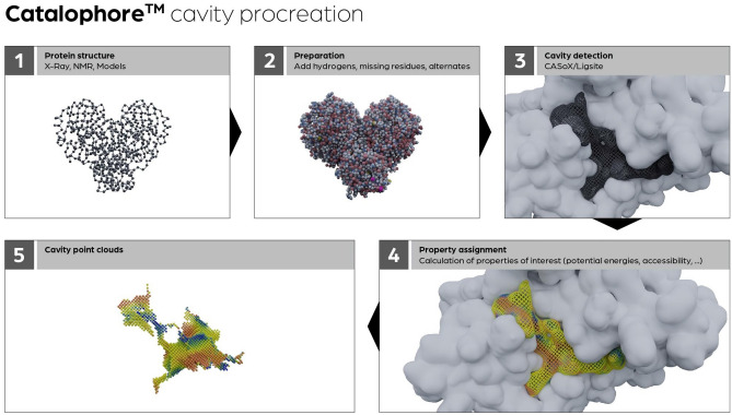
For the calculation of the cavity point clouds, the Catalophore™ technology5,6 is used. The workflow scheme to calculate protein cavities is shown in Fig. 7. The cavity calculation is based on a protein structure. This can be an experimental structure, a homology model, or a structure derived from an MD simulation. The first step is the preparation step: here, hydrogen atoms and potentially missing residues are added, and alternates are handled. For cavity detection, the ligsite algorithm52 is used. The algorithm searches for protein-non-protein–protein events on each grid point in a line on the axes. The analysis of the grid points helps identify closed cavities and points that are accessible to the solvent.
Figure 7.
Catalophore™ cavity procreation workflow scheme.
Grid settings are important to find the same cavities with different orientations in space, and the defined axis directions are usually sufficient to determine cavities appropriately. Additionally, a softness approach is employed to compensate for slight variations in grid settings and eliminate small surface cavities without altering the grid spacing or increasing computation time. After detection, a set of 19 properties using AutoGrid453 is assigned to the cavity points. These properties are derived from the three-dimensional arrangement of the amino acids and special surroundings like cofactors and or ligands within the 3D structure. For each physio-chemical property, one property point cloud is calculated. The result is a collection of pooled cavity grid points, each assigned with physico-chemical properties and additional derived values like hydrophobicity and accessibility values. These points can be compared and aligned with cavity point clouds from other proteins based on their individual physico-chemical properties.
Comparison of two cavities
For the comparison of two cavities, the point clouds from the query and select point clouds from a pre-computed database are matched. More precisely, the point clouds are superimposed and the superposition optimized. To achieve this, differences are evaluated by comparing the closest grid points between the target and the template.
For each property point-cloud pair, a “property score” is generated, representing the quality of the match. A set of weights allows for fine-tuning the influence of the different grid maps (for instance, setting the scoring factor for the H-bond point clouds to zero causes this interaction to be ignored). This scoring is combined with a geometric match into a “total score” and an iterative-closest-point algorithm is performed to optimize it and determine its final value.
Clustering and identification of cavity-cluster representatives
As described above, we ran MD simulations in order to encode the dynamics of the entire protein and, in particular, the dynamics of the active-site cavity. During each simulation, we created 1000 snapshots of each corresponding MD trajectory. The active-site point clouds were calculated from each snapshot like they would be from the origin protein structure.
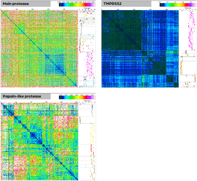
This procedure leads to a set of 1000 point clouds representing the active-site-cavity snapshots for each protein. In order to identify different representatives of the dynamic states of the active-site cavities, we compared the point clouds from all snapshots via an all-to-all match. The results from such a matrix match are visualized in the heatmaps in Fig. 8. The identification of dynamical representatives was carried out via hierarchical clustering based on the total-score difference matrix. In fact, the heatmaps in Fig. 8 are already ordered based on the clustering results.
Figure 8.
Heatmaps based on clustering of snapshot cavities for the investigated enzymes. Using the clustering, cavity representatives for different dynamic states of the cavity were detected. The heatmap for each molecule (plotted on the left side) shows matching scores of 3D point clouds with the color legend shown above each subfigure. The narrow vertical panel on the right of each heat map contains a time-resolved visualization of cluster assignments for each snapshot’s active-site point cloud. The coloring in this panel is not related to the color scheme in the heatmap but simply indicates the different clusters. The horizontal light gray lines indicate cluster boundaries and correspond to the boundaries’ location in the heatmap. While only representatives of clusters containing more than 5% of all cavities were used further in the workflow, the representatives of all clusters are marked in this figure by a light gray diamond.
As for choosing the representatives, we limited ourselves to the largest clusters, in particular those that contained more than 5% of the total number of point clouds. This emphasizes those states that are present for large enough amounts of time during the simulation in order to warrant further study. The clusters are illustrated in the side elements next to the heatmaps in Fig. 8, where snapshot location in MD-simulation time is plotted versus the clustering, each cluster is colored differently, and each representative is marked by a diamond (irrespective of cluster size).
A cluster representative was calculated for each large cluster via a minimal-distance requirement to all other cluster members. These representatives were used further in the following steps of the workflow.
Selection of potential inhibitors
Following the comparison of the cavity datasets, a selection process was undertaken to identify the most promising potential inhibitors. Each cavity in the datasets is assigned a similarity score, reflecting its resemblance to the query cavity. These similarity scores were then utilized to rank the cavities, enabling the identification of the most closely aligned cavities to the query cavity. Additionally, to ensure that only significant results for potential inhibitor binding were considered, cloud-overlap cut-offs (cloud overlap 1 and 2) were considered. These cut-offs, set at 75 and 50 percent, quantified the extent of overlap between two-point clouds during comparison. By applying these criteria, only the most relevant results were retained. Furthermore, cavities containing bound small molecules present in the crystallization conditions were discarded from further analysis. For example, small molecules like polyethylene glycol were considered irrelevant to the investigation and were therefore excluded. Following these steps, the ligands associated with the most similar cavities were investigated and carefully selected for further examination. The availability and accessibility of small molecules were taken into account, ensuring that only those small molecules that could be readily purchased for further experimentation were considered.
Cytotoxicity assay
To determine the cytotoxic effect of the substances used, Caco2 cells were seeded in 96 well plates at a density of 5000 cells/well in Gibco™ Minimum Essential Medium (MEM with 10% FCS, 1% Pen-Strep) and incubated for 48 h at 37 °C and 5% CO2. The medium was then removed, and the respective substance, diluted first in EtOH, then in the respective medium (EtOH concentration was 1% in all conditions), was added in triplicates. To mimic the conditions present in the infection assay, the FCS concentration was reduced from 10 to 2%. 48 h after substance application, cell viability was measured with resazurin (10 µM concentration) at 485/20 590/20 nm for 2 h with the synergy plate reader. Pictures were taken to determine a visible cytotoxic effect. The percent viability was calculated using the slope of the increase in fluorescence in comparison to an untreated control with GraphPadPRISM version 9.0.0. Every experiment was independently conducted twice.
Infection assay
Caco2 cells11,54 were seeded to a density of 40,000 cells/well in 48 well plates 48 h before infection, andVero-CCL81 were seeded at 30,000 cells/well, 24 h before infection. The substance dilutions were prepared in culture medium, either MEM (Gibco), supplemented with 1% penicillin–streptomycin (Sigma) and 2% FCS for Caco-2 cells, or Optipro medium supplemented with 1% penicillin–streptomycin and 1% L-glutamine for Vero CCL81 cells. Cells were then transferred to a biosafety level 3 facility (BSL-3), where they were incubated with the respective substance for 30 min. before addition of SARS-CoV-2 (Human 2019-nCoV ex China Strain: BavPat1/2020 Isolate: Germany ex China, Ref-SKU: 026V-038839) at a multiplicity of infection (MOI) of 0.825 (Caco-2) or 12 plaque-forming units (pfu) (Vero CCL81).The stock concentration was determined by TCID50. After 1 h of infection, the supernatant was removed, and cells were washed with medium 3 times. The cells were incubated at 37 °C and 5% CO2 for 48 h. Afterwards, the supernatant was harvested, and the virus was inactivated with QIAamp® Viral RNA Mini Kit Lysis buffer by Qiagen for later RNA isolation according to the manufacturer's protocol in a biosafety level 2 facility (BSL-2). The remaining supernatant was carefully removed to fix the cells with 4% buffered formalin for 20 min for immunohistochemistry staining. RNA samples were stored at − 70 °C until Quantitative Reverse Transcription PCR (qRT-PCR) analysis. Every experiment was independently conducted twice.
qRT-PCR analysis of viral RNA
The qRT-PCR was performed using the QuantiTect Multiplex RT-PCR Kit (Qiagen) in a 25 µL reaction on the Rotor Gene Q (Qiagen) according to the manufacturer's protocol. Amplification was performed for 30 min at 50 °C and 15 min at 95 °C, followed by 45 cycles (95 °C for 3 min and 55 °C for 30 s). The following probe and primer sets for detection of the N-protein gene of SARS-CoV-2 were used as listed by the CDC55.
2019-nCoV_N1-F Forward Primer 5′-GAC CCC AAA ATC AGC GAA AT-3′.
2019-nCoV_N1-R Reverse Primer 5′-TCT GGT TAC TGC CAG TTG AAT CTG-3′.
2019-nCoV_N1-P Probe 5′-FAM-ACC CCG CAT TAC GTT TGG TGG ACC-BHQ1-3′ FAM, BHQ-1.
2019-nCoV_N2-F Forward Primer 5′-TTA CAA ACA TTG GCC GCA AA-3′.
2019-nCoV_N2-R Reverse Primer 5′-GCG CGA CAT TCC GAA GAA-3′.
2019-nCoV_N2-P Probe 5′-FAM-ACA ATT TGC CCC CAG CGC TTC AG-BHQ1-3′.
Results were analyzed with Microsoft Excel and Graphpad Prism7 software.
Immunohistochemistry
After cell fixation with 4% formalin for 20 min, the cells were transferred to an BSL-2 lab for immunohistochemistry staining. First, cells were washed with PBS and then were permeabilized with Triton X-100 (Sigma) for 10 min, followed by washing with PBS (3 times for 3 min in each washing step). Then, 3% H2O2 diluted in methanol was added for 30 min to block endogenous peroxidases, followed by the PBS washing step. Afterward the cells were incubated for 1 h with 100 µL of a 1:1000 dilution of primary antibody (SARS-CoV-2 (2019-nCoV) Nucleocapsid Antibody, Rabbit Mab, Sinobiological Cat: 40143-R019) in antibody diluent (REAL Antibody diluent, Agilent Technologies, Dako Cat: S202230_2). After a washing step, cells were treated with the secondary antibody (EnVision™ + Dual Link System HRP, Agilent Technologies, Dako Cat: K5007) for 30 min, followed again by washing. Then, the substrate (AEC substrate-Chromogen, Agilent Technologies, Dako, Cat: K346430-2) was added dropwise (2 drops) on the cells and incubated until infected cells appeared red (no longer than 3 min). The reaction was stopped by washing, and cells were kept in PBS until photo documentation.
Flow cytometry
Caco2 cells were harvested by incubation in 1 × accutase for 10 min, counted, and centrifuged at 400×g for 5 min at room temperature before washing and resuspension in FACS buffer (PBS supplemented with 0.05% sodium azide and 0.5% bovine serum albumin). A total of 1 × 10⁶ cells was incubated in FACS buffer containing the primary antibodies for ACE2 (LSBio, LS-C344721, mAb, mah, 1:500) and TMPRSS2 (abcam, ab280567, mAb, rah,1:200) or IgG1 (Agilent Technologies, X0931, 1:1000) in case of the control for 30 min at 4 °C. The cells were then washed in 2 mL of FACS buffer, centrifuged at 400×g for 5 min, and incubated in FACS buffer containing the secondary antibodies Alexa Fluor 647 goat-anti-mouse IgG (Invitrogen, A21235, 1:700) and Alexa Fluor 488 goat-anti-rabbit (Invitrogen, A11034, 1:700) in the dark for 20 min at 4 °C. After washing and centrifugation, the cells were resuspended in 1.5 mL FACS buffer before adding 0.5 mL 4% PFA for 10 min for fixation. After another washing step, the cells were resuspended in 400 µL FACS buffer, transferred to FACS tubes, and kept on ice in the dark until further use. Flow cytometry was performed using a CytoFLEX S flow cytometer equipped with the CytExpert Software (both Beckman Coulter). A typical cell area was gated, and 10,000 single events per sample were acquired.
Paraphrasing was performed on some sentences using the ChatGPT language model (OpenAI). The original text was inputted into the model, and the resulting paraphrased text was compared to the original to ensure accuracy and preserve the intended meaning. Any necessary revisions were made by the authors.
OpenAI. ChatGPT, a large language model trained on the GPT-3.5 architecture. Accessed [30.03.2023]. Available from: https://openai.com/blog/chatgpt-3/.
Supplementary Information
Acknowledgements
We thank Verena Resch for her support in data visualization and Hanna Lindermuth for reading and editing the manuscript. Financial support was provided by the Austrian Centre of Industrial Biotechnology (Austrian Research Promotion Agency, FFG, Project Nr. 872161) in the Next Generation Bioproduction Project Nr. (92015) and Cell-culture system for antiviral testing (91017) and of the Austrian Research Promotion Agency General Programme funding scheme Project Nr. 41404876 “VirtualCure—Rapid Development of an Automated & Expandable In-silico High-Throughput Drug Repurposing Screening Pipeline”, MEFOgraz and BioHealth of the University of Graz. Catalphore is a registered trademark (AT 295631) of Innophore GmbH. Calculations were carried out using the software described in the methods section embedded in the CatalophoreTM Drug Solver platform with a non-commercial open-science license granted by Innophore GmbH. Initial spike models were generated within the FASTCURE consortium (https://fastcure.net/). Calculations were carried out using the software described in the methods section embedded in the CatalophoreTM Drug Solver platform with a non-commercial open-science license granted by Innophore GmbH. Catalphore is a registered trademark (AT 295631) of Innophore GmbH.
Author contributions
M.H.: Calculated, selected, and analyzed cavities; compared target cavities to database cavities; curated initial set of potential inhibitors. C.L., L.M., K.K., S.G.: Performed wet-lab analysis. M.C., V.D.: Performed molecular dynamics calculations analysis. A.K.: Performed cavity clustering and heatmap calculations. K.B.: Performed cavity analysis of AChE. K.Z..: Supervised and instructed work performed in a BSL3 laboratory. C.G., K.G., D.R., G.S.: Contributed to the design of the overall project, discussions, and interpretation of results. All authors contributed to the manuscript.
Data availability
The CavitOmiX plugin for Schrodinger’s PyMOL, a tool that allows analyzing protein cavities from any input structure, is freely available for download at https://innophore.com/cavitomix/ and https://pymolwiki.org/index.php/CavitOmiX. It enables the analysis of protein structures, Catalophore cavities, and binding sites using crystal structures and AI models from OpenFold (powered by NVIDIA’s BioNeMo service), DeepMind`s AlphaFold, and ESMFold by Meta. If required, additional supplementary data can be obtained from the authors on request.
Competing interests
M.H., K.K., M.C., V.D., and A.K. report working for Innophore. K.G., G.S., and C.G. report being shareholders of Innophore GmbH, an enzyme and drug discovery company. Additionally, G.S. and C.G. report being managing directors of Innophore. K.Z is co-founder and CEO of Zatloukal Innovations GmbH. D.R. reports working for NVIDIA.
Footnotes
Publisher's note
Springer Nature remains neutral with regard to jurisdictional claims in published maps and institutional affiliations.
Supplementary Information
The online version contains supplementary material available at 10.1038/s41598-023-39071-z.
References
- 1.Barnsley KK, Ondrechen MJ. Enzyme active sites: Identification and prediction of function using computational chemistry. Curr. Opin. Struct. Biol. 2022;74:102384. doi: 10.1016/j.sbi.2022.102384. [DOI] [PubMed] [Google Scholar]
- 2.Jumper J, Evans R, Pritzel A, Green T, Figurnov M, Ronneberger O, Tunyasuvunakool K, Bates R, Žídek A, Potapenko A, Bridgland A, Meyer C, Kohl SAA, Ballard AJ, Cowie A, Romera-Paredes B, Nikolov S, Jain R, Adler J, Back T, Petersen S, Reiman D, Clancy E, Zielinski M, Steinegger M, Pacholska M, Berghammer T, Bodenstein S, Silver D, Vinyals O, Senior AW, Kavukcuoglu K, Kohli P, Hassabis D. Highly accurate protein structure prediction with AlphaFold. Nature. 2021;596(7873):583–589. doi: 10.1038/s41586-021-03819-2. [DOI] [PMC free article] [PubMed] [Google Scholar]
- 3.Lin Z, Akin H, Rao R, Hie B, Zhu Z, Lu W, Smetanin N, Verkuil R, Kabeli O, Shmueli Y, Costa ADS, Fazel-Zarandi M, Sercu T, Candido S, Rives A. Evolutionary-scale prediction of atomic level protein structure with a language model. BioRxiv. 2022 doi: 10.1101/2022.07.20.500902. [DOI] [PubMed] [Google Scholar]
- 4.Baek M, DiMaio F, Anishchenko I, Dauparas J, Ovchinnikov S, Lee GR, Wang J, Cong Q, Kinch LN, Schaeffer RD, Millán C, Park H, Adams C, Glassman CR, DeGiovanni A, Pereira JH, Rodrigues AV, van Dijk AA, Ebrecht AC, Opperman DJ, Sagmeister T, Buhlheller C, Pavkov-Keller T, Rathinaswamy MK, Dalwadi U, Yip CK, Burke JE, Garcia KC, Grishin NV, Adams PD, Read RJ, Baker D. Accurate prediction of protein structures and interactions using a three-track neural network. Science. 2021;373(6557):871–876. doi: 10.1126/science.abj8754. [DOI] [PMC free article] [PubMed] [Google Scholar]
- 5.Gruber, K., Steinkellner, G. & Gruber, C. Determining Novel Enzymatic Functionalities Using Three-Dimensional Point Clouds Representing Physico Chemical Properties of Protein Cavities, May 30, 2014. https://patentscope.wipo.int/search/en/detail.jsf?docId=WO2014080005. Accessed 14 Jul 2022.
- 6.Steinkellner G, Gruber CC, Pavkov-Keller T, Binter A, Steiner K, Winkler C, Łyskowski A, Schwamberger O, Oberer M, Schwab H. Identification of promiscuous ene-reductase activity by mining structural databases using active site constellations. Nat. Commun. 2014;5(1):1–9. doi: 10.1038/ncomms5150. [DOI] [PMC free article] [PubMed] [Google Scholar]
- 7.Köchl K, Schopper T, Durmaz V, Parigger L, Singh A, Krassnigg A, Cespugli M, Wu W, Yang X, Zhang Y, Wang WW-S, Selluski C, Zhao T, Zhang X, Bai C, Lin L, Hu Y, Xie Z, Zhang Z, Yan J, Zatloukal K, Gruber K, Steinkellner G, Gruber CC. Optimizing variant-specific therapeutic SARS-CoV-2 decoys using deep-learning-guided molecular dynamics simulations. Sci. Rep. 2023;13(1):774. doi: 10.1038/s41598-023-27636-x. [DOI] [PMC free article] [PubMed] [Google Scholar]
- 8.Durmaz V, Köchl K, Singh A, Hetmann M, Parigger L, Krassnigg A, Nutz D, Korsunsky A, König C, Chang L, Krebs M, Bassetto R, Pavkov-Keller T, Resch V, Gruber K, Steinkellner G, Gruber CC. Structural-bioinformatics analysis of SARS-CoV-2 variants reveals higher HACE2 receptor binding affinity for omicron B.1.1.529 SPIKE RBD compared to wild-type reference. BioRxiv. 2021 doi: 10.21203/rs.3.rs-1153124/v1. [DOI] [PMC free article] [PubMed] [Google Scholar]
- 9.Fortuna S, Cespugli M, Todea A, Pellis A, Gardossi L. Criteria for engineering cutinases: Bioinformatics analysis of catalophores. Catalysts. 2021;11(7):784. doi: 10.3390/catal11070784. [DOI] [Google Scholar]
- 10.Li W, Moore MJ, Vasilieva N, Sui J, Wong SK, Berne MA, Somasundaran M, Sullivan JL, Luzuriaga K, Greenough TC, Choe H, Farzan M. Angiotensin-converting enzyme 2 is a functional receptor for the SARS coronavirus. Nature. 2003;426(6965):450–454. doi: 10.1038/nature02145. [DOI] [PMC free article] [PubMed] [Google Scholar]
- 11.Hoffmann M, Kleine-Weber H, Schroeder S, Krüger N, Herrler T, Erichsen S, Schiergens TS, Herrler G, Wu N-H, Nitsche A, Müller MA, Drosten C, Pöhlmann S. SARS-CoV-2 cell entry depends on ACE2 and TMPRSS2 and is blocked by a clinically proven protease inhibitor. Cell. 2020;181(2):271–280.e8. doi: 10.1016/j.cell.2020.02.052. [DOI] [PMC free article] [PubMed] [Google Scholar]
- 12.Glowacka I, Bertram S, Müller MA, Allen P, Soilleux E, Pfefferle S, Steffen I, Tsegaye TS, He Y, Gnirss K, Niemeyer D, Schneider H, Drosten C, Pöhlmann S. Evidence that TMPRSS2 Activates the severe acute respiratory syndrome coronavirus spike protein for membrane fusion and reduces viral control by the humoral immune response. J. Virol. 2011;85(9):4122–4134. doi: 10.1128/JVI.02232-10. [DOI] [PMC free article] [PubMed] [Google Scholar]
- 13.Matsuyama S, Nagata N, Shirato K, Kawase M, Takeda M, Taguchi F. Efficient activation of the severe acute respiratory syndrome coronavirus spike protein by the transmembrane protease TMPRSS2. J. Virol. 2010;84(24):12658–12664. doi: 10.1128/JVI.01542-10. [DOI] [PMC free article] [PubMed] [Google Scholar]
- 14.Shulla A, Heald-Sargent T, Subramanya G, Zhao J, Perlman S, Gallagher T. A transmembrane serine protease is linked to the severe acute respiratory syndrome coronavirus receptor and activates virus entry. J. Virol. 2011;85(2):873–882. doi: 10.1128/JVI.02062-10. [DOI] [PMC free article] [PubMed] [Google Scholar]
- 15.Anand K, Ziebuhr J, Wadhwani P, Mesters JR, Hilgenfeld R. Coronavirus main proteinase (3CLpro) structure: Basis for design of anti-SARS drugs. Science. 2003;300(5626):1763–1767. doi: 10.1126/science.1085658. [DOI] [PubMed] [Google Scholar]
- 16.Ziebuhr J. Molecular biology of severe acute respiratory syndrome coronavirus. Curr. Opin. Microbiol. 2004;7(4):412–419. doi: 10.1016/j.mib.2004.06.007. [DOI] [PMC free article] [PubMed] [Google Scholar]
- 17.Ho B-L, Cheng S-C, Shi L, Wang T-Y, Ho K-I, Chou C-Y. Critical assessment of the important residues involved in the dimerization and catalysis of MERS coronavirus main protease. PLoS ONE. 2015;10(12):e0144865. doi: 10.1371/journal.pone.0144865. [DOI] [PMC free article] [PubMed] [Google Scholar]
- 18.Ziebuhr J, Snijder EJ, Gorbalenya AEY. Virus-encoded proteinases and proteolytic processing in the nidovirales. J. Gen. Virol. 2000;81(4):853–879. doi: 10.1099/0022-1317-81-4-853. [DOI] [PubMed] [Google Scholar]
- 19.Hegyi A, Ziebuhr J. Conservation of substrate specificities among coronavirus main proteases. J. Gen. Virol. 2002;83(3):595–599. doi: 10.1099/0022-1317-83-3-595. [DOI] [PubMed] [Google Scholar]
- 20.Du Q-S, Wang S-Q, Zhu Y, Wei D-Q, Guo H, Sirois S, Chou K-C. Polyprotein cleavage mechanism of SARS CoV Mpro and chemical modification of the octapeptide. Peptides. 2004;25(11):1857–1864. doi: 10.1016/j.peptides.2004.06.018. [DOI] [PMC free article] [PubMed] [Google Scholar]
- 21.Thiel V, Ivanov KA, Putics Á, Hertzig T, Schelle B, Bayer S, Weißbrich B, Snijder EJ, Rabenau H, Doerr HW, Gorbalenya AE, Ziebuhr J. Mechanisms and enzymes involved in SARS coronavirus genome expression. J. Gen. Virol. 2003;84(9):2305–2315. doi: 10.1099/vir.0.19424-0. [DOI] [PubMed] [Google Scholar]
- 22.Ullrich S, Nitsche C. The SARS-CoV-2 main protease as drug target. Bioorg. Med. Chem. Lett. 2020;30(17):127377. doi: 10.1016/j.bmcl.2020.127377. [DOI] [PMC free article] [PubMed] [Google Scholar]
- 23.Zhang L, Lin D, Sun X, Curth U, Drosten C, Sauerhering L, Becker S, Rox K, Hilgenfeld R. Crystal structure of SARS-CoV-2 main protease provides a basis for design of improved α-ketoamide inhibitors. Science. 2020;368(6489):409–412. doi: 10.1126/science.abb3405. [DOI] [PMC free article] [PubMed] [Google Scholar]
- 24.Zhang L, Lin D, Kusov Y, Nian Y, Ma Q, Wang J, von Brunn A, Leyssen P, Lanko K, Neyts J, de Wilde A, Snijder EJ, Liu H, Hilgenfeld R. α-Ketoamides as broad-spectrum inhibitors of coronavirus and enterovirus replication: structure-based design, synthesis, and activity assessment. J. Med. Chem. 2020;63(9):4562–4578. doi: 10.1021/acs.jmedchem.9b01828. [DOI] [PubMed] [Google Scholar]
- 25.Hilgenfeld R. From SARS to MERS: Crystallographic studies on coronaviral proteases enable antiviral drug design. FEBS J. 2014;281(18):4085–4096. doi: 10.1111/febs.12936. [DOI] [PMC free article] [PubMed] [Google Scholar]
- 26.Barretto N, Jukneliene D, Ratia K, Chen Z, Mesecar AD, Baker SC. The papain-like protease of severe acute respiratory syndrome coronavirus has deubiquitinating activity. J. Virol. 2005;79(24):15189–15198. doi: 10.1128/JVI.79.24.15189-15198.2005. [DOI] [PMC free article] [PubMed] [Google Scholar]
- 27.Osipiuk J, Azizi S-A, Dvorkin S, Endres M, Jedrzejczak R, Jones KA, Kang S, Kathayat RS, Kim Y, Lisnyak VG, Maki SL, Nicolaescu V, Taylor CA, Tesar C, Zhang Y-A, Zhou Z, Randall G, Michalska K, Snyder SA, Dickinson BC, Joachimiak A. Structure of papain-like protease from SARS-CoV-2 and its complexes with non-covalent inhibitors. Nat. Commun. 2021;12(1):743. doi: 10.1038/s41467-021-21060-3. [DOI] [PMC free article] [PubMed] [Google Scholar]
- 28.McGleenon BM, Dynan KB, Passmore AP. Acetylcholinesterase inhibitors in Alzheimer’s disease. Br. J. Clin. Pharmacol. 1999;48(4):471–480. doi: 10.1046/j.1365-2125.1999.00026.x. [DOI] [PMC free article] [PubMed] [Google Scholar]
- 29.Costanzi S, Machado J-H, Mitchell M. Nerve agents: What they are, how they work, how to counter them. ACS Chem. Neurosci. 2018;9(5):873–885. doi: 10.1021/acschemneuro.8b00148. [DOI] [PubMed] [Google Scholar]
- 30.Crow JA, Bittles V, Borazjani A, Potter PM, Ross MK. Covalent inhibition of recombinant human carboxylesterase 1 and 2 and monoacylglycerol lipase by the carbamates JZL184 and URB597. Biochem. Pharmacol. 2012;84(9):1215–1222. doi: 10.1016/j.bcp.2012.08.017. [DOI] [PMC free article] [PubMed] [Google Scholar]
- 31.Wang M, Cao R, Zhang L, Yang X, Liu J, Xu M, Shi Z, Hu Z, Zhong W, Xiao G. Remdesivir and chloroquine effectively inhibit the recently emerged novel coronavirus (2019-NCoV) in vitro. Cell Res. 2020;30(3):269–271. doi: 10.1038/s41422-020-0282-0. [DOI] [PMC free article] [PubMed] [Google Scholar]
- 32.Touret F, de Lamballerie X. Of chloroquine and COVID-19. Antiviral Res. 2020;177:104762. doi: 10.1016/j.antiviral.2020.104762. [DOI] [PMC free article] [PubMed] [Google Scholar]
- 33.Repurposed Antiviral Drugs for Covid-19 Interim WHO solidarity trial results. N. Engl. J. Med. 2021;384(6):497–511. doi: 10.1056/NEJMoa2023184. [DOI] [PMC free article] [PubMed] [Google Scholar]
- 34.Yang Z-Y, He J-H, Lu A-P, Hou T-J, Cao D-S. Frequent hitters: Nuisance artifacts in high-throughput screening. Drug Discov. Today. 2020;25(4):657–667. doi: 10.1016/j.drudis.2020.01.014. [DOI] [PubMed] [Google Scholar]
- 35.Dutta D, Naiyer S, Mansuri S, Soni N, Singh V, Bhat KH, Singh N, Arora G, Mansuri MS. COVID-19 diagnosis: A comprehensive review of the RT-QPCR method for detection of SARS-CoV-2. Diagnostics. 2022;12(6):1503. doi: 10.3390/diagnostics12061503. [DOI] [PMC free article] [PubMed] [Google Scholar]
- 36.Dramé M, TabueTeguo M, Proye E, Hequet F, Hentzien M, Kanagaratnam L, Godaert L. Should RT-PCR Be considered a gold standard in the diagnosis of COVID-19? J. Med. Virol. 2020;92(11):2312–2313. doi: 10.1002/jmv.25996. [DOI] [PMC free article] [PubMed] [Google Scholar]
- 37.Murray IA, Cann PA, Day PJ, Derrick JP, Sutcliffe MJ, Shaw WV, Leslie AG. Steroid recognition by chloramphenicol acetyltransferase: Engineering and structural analysis of a high affinity fusidic acid binding site. J. Mol. Biol. 1995;254(5):993–1005. doi: 10.1006/jmbi.1995.0671. [DOI] [PubMed] [Google Scholar]
- 38.Fusidic Acid. DrugBankOnline. https://go.drugbank.com/drugs/DB02703. Accessed 2 Feb 2022.
- 39.Whitehouse MW. Drugs to treat inflammation: A historical introduction. Curr. Med. Chem. 2005;12(25):2931–2942. doi: 10.2174/092986705774462879. [DOI] [PubMed] [Google Scholar]
- 40.Flufenamic acid: Uses, Interactions, Mechanism of Action|DrugBank Online. https://go.drugbank.com/drugs/DB02266. Accessed 1 Feb 2022.
- 41.Zhang Y, Skolnick J. Scoring function for automated assessment of protein structure template quality. Proteins Struct. Funct. Bioinform. 2004;57(4):702–710. doi: 10.1002/prot.20264. [DOI] [PubMed] [Google Scholar]
- 42.Parigger, L. et al. Preliminary Structural Proteome of the Monkeypox Virus Causing a Multi-Country Outbreak in May 2022. 10.21203/rs.3.rs-1693803/v1.
- 43.Parigger L, Krassnigg A, Schopper T, Singh A, Tappler K, Köchl K, Hetmann M, Gruber K, Steinkellner G, Gruber CC. Recent changes in the mutational dynamics of the SARS-CoV-2 Main protease substantiate the danger of emerging resistance to antiviral drugs. Front. Med. 2022;9:1061142. doi: 10.3389/fmed.2022.1061142. [DOI] [PMC free article] [PubMed] [Google Scholar]
- 44.Jukič M, Kores K, Janežič D, Bren U. Repurposing of drugs for SARS-CoV-2 using inverse docking fingerprints. Front. Chem. 2021;9:826. doi: 10.3389/fchem.2021.757826. [DOI] [PMC free article] [PubMed] [Google Scholar]
- 45.Hemmert AC, Otto TC, Wierdl M, Edwards CC, Fleming CD, MacDonald M, Cashman JR, Potter PM, Cerasoli DM, Redinbo MR. Human carboxylesterase 1 stereoselectively binds the nerve agent cyclosarin and spontaneously hydrolyzes the nerve agent sarin. Mol. Pharmacol. 2010;77(4):508–516. doi: 10.1124/mol.109.062356. [DOI] [PMC free article] [PubMed] [Google Scholar]
- 46.Waterhouse A, Bertoni M, Bienert S, Studer G, Tauriello G, Gumienny R, Heer FT, de Beer TAP, Rempfer C, Bordoli L, Lepore R, Schwede T. SWISS-MODEL: Homology modelling of protein structures and complexes. Nucleic Acids Res. 2018;46(W1):W296–W303. doi: 10.1093/nar/gky427. [DOI] [PMC free article] [PubMed] [Google Scholar]
- 47.Bienert S, Waterhouse A, de Beer TAP, Tauriello G, Studer G, Bordoli L, Schwede T. The SWISS-MODEL repository: New features and functionality. Nucleic Acids Res. 2017;45(D1):D313–D319. doi: 10.1093/nar/gkw1132. [DOI] [PMC free article] [PubMed] [Google Scholar]
- 48.Guex N, Peitsch MC, Schwede T. Automated comparative protein structure modeling with SWISS-MODEL and Swiss-PdbViewer: A historical perspective. Electrophoresis. 2009;30(S1):S162–S173. doi: 10.1002/elps.200900140. [DOI] [PubMed] [Google Scholar]
- 49.Studer G, Rempfer C, Waterhouse AM, Gumienny R, Haas J, Schwede T. QMEANDisCo: Distance constraints applied on model quality estimation. Bioinformatics. 2020;36(6):1765–1771. doi: 10.1093/bioinformatics/btz828. [DOI] [PMC free article] [PubMed] [Google Scholar]
- 50.D. E. Shaw Research Technical Data. Molecular Dynamics Simulations Related to SARS-CoV-2 (2020). https://www.deshawresearch.com/downloads/download_trajectory_sarscov2.cgi/.
- 51.Krieger E, Vriend G. YASARA view—Molecular graphics for all devices—from smartphones to workstations. Bioinformatics. 2014;30(20):2981–2982. doi: 10.1093/bioinformatics/btu426. [DOI] [PMC free article] [PubMed] [Google Scholar]
- 52.Hendlich M, Rippmann F, Barnickel G. LIGSITE: Automatic and efficient detection of potential small molecule-binding sites in proteins. J. Mol. Graph Model. 1997;15(6):359–363. doi: 10.1016/s1093-3263(98)00002-3. [DOI] [PubMed] [Google Scholar]
- 53.Huey R, Morris GM, Olson AJ, Goodsell DS. A semiempirical free energy force field with charge-based desolvation. J. Comput. Chem. 2007;28(6):1145–1152. doi: 10.1002/jcc.20634. [DOI] [PubMed] [Google Scholar]
- 54.Mautner L, Hoyos M, Dangel A, Berger C, Ehrhardt A, Baiker A. Replication kinetics and infectivity of SARS-CoV-2 variants of concern in common cell culture models. Virol. J. 2022;19(1):76. doi: 10.1186/s12985-022-01802-5. [DOI] [PMC free article] [PubMed] [Google Scholar]
- 55.Lu X, Wang L, Sakthivel SK, Whitaker B, Murray J, Kamili S, Lynch B, Malapati L, Burke SA, Harcourt J, Tamin A, Thornburg NJ, Villanueva JM, Lindstrom S. US CDC real-time reverse transcription PCR panel for detection of severe acute respiratory syndrome coronavirus 2. Emerg. Infect. Dis. 2020;26(8):1654–1665. doi: 10.3201/eid2608.201246. [DOI] [PMC free article] [PubMed] [Google Scholar]
Associated Data
This section collects any data citations, data availability statements, or supplementary materials included in this article.
Supplementary Materials
Data Availability Statement
The CavitOmiX plugin for Schrodinger’s PyMOL, a tool that allows analyzing protein cavities from any input structure, is freely available for download at https://innophore.com/cavitomix/ and https://pymolwiki.org/index.php/CavitOmiX. It enables the analysis of protein structures, Catalophore cavities, and binding sites using crystal structures and AI models from OpenFold (powered by NVIDIA’s BioNeMo service), DeepMind`s AlphaFold, and ESMFold by Meta. If required, additional supplementary data can be obtained from the authors on request.


