Abstract
By definition, supplemental teeth are supernumerary teeth (ST) resembling adjacent teeth at the end of a tooth series and are well aligned in the arch. A case of the non-syndrome, supplemental type of supernumerary lateral incisor is presented, along with an unusual habit that was noted accidentally in the same child. In the present case, of the two lateral incisors, the mesial supplemental lateral incisor was causing an impaction of 11. In addition, the patient was aesthetically concerned. So, the decision was made to extract the supplemental tooth with altered morphology (mesial one) under local anesthesia even though, as per definition, the distal one is the supplementary tooth. And finally, to align the incisors by orthodontic treatment.
Aim
A case of the non-syndrome, supplemental type of supernumerary lateral incisor is presented, along with an unusual habit of that was noted accidentally in the same child.
Background
By definition, supplemental teeth are supernumerary teeth (ST) resembling adjacent teeth at the end of a tooth series and are well aligned in the arch.
Case description
In the present case, of the two lateral incisors, the mesial supplemental lateral incisor was causing impaction of 11. In addition, the patient was aesthetically concerned.
Conclusion
Decision was made to extract the supplemental tooth with altered morphology (mesial one) under local anesthesia even though, as per definition, the distal one is the supplementary tooth. And finally, to align the incisors by orthodontic treatment.
Clinical significance
Decision-making regarding removal of tooth is quite baffling as a selective removal of the normal or the supplementary tooth may be required and it should be made after analyzing multiple factors.
How to cite this article
Sojan M, Thakur S. An Unusual Case of Mesial Supplementary Lateral Incisor; A Case Report. Int J Clin Pediatr Dent 2023;16(3):518-521.
Keywords: Supernumerary tooth, Teeth, Tooth, Unerupted
Introduction
Odontostomatologic anomaly characterized by an additional number of the tooth in any region of the dental arch is known as ST.1 ST, which are eumorphic to adjacent teeth at the end of a tooth series and well aligned in the arch is known as supplementary teeth.2,3 The majority of supernumeraries found in the primary dentition are of the supplemental type and seldom remain impacted. Even though supernumerary canines and lateral incisors are rare as such with a low frequency of occurrence (2.8%),4 the most common supplemental tooth in the permanent dentition is the maxillary lateral incisor.5
The prevalence of ST in permanent dentition for the Asian population is reported between 2.7 and 3.4%,6 while for the caucasian general population, it has been reported between 0.1 and 3.8%.7 Lower prevalence rates were reported in an Indian population (0.05%), while Nigerian, Turkish, and Western Romanian had high prevalence rates (0.5 and 1.59%).8 A study conducted in South India reported male:female ratio of 3:1,8 with the maxillary posterior region (53.12%) being the most common location.9
The most supported theory to explain the etiology of ST is the local, independent, and conditioned hyperactivity of the dental lamina. Another theory suggests the dichotomy of the tooth bud. According to some, it can be hereditary but does not follow a simple Mendelian pattern.10
Wedrychowska-Szulc et al. reported that it is difficult to distinguish between normal teeth and their supplemental twin and stated that supernumerary lateral incisors are usually smaller than the adjacent lateral incisors; however, the normal lateral incisors next to ST are smaller than the lateral incisors on the opposite side.11
Even though extraction is not always the treatment of choice for ST,12–13 it should be removed if associated with any complications.14 Impaction, displacement or rotation, root resorption, and malformation of the adjacent teeth are the commonly reported complications. In addition, aesthetic disturbances, including diastema, midline shift, crowding, or ectopic eruption, are also reported. In a later stage, an impacted tooth can even result in a dentigerous cyst.4,8,11
Case Description
A 10-year-old girl reported to the pediatric dental clinic with a chief complaint of a missing front tooth and gave a history of exfoliation of a milk tooth in the same region one year back. There was no history of trauma. Family history and medical history were noncontributory. The patient had good general health, and their parents did not report any history of systemic disease or immune-compromised status. Intraoral features are mixed dentition with class I molar relation. Partially erupted lateral incisor, almost occupying half of the space for 11 (Fig. 1). Distal to it, the incisal tip of its supplemental twin was present. Radiographic examination depicted an unerupted and mesiobuccally rotated right maxillary permanent central incisor with an immature root (Fig. 2). Among the two lateral incisors, the mesial one showed a slightly curved root with some evidence of root resorption. A routine hematological examination did not reveal any abnormal findings.
Figs 1A to C.
Preoperative clinical photographs of the patient. (A) Maxillary occlusal view; (B) Mandibular occlusal view; (C) Frontal view
Figs 2A to C.
Radiographic records of the patient; (A) Intraoral periapical radiograph; (B) Maxillary occlusal view; (C) Orthopantomagram
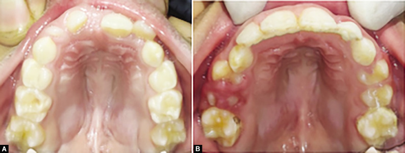
On detailed examination, the presence of a high-arched palate and hypertrophy of mid-palatine raphe was noted. It was hypothesized that this could be a case of an unusual habit of tongue-sucking. On detailed inquiry, the parent reported that the child performs tongue-sucking by holding the tongue as if sucking on a lozenge, especially during sleeping hours. The parent noticed the habit because of the sucking sound during sleep and less frequently during waking hours. This habit was dated by the parent as being definitely practiced in the last 2 years at a minimum and probably even earlier (Figs 3 and 4).
Figs 3A to C.
Intraoperative and postoperative records of the patient. (A) Supplementary LI; (B) A 2 × 4 appliance for alignment; (C) Aligned 12, 11, 21, 22
Fig. 4.
Pre and postoperative palatal contour
Discussion
The anomalous surplus tooth is defined as “supplementary” if they present a normal morphology and as “supernumerary” if they present morphologic and volumetric anomalies.14–18
Types of Supernumerary Tooth
Di Biase et al.19
Supplemental (eumorphic/incisiform)
Rudimentary (dysmorphic)
Conical
Molariform
Tuberculate
Primosch et al.20
Eumorphic (supplemental/incisiform)—ST with normal shapes and sizes.
Dysmorphic (rudimentary/heteromorphic)—ST abnormal shapes and smaller sizes.
Conical or pin
Tuberculate
Infundibular
Molariform
Fernández-Montenegro (according to morphology)12
| Supernumerary tooth | Frequency of occurrence |
|---|---|
| Mesiodens | 46.9% |
| Supernumerary premolars | 24.1% |
| Distomolars | 18% |
As the coronal dimension and morphology of the ST in the present case closely matched those of the lateral incisor, it is considered a supplemental tooth.17 Supplemental type lateral incisor is rare in comparison to conical type, and only a few cases have been reported in the literature.15 Most of the supplemental teeth remain unerrupted16 and can cause numerous complications.18 Nonetheless, in the present case, the supplemental lateral incisor erupted and resulted in impaction and rotation 11.
Usually, the supplemental teeth were extracted to facilitate the eruption of the permanent dentition.21 But most of the time, decision-making is quite baffling as a selective removal of normal or supplementary may be required. In such circumstances, when both teeth are healthy, it has been suggested to extract the tooth/teeth that are most displaced from the line of the arch so as to relieve crowding.18 In this case, both lateral incisors are almost on the line of the arch, but the one which causes impaction of 11 is the tooth very next to it. In addition, its root morphology was altered and showed slight saucerization indicating a chance of resorption.
In case of delayed eruption of adjacent teeth due to the presence of ST, there are two treatment options. Either removal of the ST and allowing it to erupt to the available space or removal of ST followed by a surgical-orthodontic treatment to obtain space for the impacted tooth.22
In the majority of cases, a spontaneous eruption occurs after the removal of the obstacle ST23,24 or by the extraction of primary canines along with ST removal.23 One study reported that nearly 73% of incisors with an immature root erupted spontaneously after the removal of an associated ST.25 Several authors have reported cases of ST removal and orthodontic traction of impacted permanent maxillary incisors simultaneously.26
Das et al. reported a case of impact of 11 and 21 where 21 normally erupted as the root was not completed during the time of surgery. Orthodontic extrusion for 11 was carried out later as it lacked the eruptive forces (failed to erupt along with 21 even after 4 months) because the root was almost completed at the time of surgery. They also reported that the following complications could be encountered in the case of combined surgical/orthodontic therapy,
Ankylosis
Nonvital pulps
Root resorptions27
In the present case, the root formation was not complete compared to the adjacent central incisor, depicting that there is a chance for remaining the eruption potential.
So, the treatment plan established for this case is mentioned below.
Removal of the one supplementary lateral incisor (mesial one) that is an obstruction for the eruption of 11.
Waiting for the eruption of 11 and 12 (erupted in <2 months) and to align it orthodontically.
To achieve good gingival attachment and symmetrical gingival margins for both the central incisors.
Esthetical management of peg lateral (light cure build until an eruption of permanent canine and permanent restoration in the future).
Help in the establishment of final stable, functional occlusion.
Regarding tongue-sucking habit, a similar case was reported where the child had an ovoid, elevated, tender erythematous swelling on the tongue.28 In the present case, the patient doesn't have any such lesions, and the habit was detected accidentally. So, regarding the habit, it was planned to provide the patient with a habit reminder appliance. During the course of treatment, the patient discontinued the habit, and palatal contour improved significantly along with tooth alignment.
Footnotes
Source of support: Nil
Conflict of interest: None
Patient consent statement: The author(s) have obtained written informed consent from the patient's parents/legal guardians for publication of the case report details and related images.
References
- 1.Nirmala SV, Sandeep C, Nuvvula S, et al. Mandibular hypo-hyperdontia: a report of three cases. J Int Soc Prev Community Dent. 2013;3(2):92–96. doi: 10.4103/2231-0762.122451. [DOI] [PMC free article] [PubMed] [Google Scholar]
- 2.Meighani G, Pakdaman A. Diagnosis and management of supernumerary (mesiodens): a review of the literature. J Dent (Tehran) 2010;7(1):41–49. [PMC free article] [PubMed] [Google Scholar]
- 3.Yildirim G, Bayrak S. Early diagnosis of bilateral supplemental primary and permanent maxillary lateral incisors: a case report. Eur J Dent. 2011;5(2):215–219. [PMC free article] [PubMed] [Google Scholar]
- 4.Wedrychowska-Szulc B, Janiszewska-Olszowska J. The supernumerary lateral incisors—morphology and concomitant abnormalities. Ann Acad Med Stetin. 2007;53(3):107–112. [PubMed] [Google Scholar]
- 5.Garvey MT, Barry HJ, Blake M. Supernumerary teeth—an overview of classification, diagnosis and management. J Can Dent Assoc. 1999;65(11):612–616. [PubMed] [Google Scholar]
- 6.Davis PJ. Hypodontia and hyperdontia of permanent teeth in Hong Kong schoolchildren. Community Dent Oral Epidemiol. 1987;15(4):218–220. doi: 10.1111/j.1600-0528.1987.tb00524.x. [DOI] [PubMed] [Google Scholar]
- 7.Rajab LD, Hamdan MA. Supernumerary teeth: review of the literature and a survey of 152 cases. Int J Paediatr Dent. 2002;12(4):244–254. doi: 10.1046/j.1365-263x.2002.00366.x. [DOI] [PubMed] [Google Scholar]
- 8.Khandelwal P, Rai AB, Bulgannawar B, et al. Prevalence, characteristics, and morphology of supernumerary teeth among patients visiting a dental institution in Rajasthan. Contemp Clin Dent. 2018;9(3):349–356. doi: 10.4103/ccd.ccd_31_18. [DOI] [PMC free article] [PubMed] [Google Scholar]
- 9.Kashyap RR, Kashyap RS, Kini R, et al. Prevalence of hyperdontia in nonsyndromic South Indian population: an institutional analysis. Indian J Dent. 2015;6(3):135–138. doi: 10.4103/0975-962X.163044. [DOI] [PMC free article] [PubMed] [Google Scholar]
- 10.Arandi NZ. Supernumerary lateral incisors: a narrative review. J Int Oral Health. 2020;12(4):299–304. doi: 10.4103/jioh.jioh_93_20. [DOI] [Google Scholar]
- 11.Nuvvula S, Pavuluri C, Mohapatra A, et al. Atypical presentation of bilateral supplemental maxillary central incisors with unusual talon cusp. J Indian Soc Pedod Prev Dent. 2011;29(2):149–154. doi: 10.4103/0970-4388.84689. [DOI] [PubMed] [Google Scholar]
- 12.Dhote VS, Thosar N. Supplemental permanent maxillary lateral incisor: a rare case. J Datta Meghe Inst Med Sci Univ. 2013;8(2):134–136. [Google Scholar]
- 13.Fernández Montenegro P, Valmaseda Castellón E, Berini Aytés L, et al. Retrospective study of 145 supernumerary teeth. Med Oral Patol Oral Cir Bucal. 2006;11(4):E339–E344. [PubMed] [Google Scholar]
- 14.Balaji K, Balaji N, Nirupama C. Nonsyndromic multiple supernumerary teeth: a case report of 11 supernumerary teeth. J Indian Acad Oral Med Radiol. 2012;24(4):296–299. doi: 10.5005/jp-journals-10011-1317. [DOI] [Google Scholar]
- 15.Jana S, Chakraborty A, Dey B, et al. Bilateral supplemental maxillary lateral incisors in the permanent dentition - a rare case report. Int J Oral Health Med Res. 2017;4(1):73–75. [Google Scholar]
- 16.Stafne EC. Supernumerary upper central incisors. Dent Cosmos. 1931;73:976–998. [Google Scholar]
- 17.Hans MK, Shetty S, Shekhar R, et al. Bilateral supplemental maxillary central incisors: a case report. J Oral Sci. 2011;53(3):403–405. doi: 10.2334/josnusd.53.403. [DOI] [PubMed] [Google Scholar]
- 18.Rock WP. A case of bilateral supplemental maxillary central incisors. Int J Paediatr Dent. 1991;1(3):155–158. doi: 10.1111/j.1365-263x.1991.tb00336.x. [DOI] [PubMed] [Google Scholar]
- 19.Di Biase DD. Midline supernumeraries and eruption of the maxillary central incisor. Dent Pract Dent Rec. 1969;20(1):35–40. [PubMed] [Google Scholar]
- 20.Primosch RE. Anterior supernumerary teeth—assessment and surgical intervention in children. Pediatr Dent. 1981;3(2):204–15. [PubMed] [Google Scholar]
- 21.Lo Giudice G, Nigrone V, Longo A, et al. Supernumerary and supplemental teeth: case report. Eur J Paediatr Dent. 2008;9(2):97–101. [PubMed] [Google Scholar]
- 22.Scheiner MA, Sampson WJ. Supernumerary teeth: a review of the literature and four case reports. Aust Dent J. 1997;42(3):160–165. doi: 10.1111/j.1834-7819.1997.tb00114.x. [DOI] [PubMed] [Google Scholar]
- 23.Bryan RA, Cole BO, Welbury RR. Retrospective analysis of factors influencing the eruption of delayed permanent incisors after supernumerary tooth removal. Eur J Paediatr Dent. 2005;6(2):84–89. [PubMed] [Google Scholar]
- 24.Leyland L, Batra P, Wong F, et al. A retrospective evaluation of the eruption of impacted permanent incisors after extraction of supernumerary teeth. J Clin Pediatr Dent. 2006;30(3):225–231. doi: 10.17796/jcpd.30.3.60p6533732v56827. [DOI] [PubMed] [Google Scholar]
- 25.Foley J. Surgical removal of supernumerary teeth and the fate of incisor eruption. Eur J Paediatr Dent. 2004;5(1):35–40. [PubMed] [Google Scholar]
- 26.Yaqoob Omar, O’Neill Julian, Gregg Terry, Noar Joe, Cobourne Martyn, Morris David. Management of unerupted maxillary incisors. Royal College of Sugeons England Publication; 2010. [Google Scholar]
- 27.Das D, Misra J. Surgical management of impacted incisors in associate with supernumerary teeth: a combine case report of spontaneous eruption and orthodontic extrusion. J Indian Soc Pedod Prev Dent. 2012;30(4):329–332. doi: 10.4103/0970-4388.108932. [DOI] [PubMed] [Google Scholar]
- 28.Swamy DF, Barretto ES, D’Souza KM, et al. Suctional hyperplasia of tongue as a consequence of tongue-sucking habit. Int J Prev Clin Dent Res. 2017;4(3):246–248. [Google Scholar]


