Background:
The A2 and A4 pulleys are fibro-osseous structures that support the flexor tendon function. Injury to these pulleys can result in bowstringing and limited tendon excursion. Thus, having an understanding of the skin surface landmark of the A2 pulley is crucial to safeguard it during hand surgery.
Methods:
We performed cadaveric dissection of 62 hands. For 248 fingers, the measurement of distance A, which is half the distance between the palmar digital crease and proximal interphalangeal crease reflected in the palm, and distance B, which is the distance between the A2 pulley’s starting point and the palmar digital crease, were taken by a caliber. Statistical analysis was performed using the paired sample t test to determine whether there was a significant difference between distances A and B.
Results:
Our study revealed that there was no significant difference (p>0.05) between the measured starting point of the A2 pulley and its proposed surface landmark for the index, middle, and small fingers. Conversely, the ring finger showed a statistically significant difference of 1 mm more proximal.
Conclusions:
By measuring the distance between the palmar digital crease and proximal interphalangeal crease and reflecting it proximally in the palms, one can anticipate the location of the A2 pulley’s starting point for each digit, except for the ring finger. The ring finger’s starting point is 1 mm more proximal than the other digits. Knowing the starting point of the A2 pulley will help hand surgeons limit incisions and avoid accidental injury during hand surgery.
Takeaways
Question: How to identify the A2 pulley.
Findings: By measuring the distance between the palmar digital crease and proximal interphalangeal crease and reflecting it proximally in the palms, one can anticipate the location of the A2 pulley’s starting point for each digit, except for the ring finger. The ring finger’s starting point is 1 mm more proximal than the other digits.
Meaning: Knowing the starting point of the A2 pulley will help hand surgeons limit incisions and avoid accidental injury during hand surgery.
INTRODUCTION
Attention towards the pulley system has increased since Barton’s experimental cadaveric study in 1969.1 These fibro-osseous structures support flexor tendons, improving biomechanical efficiency, and are categorized into annular and cruciate types. The A2 and A4 pulleys are crucial biomechanically and provide the strongest support for tendon excursion.2 As a result, protecting them during hand surgery is crucial to prevent bowstringing of the flexor tendon and reduce tendon excursion.2 Knowledge of a surface landmark for the A2 pulley will help guide hand surgeons to protect it during procedures like trigger finger A1 release. Similarly, a surface landmark ratio for the proximal end of the A1 pulley has been reported by Welhelmi and Fiorini to assist surgeons during A1 trigger release.3,4
Injuries to the flexor pulley system are uncommon. However, the literature reported that these injuries are commonly found among different kinds of sports players (eg, rock climbers and baseball players). The incidence of injury of the A2 pulley can reach up to 26% in rock climbers.5 A study conducted by Zafonte et al reported a 33% incidence of flexor tendon injuries and an 8% incidence of A2 pulley injury.6
Gordon et al conducted a study in 2012 after dissecting 12 cadaveric hands. They divided the palm and finger into four zones and used palmar skin creases to measure the location of each pulley.7 Their report concludes that skin creases can accurately locate the underlying structures.
This study aimed to identify a skin surface landmark to locate the A2 pulley accurately. This landmark will guide hand surgeons to locate the A2 pulley optimally during hand surgery, minimize inadvertent injury to this critical structure during A1 trigger release, and reduce the extent of the incision needed.
MATERIALS AND METHODS
Study Design
Sixty-two hands from 31 cadavers were dissected using the same technique described by Fiorini et al.4 A total of 248 fingers were examined.
ETHICAL CONSIDERATIONS
The study was conducted in accordance with the Declaration of Helsinki, and approval was obtained from the institutional review board and research ethics committee of McGail University. Patient medical records were collected following ethical guidelines.
DATA COLLECTION
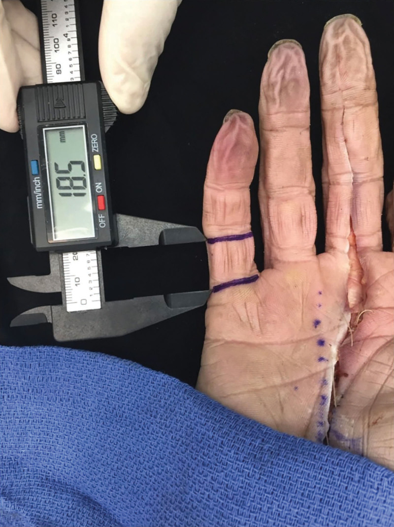
Each hand was placed flat on a table, with fingers extended. The proximal interphalangeal joint crease and palmar digital crease were marked on each finger. The width of the proximal and middle phalanx was measured using a mechanical caliber from the radial to the ulnar side, and the midpoint of each marked crease was located. A straight line was marked connecting the midpoints to represent the longitudinal axis of the fingers. Using magnification loupes or a microscope and the mechanical caliber, the distance between the palmar digital crease and the proximal interphalangeal crease was measured (Fig. 1). Half of this distance was reflected proximally and marked along the longitudinal axis of each finger between the palmar digital crease and the distal palmar crease, resulting in distance A (Fig. 2). A needle was transfixed perpendicularly to the skin until it reached the proximal phalanx cortex at the level of the palmar digital crease mark (Fig. 3). Dissection of each finger using a 15-blade, Steven scissors, Ragnell, and self-retaining retractors was carried out from the proximal palmar crease to the proximal interphalangeal crease. The proximal edge of the A2 pulley was identified, and the distance from the proximal edge of the A2 pulley to the needle transfixed at the palmar digital crease was measured using a mechanical caliber, resulting in distance B (Fig. 4). Another needle was transfixed at the level of the proximal edge of the A2 pulley to assess its proximity to the proposed surface landmark (Fig. 5).
Fig. 1.
Measuring the distance between the palmar digital crease and proximal interphalangeal crease.
Fig. 2.
Marking a point located half the distance between the palmar digital crease and proximal interphalangeal crease reflected in the palm; distance A represents the distance from this point to the palmar digital crease.
Fig. 3.
A needle is transfixed perpendicular to the skin until it reaches the proximal phalanx cortex at the level of the palmar digital crease mark.
Fig. 4.
Dissection of the finger and measuring distance B, which is the distance between the A2 pulley starting point and the palmar digital crease.
Fig. 5.
A picture showing the proximity of a needle transfixed at the level of the A2 proximal edge and the skin land mark located half the distance of the palmar digital crease and proximal interphalangeal crease in the palm (black arrow).
STATISTICAL ANALYSIS
The data were checked for completeness and correctness. The analysis was performed in a 95% confidence interval using the Statistical Package for Social Science, version 23.0 (IBM, Armonk, N.Y.). Student t test was used to assess if there was any significant difference between distance A and distance B. A P value of 0.05 level of statistical significance was considered significant. A power analysis of 240 fingers is needed for a power of 90% with an alpha error of 0.05.
RESULTS
The mean distance A for the index finger was 11.2 mm, and distance B was 11 mm, with no significant difference observed (P > 0.05). Similarly, for the middle finger, the mean distance A was 12 mm, and distance B was 12.1 mm, with no significant difference observed (P > 0.05). However, for the ring finger, there was a significant difference (P < 0.05) between the mean distance A of 10.9 mm and the mean distance B of 11.9 mm, with a mean difference of 1 mm. For the small finger, the mean distance A was 8.8 mm, and the mean distance B was 8.8 mm, with no significant difference observed (P > 0.05). In 24 fingers, the A1 pulley was continuous with the A2 pulley, and we had to raise the single pulley as a flap to define the A2 by exploring the dorsal surface. The mean and standard deviation of distance A and distance B for each finger are recorded in Table 1. Therefore, half the distance from the palmar digital crease to the proximal interphalangeal crease can predict the location of the starting point of the A2 pulley if reflected proximally in the palm, except for the ring finger, where the A2 starting point is located 1 mm proximal to the measured surface landmark.
Table 1.
Mean, Standard Deviation of Distance A* and Distance B† in mm for Index, Middle, Ring, and Small Fingers
| Index | Middle | Ring | Small | |||||
|---|---|---|---|---|---|---|---|---|
| Distance A | Distance B | Distance A | Distance B | Distance A | Distance B | Distance A | Distance B | |
| Mean (mm) | 11.2 | 11 | 12 | 12.1 | 11.9 | 10.9 | 8.8 | 8.8 |
| SD (mm) | 0.8 | 1.04 | 2.1 | 1.8 | 0.9 | 0.7 | 1.04 | 1.04 |
| P | P > 0.05 | P > 0.05 | P < 0.05 | P > 0.05 | ||||
*Half the distance between the palmar digital crease and proximal interphalangeal crease reflected in the palm); †The distance between the A2 pulley’s starting point and the palmar digital crease.
Statistical difference among these values is represented by the P value. P < 0.05 is considered statistically significant difference.
DISCUSSION
The A2 pulley is a crucial structure that can be inadvertently injured during the percutaneous or open release of the A1 pulley for trigger finger, leading to bowstringing and reduced tendon excursion.8,9 The location of the A2 pulley has been described variably at the proximal third or middle third of the proximal phalanx.10 To guide surgeons in planning incisions for trigger finger release and A2 reconstruction procedures, Gordon located the A2 pulley on 48 fingers relative to palm creases, rather than to bone anatomy.11 His rule of thirds is meant to be a practical guide, rather than absolute numbers.7 Our study of 248 finger dissections consistently found that the A2 pulley is located more proximally at the midpoint of the distance from the palmar digital crease to the proximal interphalangeal joint crease in the index, middle, and small fingers. In fact, it is even 1 mm further proximal than the midpoint of the same distance on the ring finger. Tomaino et al and Mitsionis et al found that isolated A2 partial release of up to 25% does not affect flexion excursion.8,12 However, Chow et al found a significant decrease in overall finger motion with the partial proximal release of either the A2 or A4 pulley compared with partial release distally.13 On the other hand, Leeflang and Coert found significant bowstringing with the partial distal release of the A2 pulley.14 Despite the controversy in the literature, our findings should alert hand surgeons to the proximity of the A2 pulley during A1 release to avoid inadvertent injury.6,15–17 In our study, we found that the proximal edge of the A2 pulley can be located at the midpoint of a distance equal to the distance between the palmar digital crease and the proximal interphalangeal crease in the palm of the index, middle, and long fingers (Fig. 6). The proximal edge of the ring finger A2 pulley lies 1 mm proximal to this landmark. This surface landmark will aid hand surgeons in avoiding injury to the A2 pulley during the open or percutaneous release of the A1 pulley for trigger finger and in planning skin incisions during A2 reconstruction procedures.
Fig. 6.
Sketch drawing demonstrating the PDC, PIPC, and the proximate location of the A1 and the A2 pulley starting edge based on the study results. *PIPC: proximal interphalangeal crease. PDC: palmar digital crease.
Limitations of our study are our sample size (which was relatively small), and we only examined cadaveric specimens. Therefore, the generalizability of our findings to living patients may be limited. Additionally, we did not investigate the potential influence of other factors that could affect our outcomes, such as the patient’s age, gender, or comorbidities. Future research should include larger-scale clinical studies that can evaluate the effectiveness of our proposed technique in a more diverse patient population.
CONCLUSIONS
In our study, we discovered that a specific landmark on the surface of the palm can serve as a valuable indicator for predicting the proximal edge of the A2 pulley. Our findings indicate that the proximal edge of the A2 pulley can be precisely located at the midpoint of a distance equivalent to the measurement between the palmar digital crease and the proximal interphalangeal crease in the palm. This observation holds true for the index, middle, and long fingers. Additionally, we observed that the starting point of the A2 pulley for the ring finger is positioned 1 mm more proximally compared with the other digits. Identifying the precise location of the A2 pulley can greatly assist hand surgeons in minimizing incisions and avoiding inadvertent injuries during hand surgery.
DISCLOSURE
The authors have no financial interest to declare in relation to the content of this article.
Footnotes
Published online 25 July 2023.
Disclosure statements are at the end of this article, following the correspondence information.
REFERENCES
- 1.Barton NJ. Experimental study of optimal location of flexor tendon pulleys. Plast Reconstr Surg. 1969;43:125–129. [DOI] [PubMed] [Google Scholar]
- 2.Lin GT, Amadio PC, An KN, et al. Functional anatomy of the human digital flexor pulley system. J Hand Surg Am. 1989;14:949–956. [DOI] [PubMed] [Google Scholar]
- 3.Wilhelmi BJ, Snyder N, Verbesey JE, et al. Trigger finger release with hand surface landmark ratios: an anatomic and clinical study. Plast Reconstr Surg. 2001;108:908–915. [DOI] [PubMed] [Google Scholar]
- 4.Fiorini HJ, Santos JB, Hirakawa CK, et al. Anatomical study of the A1 pulley: length and location by means of cutaneous landmarks on the palmar surface. J Hand Surg Am. 2011;36:464–468. [DOI] [PubMed] [Google Scholar]
- 5.Berrigan W, White W, Cipriano K, et al. Diagnostic imaging of A2 pulley injuries: a review of the literature. J Ultrasound Med. 2022;41:1047–1059. [DOI] [PMC free article] [PubMed] [Google Scholar]
- 6.Zafonte B, Rendulic D, Szabo RM. Flexor pulley system: anatomy, injury, and management. J Hand Surg Am. 2014;39:2525–2532; quiz: 2533. [DOI] [PubMed] [Google Scholar]
- 7.Gordon JA, Stone L, Gordon L. Surface markers for locating the pulleys and flexor tendon anatomy in the palm and fingers with reference to minimally invasive incisions. J Hand Surg Am. 2012;37:913–918. [DOI] [PubMed] [Google Scholar]
- 8.Mitsionis G, Bastidas JA, Grewal R, et al. Feasibility of partial A2 and A4 pulley excision: effect on finger flexor tendon biomechanics. J Hand Surg Am. 1999;24:310–314. [DOI] [PubMed] [Google Scholar]
- 9.Mitsionis G, Fischer KJ, Bastidas JA, et al. Feasibility of partial A2 and A4 pulley excision: residual pulley strength. J Hand Surg Br. 2000;25:90–94. [DOI] [PubMed] [Google Scholar]
- 10.Manske PR, Lesker PA. Palmar aponeurosis pulley. J Hand Surg Am. 1983;8:259–263. [DOI] [PubMed] [Google Scholar]
- 11.Doyle JR. Anatomy and function of the palmar aponeurosis pulley. J Hand Surg Am. 1990;15:78–82. [DOI] [PubMed] [Google Scholar]
- 12.Tomaino M, Mitsionis G, Basitidas J, et al. The effect of partial excision of the A2 and A4 pulleys on the biomechanics of finger flexion. J Hand Surg Br. 1998;23:50–52. [DOI] [PubMed] [Google Scholar]
- 13.Chow JC, Sensinger J, McNeal D, et al. Importance of proximal A2 and A4 pulleys to maintaining kinematics in the hand: a biomechanical study. Hand (N Y). 2014;9:105–111. [DOI] [PMC free article] [PubMed] [Google Scholar]
- 14.Leeflang S, Coert JH. The role of proximal pulleys in preventing tendon bowstringing: pulley rupture and tendon bowstringing. J Plast Reconstr Aesthet Surg. 2014;67:822–827. [DOI] [PubMed] [Google Scholar]
- 15.Elliot D, Lalonde DH, Tang JB. Commentaries on Clinical results of releasing the entire A2 pulley after flexor tendon repair in zone 2C. K. Moriya, T. Yoshizu, N. Tsubokawa, H. Narisawa, K. Hara and Y. Maki. J Hand Surg Eur . 2016, 41: 822.– . J Hand Surg Eur Vol. 2016;41: 829–830. [Google Scholar]
- 16.Moriya K, Yoshizu T, Tsubokawa N, et al. Clinical results of releasing the entire A2 pulley after flexor tendon repair in zone 2C. J Hand Surg Eur Vol. 2016;41:822–828. [DOI] [PubMed] [Google Scholar]
- 17.Tang JB. New developments are improving flexor tendon repair. Plast Reconstr Surg. 2018;141:1427–1437. [DOI] [PubMed] [Google Scholar]


