Abstract
Background
Thermoxidation of edible oil through deep fat frying results in the generation of several oxidized products that promote lipid peroxidation and ROS production when eaten. Consumption of thermoxidized oil in post-menopausal conditions where the estrogen level is low contributes to cardiovascular disease. This study evaluates the role of estradiol and antihyperlipidemic agents (AHD) in restoring the vascular health of ovariectomized (OVX) rats fed with thermoxidized palm oil (TPO) and thermoxidized soya oil (TSO) diets.
Method
A total of 10 groups of rats (n = 6) were set up for the experiment. Group I (normal control) rats were sham handled while other groups were OVX to bring about estrogen deficient post-menopausal state. Group II (OVX only) was not treated and received normal rat chow. Groups III-X were fed with either TPO or TSO diet for 12 weeks and treated with estradiol (ETD) 0.2 mg/kg/day, atorvastatin (ATV) 10 mg/kg/day, and a fixed-dose combination of ezetimibe and ATV (EZE 3 mg/kg/day + ATV 10 mg/kg/day).
Results
Pro-atherogenic lipids levels were significantly elevated in untreated TSO and TPO groups compared to OVX and sham, resulting in increased atherogenic and Coronary-risk indices. Treatment with Estradiol and AHDs significantly reduced the total cholesterol, triglycerides, low-density lipoprotein cholesterol as well as AI and CRI compared to untreated TSO and TPO groups, whereas TSO and TPO groups showed significant elevation in these parameters compared to Group I values. Moreover, aortic TNF-α levels were extremely elevated in the untreated TSO and TPO compared to Group I. TNF-α levels were significantly reduced in rats treated with AHDs and ETD. Localized oxidative stress was indicated in the aortic tissues of TSO and TPO-fed OVX rats by increased malondialdehyde and decreased glutathione, catalase, and superoxide dismutase levels. This contributed to a depletion in aortic nitric oxide. AHDs and ETD replenished the nitric oxide levels significantly. Histological evaluation of the aorta of TSO and TPO rats revealed increased peri-adventitia fat, aortic medial hypertrophy, and aortic recanalization. These pathologic changes were less seen in AHDs and ETD rats.
Conclusion
This study suggests that ETD and AHDs profoundly attenuate oxidized lipid-induced vascular inflammation and atherogenesis through oxidative-stress reduction and inhibition of TNF-α signaling.
Keywords: Oxidized lipids, Vascular inflammation, Atherogenesis, Estrogen, Menopause, Antihyperlipidemic drugs
Background
Intermittent deep frying of edible oils is a common food processing practice in homes, restaurants, food stalls, and industries adopted to reduce cost and improve taste [1, 2]. In deep-fat frying, the oil is usually heated to an extreme temperature of 180 °C and higher in the presence of moisture and air, resulting in an array of chemical reactions known as lipid oxidation [3]. Thermal oxidation of dietary oils destroys essential fatty acids and generates products such as peroxides, hydroperoxides, aldehydes, and several other potentially hazardous non-volatile polar compounds that are retained in the thermoxidised oil and when consumed repeatedly increase the risk for cardiovascular diseases [1, 4, 5].
Atherosclerosis is an inflammatory disorder of large and medium-sized arteries. Endothelial dysfunction, vascular inflammation, and lipid deposition in the tunica intima are some of the features of atherosclerotic development and progression [6]. Oxidized-low density lipoprotein (Ox-LDL), a product of LDL-c oxidation, is associated with several pathological manifestations including atherosclerosis [5, 7]. In the early phase of atherosclerosis, LDL-c is deposited in the intima of the arterial wall and recruits inflammatory cells to the sub-endothelium. It is also involved in the formation of Ox-LDL, whose interaction with inflammatory cells results in the generation of reactive oxygen species (ROS) and depletion of Nitric oxide (NO) [7, 8]. Endothelial dysfunction and impaired NO release disrupt the regulation of vascular tone, creating an imbalance between vasoconstriction and vasodilation, resulting in atherogenesis [7, 9]. Structural changes observed in atherosclerotic arterial tissues include accumulation of blood cells in the luminal aspect of the aortic wall, leukocyte infiltration, aortic medial hypertrophy, and intimal plaques [6, 10].
There is a noticeable increase in risk for cardiovascular diseases (CVDs) including atherosclerosis in postmenopausal women when estrogen levels decline. While CVD is not prevalent in young women, it is the leading cause of morbidity and mortality in women above 50 years and accounts for about 75% of deaths in postmenopausal women [11]. Some mechanisms have been proposed to delineate the cardioprotective role of estrogen in premenopausal women, namely, estrogen administration positively impacts plasma lipid profiles, anti-platelet, and antioxidant status [12]. A Previous study revealed greater arterial stiffening, a known indicator of vascular aging in postmenopausal women when compared with pre-menopausal women alongside estrogen-linked endothelial-dependent vasodilatation [13]. The role of estradiol in the inhibition of monocyte adhesion to vascular endothelium, which is a critical step in the development of atheromas and arteriosclerosis has been established [14]. A synergy of these mechanisms which confers cardioprotective prowess to premenopausal women in the presence of estrogen invariably leads to a myriad of cardiac pathologies in postmenopausal women with very low levels of estrogen.
Women are around 10 years older than men at the first presentation of atherosclerotic cardiovascular disease, and this is related to the detrimental effect of estrogen withdrawal on cardiovascular functions and metabolism [15–17]. Several risk factors of cardiovascular disease are altered following the menopausal transition, resulting in a significant increase in the risk for myocardial infarction and cerebrovascular disease. These factors include dyslipidemia, hypertension, inflammation, and hemostatic factors [18, 19]. While post-menopausal status decreases vascular protection due to reduced estrogen levels, the consumption of thermoxidized oil further diminishes vascular integrity via oxidative stress and increased levels of oxidized lipids [3, 4].
Dyslipidemia is prominent among the surrogate markers of increased cardiovascular risks in postmenopausal women [16]. Despite the availability of various lipid-lowering drugs, reduction in cardiovascular risk and prevention of atherosclerosis in postmenopausal females has proven to be a tough challenge [2, 16]. Epidemiological studies clearly show that cardiovascular disease is the major cause of mortality in women in developed countries accounting for deaths of 75% or greater in postmenopausal women [11, 16].
This study examines estradiol and selected antihyperlipidemic drugs used singly or in combination for their athero-protective potentials in ovariectomized rats fed with thermoxidized oil diet.
Methods
Preparation of thermoxidised oil diet
Five liters of palm oil (Ace Products and Services Ltd., Nigeria) and soya oil (Sunola Foods Ltd., Plot 122–132, Oshodi-Apapa Expressway, Lagos State, Nigeria) were processed by repeated heating on a hot plate at 180°C for 20 min [20]. One and a half liters of oil was used to fry 2 kg of sweet potatoes in five cycles. At the end of each frying batch, the oil was cooled to room temperature (25° C) for 5 h and used to fry a fresh batch of potatoes for the second time without replacing lost oils. The cycle was repeated five times to make five times heated oil for the thermoxidized oil diet. Thermoxidized palm/soya oil (15% w/w) was formulated into diets with standard rat chow (Ladokun Feeds, Ibadan, Oyo State, Nigeria).
Experimental animals
Healthy female Wistar rats (200–250 g) obtained from a private breeder (McTemmy Farms, Ogbomosho, Oyo State, Nigeria) were used for this study. The animals were housed individually in plastic cages at room temperature of 27 ± 2°C at the University of Ilorin, Central Laboratory Animal House under a 12-hour light/dark cycle and had ad libitum access to food and water. Institutional ethical approval for this study was obtained from the Ethical Review Committee of the University of Ilorin, with approval number UERC/ASN/2021/2038.
Ovariectomy procedure and treatment
Ovariectomy was performed under ketamine (43.5 mg/kg) /xylazine (6.5 mg/kg) anesthesia injected intraperitoneally [21]. Skin and muscle layers were breached with incisions to gain access to the abdominal cavity, after which the fat bed embedding the ovary was identified and externalized. To initiate an estrogen-deficient postmenopausal state, the ovary was excised while the uterus and the abdominal fat bed were placed back into the cavity. Incisions were closed with a 4/0 absorbable suture. These steps were repeated to remove the contralateral ovary. Penicillin ointment was applied to the incision site to minimize infection while ofloxacin 1000 mg/kg was administered orally to the rats for seven days [22]. The rats were allowed free access to food and water for two weeks to recover from surgery and attain a post-menopausal state before drug treatments and formulated thermoxidized oil diets were instituted.
The experimental animals were randomly divided into ten groups (n = 6). Group I was sham-handled to simulate surgical stress, group II was ovariectomized and both groups were fed with standard rat chow while the other eight groups (Groups III-X) were ovariectomized (OVX) and fed with either thermoxidized palm oil (TPO) or thermoxidized Soya oil (TSO) diets. Estradiol and antihyperlipidemic drugs (AHDs) were orally administered as follows: estradiol valerate (ETD) 0.2 mg/kg/day, atorvastatin (ATV) 10 mg/kg/day and fixed-dose combination of ezetimibe and ATV (EZE 3 mg/kg/day + ATV 10 mg/kg/day). The animals were treated for 12 weeks at the end of which aortic tissues were harvested for biochemical and histological analysis.
Sacrifice of experimental animals
At the end of treatment for a period of 12 weeks, the rats were fasted for 12 h and anesthetized with diethyl ether, and blood was obtained through cardiac puncture into heparinized tubes. The aortas were identified and harvested, rinsed in Tris-KCl buffer (0.15 M, pH 7.4) and frozen. Whole blood in heparin tubes was left standing at room temperature for 30 min and centrifuged at 800 g for 15 min to obtain plasma. The tissues were homogenized using a mechanical homogenizer with Teflon at 4 °C in sodium phosphate buffer (0.1 M, pH 7.4). The tissue homogenates were centrifuged at 2000 g for 10 min at 4 °C. The supernatants were aliquoted and stored at -20 °C for biochemical assays.
Plasma lipid profile analyses
Total cholesterol (TC), Triglycerides (TG) and high-density lipoprotein-cholesterol (HDL-c) were assayed in plasma using Randox kits (Randox Laboratories Limited, United Kingdom). Very low-density lipoprotein cholesterol (VLDL-c) and low-density lipoprotein cholesterol (LDL-c) levels [23], atherogenic index (AI) [24], coronary risk index (CRI) [25] and HDL-c/LDL-c ratio [23] were calculated from the lipid profile.
Determination of aortic nitric oxide and tumor necrosis factor-alpha (TNF-α)
Nitrite was measured as an indicator of nitric oxide (NO) production using the Griess method as previously reported [26]. Aortic TNF- α level was determined using the BioLegend ELISA kit (BioLegend Biotechnology Research, San Diego, California, U.S.A.) according to the manufacturer’s instructions.
Aortic tissue oxidative stress assessment
Aortic tissue homogenates supernatants were used for oxidative stress assessment. Malondialdehyde (MDA) was measured as an index of lipid peroxidation using the assay of thiobarbituric reacting substances (TBARs) [27]. Reduced glutathione (GSH) as a non-enzymic antioxidant maker was measured using the method reported in [28]. Catalase (CAT) activity in the supernatants was determined using the colorimetric assay based on the yellow complex with molybdate and H2O2 as described by [29]. Superoxide dismutase (SOD) activity was determined using the method previously reported by Misra and Fridovich [30].
Histological analysis
Ascending and thoracic aorta tissues were dehydrated and embedded in paraffin wax. Thin sections (5 μm) of the aortic tissues were cut and stained with hematoxylin and eosin (H &E) stain for light microscopy and viewed at x100 magnification.
Statistical analysis
All data were presented as mean ± standard error mean (SEM). Normally distributed data were analyzed by parametric test using One-way analysis of variance (ANOVA) and Bonferroni’s multiple comparisons test. The level of significance was set at p < 0.05. Statistical analysis was performed by using GraphPad Prism 8 software.
Results
Effect of selected antihyperlipidemic drugs and estradiol on plasma lipid profile and atherogenic and coronary artery risk indices of ovariectomized rats fed with TPO and TSO
TC, TG, and LDL-c were significantly increased in untreated OVX + TSO (Table 1) and OVX + TPO rats (Table 2) compared to sham. Treatment with ETD and ATV significantly (p < 0.05) reduced these parameters compared to untreated OVX + TSO (Table 1). In OXV + TPO rats (Table 2), ETD, ATV and EZE + ATV treatments decreased TC and TG levels significantly (p < 0.05), and LDL-c levels were decreased by ATV treatment.
Table 1.
Effects of estradiol and selected antihyperlipidemic drugs on plasma lipid profile of ovariectomized Wistar rats fed with thermoxidized soya oil diet
| Treatment | Plasma Lipid profile parameters (mg/dL) | ||||
|---|---|---|---|---|---|
| TC | TG | HDL-c | LDL-c | VLDL-c | |
| Sham (Group I) | 77.72 ± 1.29 | 96.23 ± 10.22 | 22.08 ± 0.77 | 36.40 ± 2.24 | 19.25 ± 2.04 |
| OVX | 93.58 ± 4.76 | 104.12 ± 2.01 | 21.18 ± 0.79 | 49.62 ± 0.86 | 21.3 ± 1.34 |
| OVX + TSO | 107.89 ± 1.68# | 139.72 ± 21.97# | 19.88 ± 1.21 | 60.07 ± 7.01# | 27.94 ± 4.39# |
| OVX + TSO + ATV | 82.35 ± 2.07*** | 103.20 ± 10.59*** | 24.53 ± 4.08** | 37.17 ± 7.69*** | 20.64 ± 2.12* |
| OVX + TSO + EZE + ATV | 88.68 ± 2.13*** | 97.22 ± 18.33*** | 20.67 ± 1.61 | 57.21 ± 10.10 | 19.44 ± 3.67** |
| OVX + TSO + ETD | 76.05 ± 0.94*** | 104.26 ± 14.06*** | 20.88 ± 1.64 | 34.31 ± 10.02*** | 20.85 ± 2.81* |
Values are expressed as mean ± SEM (n = 6)
#p < 0.05 compared to Group I values accepted as significant
*p < 0.05 compared to OVX + TPO
**p < 0.01 compared to OVX + TPO
***p < 0.001 compared to OVX + TPO
OVX – ovariectomized; TSO – thermoxidized soya oil; ATV – atorvastatin; EZE – ezetimibe; ETD – estradiol; TC – total cholesterol; TG – triglycerides; HDL-c – high density lipoprotein cholesterol; LDL-c – low density lipoprotein cholesterol; VLDL-c – very low density lipoprotein cholesterol
Table 2.
Effects of estradiol and selected antihyperlipidemic drugs on plasma lipid profile of ovariectomized Wistar rats fed with thermoxidized palm oil diet
| Treatment | Plasma Lipid profile parameters (mg/dL) | ||||
|---|---|---|---|---|---|
| T-CHOL | TRIG | HDL | LDL | VLDL | |
| Sham (Group I) | 77.72 ± 1.29 | 96.23 ± 10.22 | 22.08 ± 0.77 | 36.40 ± 2.24 | 19.25 ± 2.04 |
| OVX | 93.58 ± 4.76 | 104.12 ± 2.01 | 21.18 ± 0.79 | 49.62 ± 0.86 | 21.3 ± 1.34 |
| OVX + TPO | 106.95 ± 2.70# | 163.95 ± 16.53# | 20.92 ± 0.60 | 55.66 ± 4.73# | 30.37 ± 3.63# |
| OVX + TPO + ATV | 82.49 ± 2.25*** | 110.45 ± 18.34*** | 21.48 ± 1.54 | 38.92 ± 4.62*** | 22.09 ± 3.67** |
| OVX + TPO + EZE + ATV | 98.54 ± 0.66 | 114.89 ± 6.68*** | 20.73 ± 0.69 | 54.83 ± 1.95 | 22.98 ± 1.34* |
| OVX + TPO + ETD | 95.33 ± 1.69** | 126.19 ± 12.78* | 19.54 ± 1.25 | 50.55 ± 5.32 | 25.24 ± 2.56 |
Values are expressed as mean ± standard error of mean (n = 6)
#p < 0.05 compared to Sham accepted as significant
*p < 0.05 compared to OVX + TPO
**p < 0.01 compared to OVX + TPO
***p < 0.001 compared to OVX + TPO
OVX – ovariectomized; TPO – thermoxidized palm oil; ATV – atorvastatin; EZE – ezetimibe; ETD – estradiol; TC – total cholesterol; TG – Triglycerides; HDL-c – high density lipoprotein cholesterol; LDL-c – low density lipoprotein cholesterol; VLDL-c – very low density lipoprotein cholesterol.
AI and CRI levels were significantly (p < 0.05) increased in OVX + TSO (Table 3) and OVX + TPO (Table 4) rats when compared with sham. ETD, ATV and EZE + ATV treatments lowered AI significantly (p < 0.05) in OVX + TSO and OVX + TPO diet groups. Similarly, CRI level was significantly (p < 0.05) decreased by ATV treatment in both diet groups when compared with sham. ETD treatment equally decreased the CRI, albeit in OVX + TSO diet group only (Table 3).
Table 3.
Effects of estradiol and selected antihyperlipidemic drugs on plasma atherogenic indices of ovariectomized Wistar rats fed with thermoxidized soya oil diet
| Treatment | Plasma Atherogenic indices | ||
|---|---|---|---|
| AI | CRI | HDL-c/LDL-c | |
| Sham (Group I) | 3.44 ± 0.59 | 3.55 ± 0.22 | 0.62 ± 0.05 |
| OVX | 4.71 ± 0.55 | 4.65 ± 0.33# | 0.3 ± 0.04 |
| OVX + TSO | 6.03 ± 1.02# | 5.57 ± 0.49# | 0.36 ± 0.06 |
| OVX + TSO + ATV | 3.43 ± 0.41*** | 3.74 ± 0.52*** | 1.25 ± 0.32*** |
| OVX + TSO + EZE + ATV | 3.86 ± 0.94*** | 4.90 ± 0.67 | 0.45 ± 0.11 |
| OVX + TSO + ETD | 4.04 ± 0.64** | 3.64 ± 0.38*** | 0.18 ± 0.31 |
Values are expressed as mean ± SEM (n = 6)
#p < 0.05 compared to Group I values accepted as significant
*p < 0.05 compared to OVX + TSO
**p < 0.01 compared to OVX + TSO
***p < 0.001 compared to OVX + TSO
OVX – ovariectomized; TSO – thermoxidized soya oil; ATV – atorvastatin; EZE – ezetimibe; ETD – estradiol; HDL-c – high density lipoprotein cholesterol; LDL-c – low density lipoprotein cholesterol; AI – atherogenic index; CRI – coronary risk index
Table 4.
Effects of estradiol and selected antihyperlipidemic drugs on plasma atherogenic indices of ovariectomized Wistar rats fed with thermoxidized palm oil diet
| Treatment | Plasma Atherogenic indices | ||
|---|---|---|---|
| AI | CRI | HDL-c/LDL-c | |
| Sham (Group I) | 3.44 ± 0.59 | 3.55 ± 0.22 | 0.62 ± 0.05 |
| OVX | 4.71 ± 0.55 | 4.65 ± 0.33# | 0.3 ± 0.04 |
| OVX + TPO | 6.29 ± 0.92# | 5.09 ± 0.21# | 0.38 ± 0.02 |
| OVX + TPO + ATV | 4.21 ± 0.81*** | 3.88 ± 0.20** | 0.57 ± 0.06 |
| OVX + TPO + EZE + ATV | 4.58 ± 0.39* | 4.78 ± 0.17 | 0.38 ± 0.02 |
| OVX + TPO + ETD | 4.39 ± 0.29** | 5.03 ± 0.52 | 0.42 ± 0.06 |
Values are expressed as mean ± standard error of mean (n = 6)
# - p < 0.05 compared to Sham accepted as significant
* - p < 0.05 compared to OVX + TPO
** - p < 0.01 compared to OVX + TPO
*** - p < 0.001 compared to OVX + TPO
OVX – ovariectomized; TPO – thermoxidized palm oil; ATV – atorvastatin; EZE – ezetimibe; ETD – estradiol; HDL-c – high density lipoprotein cholesterol; LDL-c – low density lipoprotein cholesterol; AI – atherogenic index; CRI – coronary risk index.
Effect of selected antihyperlipidemic drugs and estradiol on aortic nitrites of ovariectomized rats fed with TPO and TSO
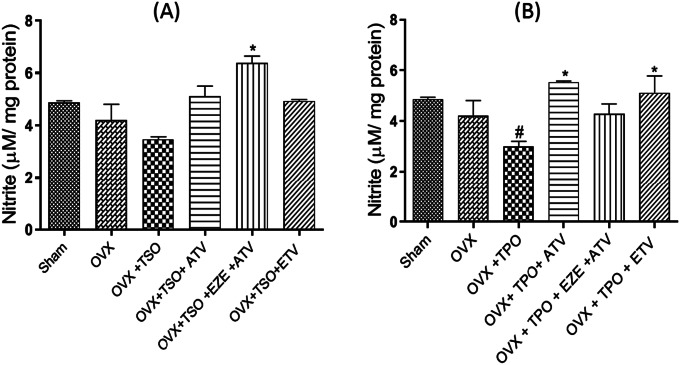
Aorta tissue nitrites level was measured as an index for nitric oxide (NO) as shown in Fig. 1a and lb. There was a significant reduction in aortic nitrites levels in OVX + TPO groups when compared with Group I rats. Treatment with ATV and ETD significantly increased aortic nitrite levels when compared with OVX + TPO (Fig. 1b). In OVX + TSO diet groups, nitrite level was significantly increased in the EZE + ATV treated group (Fig. 1a).
Fig. 1.
Effects of estradiol and selected antihyperlipidemic drugs on aortic nitric oxide levels in ovariectomized Wistar rats fed with thermoxidized soya oil diet (A) and thermoxidized palm oil diet (B)
Values are expressed as mean ± standard error of mean (n = 3)
#p < 0.05 compared to Sham accepted as significant
*p < 0.05 compared to OVX + TPO/OVX + TSO
OVX – ovariectomized; TPO – thermoxidized palm oil; TSO – thermoxidized soya oil; ATV – atorvastatin; EZE – ezetimibe; ETD – estradiol
Effect of selected antihyperlipidemic drugs and estradiol on aortic TNF-α levels of ovariectomized rats fed with TPO and TSO
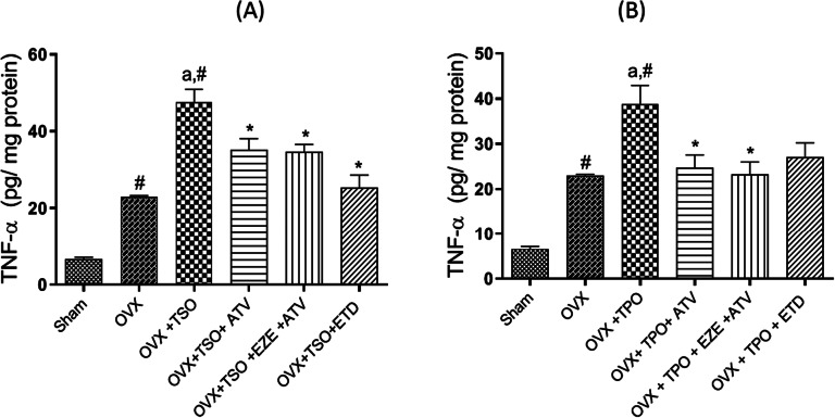
As shown in Fig. 2a, TNF-α levels were significantly (p < 0.05) increased in OVX + TSO rats by about 80% compared to sham. However, treatment with ATV, EZE + ATV and ETD significantly (p < 0.05) decreased the TNF-α levels. Similarly, as shown in Fig. 2b, TNF-α levels were significantly (p < 0.05) elevated in OVX + TPO rats compared to sham. Treatment with ATV, EZE + ATV and ETD significantly decreased the aortic TNF-α concentrations in comparison to the untreated OVX + TPO.
Fig. 2.
Effects of estradiol and selected antihyperlipidemic drugs on aortic TNF-α levels in ovariectomized Wistar rats fed with thermoxidized soya oil diet (A) and thermoxidized palm oil diet (B)
Values are expressed as mean ± standard error of mean (n = 3)
#p < 0.05 compared to Sham accepted as significant
ap < 0.05 compared to OVX
*p < 0.05 compared to OVX + TPO/OVX + TSO
OVX – ovariectomized; TPO – thermoxidized palm oil; TSO – thermoxidized soya oil; ATV – atorvastatin; EZE – ezetimibe; ETD – estradiol; TNF-α – tumor necrosis factor-alpha
Effect of selected antihyperlipidemic drugs and estradiol on aortic markers of oxidative stress in ovariectomized rats fed with TPO and TSO
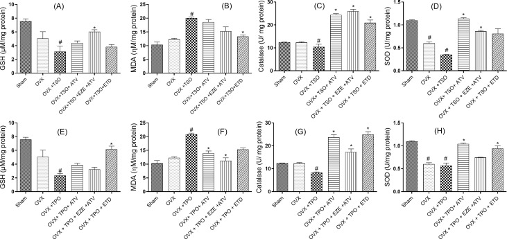
Repeated oral treatments of ovariectomized rats with TSO significantly (p < 0.05) reduced GSH levels when compared to Group I values (Fig. 3). Among the treatment groups, ATV + EZE treated rats showed a significant increase in their GSH levels in comparison with the untreated ovariectomized rats treated with TSO. Similarly, the MDA levels were increased (p < 0.05) by two folds in the ovariectomized rats treated with TSO when compared with sham. ETD decreased (p < 0.05) the MDA levels in comparison with OVX + TSO. CAT levels were significantly decreased in the OVX + TSO group compared to sham. Treatment with ATV, EZE + ATV and ETD all increased (P < 0.05) the CAT levels when compared to rats in the untreated OVX + TSO group. In the EZE + ATV treated rats, there was a 150% increase in CAT levels. SOD levels decreased significantly (p < 0.05) in both OVX and OVX + TSO rats compared with sham rats, however, SOD was decreased more in OVX + TPO group compared to the OVX group, although this difference was not significant (p > 0.05). All the treatment groups showed significantly elevated SOD levels when compared with the untreated OVX + TSO. ATV treated rats showed a profound (p < 0.05) increase in the SOD levels of more than three folds when compared with OVX + TSO. GSH concentration in the untreated OVX + TPO was significantly reduced compared to sham. ETD treatment increased the GSH concentration significantly (p < 0.05) when compared with the untreated OVX + TPO group. MDA concentration increased significantly in the OVX + TPO group by 100% when compared with Group I values. However, all the treatment groups ATV, EZE + ATV and ETD decreased MDA concentration significantly (p < 0.05) when compared to the untreated TSO-fed ovariectomized rats. CAT concentration decreased significantly (p < 0.05) in the untreated TSO-fed ovariectomized rats when compared with Group I values. Conversely, ATV, EZE + ATV and ETD treatment increased CAT levels significantly when compared with the untreated TPO-fed ovariectomized rats. In ETD treated rats, there was a threefold increase in CAT levels. SOD levels decreased significantly (p < 0.05) in the untreated OVX as well as OVX + TPO rats when compared to sham. However, SOD was further decreased in OVX + TPO group compared to the OVX group albeit insignificantly (p > 0.05). Treatment groups ATV and ETD raised the SOD levels significantly (p < 0.05) when compared with the untreated TPO-fed ovariectomized rats.
Fig. 3.
Effects of estradiol and selected antihyperlipidemic drugs on aortic oxidative stress markers in ovariectomized Wistar rats fed with thermoxidized soya oil (A-D) and thermoxidized palm oil diets (E-H)
Values are expressed as mean ± standard error of mean (n = 3)
#p < 0.05 compared to Sham accepted as significant
*p < 0.05 compared to OVX + TSO/ OVX + TPO
**p < 0.01 compared to OVX + TSO/ OVX + TPO
***p < 0.001 compared to OVX + TSO/ OVX + TPO
OVX – ovariectomized; TSO – thermoxidized soya oil; TPO – thermoxidized palm oil; ATV – atorvastatin; EZE – ezetimibe; ETD – estradiol; GSH – glutathione; MDA – Malondialdehyde; CAT – catalase; SOD – superoxide dismutase
Effect of selected antihyperlipidemic drugs and estradiol on the histology of ascending and thoracic aorta of ovariectomized rats fed with TPO and TSO
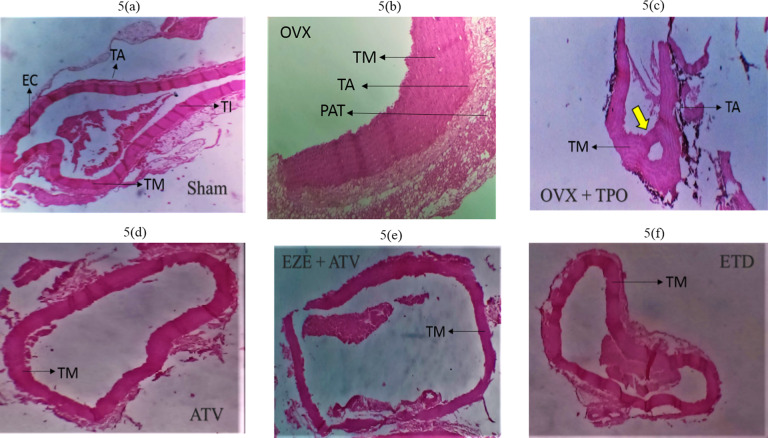
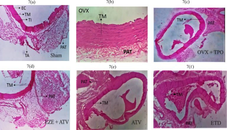
Histopathological examination of the ascending aorta of untreated TSO-fed ovariectomized rats showed a general disruption in the histoarchitecture of the aorta coupled with a large amount of red blood cell deposits in the luminal part of the arterial wall (Fig. 4b). These disruptions included associated tunica media (TM) hypertrophy and increased peri-adventitia fat (PAT) (Fig. 4b and c) when compared to normal histoarchitecture in the Group I rats (Fig. 4a). ATV, EZE + ATV and ETD treatment groups all showed essentially normal histoarchitecture of the aorta (Fig. 4d–f). Similarly, the aortas of the untreated TPO-fed ovariectomized rats showed deposition of red blood cells in the luminal region of the arterial wall as well as hypertrophy of the peri-adventitia adipose tissue (Fig. 5c) when compared to normal histoarchitecture in the Group I rats (Fig. 5a). The Aorta of ATV, EZE + ATV and ETD treated rats fed with TPO diet all showed essentially normal histology (Fig. 5d–f).
Fig. 4.
Photomicrographs of the effects of estradiol and selected antihyperlipidemic drugs on the ascending aorta of ovariectomized Wistar rats fed with thermoxidized soya oil diet. (4a) Sham (4b) OVX (4c) OVX + TSO (4d) ATV (4e) EZE + ATV (4f) ETD
Note the thickening of the tunica media (TM) indicated by the small yellow arrow and aortic recanalization indicated by the big yellow arrow in OVX + TSO (c). Note the reduced amount of red blood cells in the luminal aspect of the aortic wall and the preservation of the elastic laminae of the tunica media indicated by TM in ATV, EZE + ATV and ETD treatment groups. H&E staining of the rat aorta (x100 magnification)
OVX – ovariectomy; TSO – thermoxidised soya oil; EC – endothelial cells; TM – tunica media; TI – tunica intima; TA – tunica adventitial; PAT – peri-adventitial fat; ATV – atorvastatin; EZE – ezetimibe; ETD – estradiol
Fig. 5.
Photomicrographs of the effects of estradiol and selected antihyperlipidemic drugs on the ascending aorta of ovariectomized Wistar rats fed with thermoxidized palm oil diet. (5a) Sham (5b) OVX (5c) OVX + TPO (5d) ATV (5e) EZE + ATV (5f) ETD
Note the thickening of the tunica media indicated by TM and aortic recanalization indicated by the yellow arrow in OVX + TPO. Note the reduced amount of red blood cells in the luminal aspect of the aortic wall and the preservation of the elastic laminae of the tunica media indicated by TM in ATV, EZE + ATV and ETD treatment groups. H&E staining of the rat aorta (x100 magnification)
OVX – ovariectomy; TPO – thermoxidised palm oil; EC – endothelial cells; TM – tunica media; TI – tunica intima; TA – tunica adventitial; PAT – peri-adventitial fat; ATV – atorvastatin; EZE – ezetimibe; ETD – estradiol
The thoracic aorta of OVX + TSO rats (Fig. 6b) revealed recanalization with tunica hypertrophy. Sham aorta revealed normal histoarchitecture (Fig. 6a). The endothelium, tunica intima, tunica media and tunica adventitia were observed to be normal (Fig. 6a). ATV, EZE + ATV and ETD treatment all showed essentially normal histology of the aorta (Fig. 6d–f). Thoracic Aorta of OVX + TPO (Fig. 7c) rats showed recanalization with marked endothelial disruption, tunica media hypertrophy, tunica adventitia disruption, as well as increased peri-adventitia fat when compared to normal histoarchitecture in the Group I rats (Fig. 7a). Thoracic aortas of ATV, EZE + ATV and ETD-treated rats fed showed essentially normal histoarchitecture (Fig. 7d–f).
Fig. 6.
Photomicrographs of the effects of estradiol and selected antihyperlipidemic drugs on the thoracic aorta of ovariectomized Wistar rats fed with thermoxidized soya oil diet. (6a) Sham (6b) OVX (6c) OVX + TSO (6d) ATV (6e) EZE + ATV (6f) ETD
Note the accumulation of large amount of red blood cells indicated by the yellow arrow; thickness of tunica media and complete disruption of the elastic laminae indicated by the white arrow and increased deposition of peri-adventitial fat (PAT) indicated by the red arrow in (c). Note the preservation of the elastic laminae of the tunica media, indicated by TM in (d-f). H&E staining of the rat aorta (x100 magnification)
OVX – ovariectomy; TSO – thermoxidized soya oil; EC – endothelial cells; TM – tunica media; TI – tunica intima; TA – tunica adventitia; PAT – peri-adventitial fat; ATV – atorvastatin; EZE – ezetimibe; ETD – estradiol
Fig. 7.
Photomicrographs of the effects of estradiol and selected antihyperlipidemic drugs on the thoracic aorta of ovariectomized Wistar rats fed with thermoxidized palm oil diet. (7a) Sham (7b) OVX (7c) OVX + TPO (7d) EZE+ATV (7e) ATV (7f) ETD
Note hypertrophy of the peri-adventitial adipose tissue indicated by the black arrow in (OVX + TPO). Note the preservation of the elastic laminae of the tunica media, indicated by TM in all treatment groups. H&E staining of the rat aorta (x100 magnification)
OVX – ovariectomy; TPO – thermoxidized palm oil; EC – endothelial cells; TM – tunica media; TI – tunica intima; TA – tunica adventitia; PAT – peri-adventitial fat; ATV – atorvastatin; EZE – ezetimibe; ETD – estradiol
Discussion
Selected antihyperlipidemic drugs and estrogen prevented hyperlipidemia, aortic tissue inflammation and oxidative stress, in ovariectomized rats fed with thermoxidized oil diets. Deep fat frying with palm and Soybean oil initiated chemical reactions that resulted in the generation of oxidized products while also reducing the antioxidant contents of the oil [11]. These reactive products can activate inflammatory processes that culminate in diminished vascular integrity and cardiovascular health. These effects, coupled with estrogen withdrawal in postmenopausal women significantly increase the risk for CVD [31–34].
Chronic consumption of thermoxidized palm/soya oil in postmenopausal rats has been reported to cause atherogenic dyslipidemia through the elevation of total cholesterol, triglycerides, LDL-c, and decreased the HDL-c [35–37]. In this study, palm and soya oils supplemented diet fed to ovariectomized rats significantly increased atherogenic dyslipidemia. Treatment with antihyperlipidemic drugs and estradiol reversed indices of atherogenic dyslipidemia in ovariectomized rats fed with palm and soya oil-supplemented diets. The association between hyperlipidemia and atherosclerosis including the ensuing incidence of cardio-cerebrovascular diseases is well established in the literature [35, 38, 39]. ETD, a major component of the therapeutic regimen is widely used in the treatment of postmenopausal hormonal deficiency syndrome [40]. The effect of ETD on serum and lipoprotein lipids is characterized by an increase in the lipid constituents of high-density lipoproteins (HDL) and, usually, a decrease in low-density lipoproteins (LDL). LDL-c reduction by these treatments shows that they offer significant protection against atherosclerosis [40–42]. According to the European Society of Cardiology (ESC) and European atherosclerosis society (EAS) guidelines for the management of dyslipidemia, 1 mmol/L reduction in LDL cholesterol level corresponds to a one-fifth reduction in the risk of atherosclerotic cardiovascular disease (ASCVD) [43]. When evaluated for other lipid parameters, ATV, EZE + ATV and ETD treatments decreased the TG levels, while ATV increased the HDL-c levels. The mechanisms underlying the lipid-controlling effect of these antihyperlipidemic drugs have been widely described in the literature [43]. ATV inhibits HMG-CoA reductase while EZE inhibits dietary and biliary cholesterol uptake in the intestine [43]. In OVX + TPO rats, ATV decreased the TC levels significantly while ATV and EZE + ATV significantly decreased TG and VLDL-c levels. Estradiol reduces LDL cholesterol levels and increases HDL cholesterol levels in postmenopausal women with normal or elevated baseline lipid levels [44, 45]. The reduction in LDL cholesterol levels is probably a result of the accelerated conversion of hepatic cholesterol to bile acids and increased expression of LDL receptors on cell surfaces, resulting in augmented clearance of LDL from the plasma [44, 45].
A search of the literature shows evidence of increased cardiovascular risk and incidence of adverse events associated with a decrease in the HDL/LDL cholesterol ratio [46, 47]. TSO and TPO decreased the HDL/LDL ratio albeit insignificantly while also elevating the AI and CRI significantly (p < 0.05). The atherogenic index is a strong marker for assessing the risk of atherosclerosis and coronary heart disease in vessels. As AI increases, the risk of atherogenesis increases [48, 49]. Estradiol and the antihyperlipidemic drugs employed in this study significantly decreased the AI and CRI. ATV, EZE + ATV and ETD administration significantly decreased the AI and/or CRI. This decrease in the atherogenic indices demonstrates the beneficial effect of early administration of antihyperlipidemic drugs and estrogen in the reduction or prevention of atherogenesis. In rats fed with TSO diet, ATV treatment significantly increased the HDL/LDL cholesterol ratio by approximately three folds. An increase in the HDL/LDL cholesterol ratio is an indicator of low circulating LDL in the plasma, which is beneficial in the prevention of atherosclerotic vascular disease [43, 50]. In rats fed with TPO diet, ATV administration significantly decreased the AI and CRI, while ETD and EZE + ATV decreased AI significantly.
Aortic nitric oxide was examined as an indicator of vascular integrity in this study. In both TPO and TSO-fed ovariectomized rats, aortic nitric oxide levels were reduced. This effect was reversed in antihyperlipidemic, and estrogen treated rats. Diminished NO is associated with endothelial dysfunction and predisposes the vessel to atherosclerosis [51, 52]. The elevation of NO through the upregulation of endothelial nitric oxide synthase (eNOS) plays an important role in preventing or reversing endothelial dysfunction associated with hypertension, atherosclerosis and other cardiovascular diseases [53, 54]. Estrogen and the combination of ATV and EZE reportedly increased aortic nitric oxide levels, possibly via the up-regulation of eNOS as earlier demonstrated for the individual drugs [55, 56].
TNF-α-activated signaling contributes to vascular dysfunction, development and progression of atherosclerosis [9]. The pro-atherogenic effects of TNF-α on the endothelium include reactive oxygen species (ROS) production while increased TNF-α levels impair endothelium-dependent vasodilation in a NO-dependent manner [9]. Evaluation of TNF-α levels in the tissues of the thoracic aorta revealed a significant increase in TNF-α levels of TSO and TPO fed rats. This indicates that chronic consumption of thermoxidised oils results in diminished endothelial and, by extension, vascular integrity. Prolonged increase in aortic TNF-α concentration initiates expression of pro-inflammatory, pro-atherogenic proteins that contribute to increased recruitment and transmigration of circulating leukocytes into the vascular wall. These processes are deleterious to vascular health and contribute to atherogenesis [9]. In both TSO and TPO fed rats, the antihyperlipidemic drugs and estradiol decreased TNF-α levels significantly. Results from this study suggest that inhibition of TNF-α signaling is a possible mechanism for the prevention of endothelial dysfunction and atherosclerosis progression by these drugs.
The role of oxidative stress in aortic damage progression has been widely described in the literature [57, 58]. It has been reported to involve the generation of ROS and reactive nitrogen species (RNS) through different pathways such as mitochondrial xanthine oxidase, NADPH oxidase (Nox) or endothelial nitric oxide synthase (eNOS). The thermal oxidation of edible oils decreases the antioxidant content and increases lipid peroxidation and generation of ROS [35]. Oxidative stress occurs when the cellular antioxidant system is overwhelmed by pro-oxidant molecules. Vascular oxidative stress is one of the leading causes of cardiovascular diseases, including atherosclerosis. Elevated oxidative stress results in vasoconstriction, vascular remodeling, inflammation, and fibrosis [59, 60]. MDA is a product of lipid peroxidation and a marker of oxidative stress while GSH, catalase and SOD are important components of the antioxidant system. Lipid peroxidation is associated with the endothelial dysfunction involved in atherosclerosis and plays an important role in the mechanism of immune response to vascular injury [61]. Results from this study suggest that the chronic consumption of TSO and TPO diet elevates oxidative stress through the generation of ROS and the deficiency of the antioxidant system, indicated by the significant increase in aortic MDA levels, as well as the significant decrease in the GSH, SOD and catalase in the untreated OVX + TSO and OVX + TPO rats. ETD appears to reduce the levels of lipid peroxidation and generation of ROS in both TSO and TPO fed rats, as shown by a significant decrease in MDA levels. On the other hand, Catalase and SOD levels were significantly increased by these treatments as well as the ATV and EZE + ATV treatments. Similarly, aortic GSH level was significantly increased by EZE + ATV treatment. Estrogen modulates genes involved in the regulation of vascular tone and response to vascular injury. Estrogen binding to the vascular receptor stimulates the expression of genes for enzymes that participate in vasodilation such as Nitric oxide synthase and prostacyclin synthase [62]. This result indicates a replenishment of the antioxidant system by the respective treatments. Replenishing the antioxidant system helps to combat oxidative stress, thus reducing or preventing tissue damage. An increase in the expression of SOD has been reported to offer vascular protection including the inhibition of vascular hypertrophy [63].
In rats fed with TPO diet, ATV, EZE + ATV and ETD treatments offered protection against oxidative stress, as shown by the significant decrease in aortic tissue MDA levels while ATV, EZE + ATV and ETD treatments replenished the antioxidant system as shown by the significant increases in one or more of aortic tissue concentrations of GSH, CAT, and SOD.
Histological evaluation of the ascending aorta of the untreated OVX + TSO rats showed evidence of atherosclerotic changes. Accumulation of numerous blood cells were observed in the luminal aspect of the aortic wall, indicating inflammation in the aorta. The process of vascular inflammation includes stasis of blood flow and vascular congestion in the lumen of the aorta, leukocyte margination and adhesion along the endothelium, diapedesis and phagocytosis [44, 64–66].
Further examination revealed hypertrophy of the tunica media and increased peri-adventitial fat in the aorta of the untreated OVX + TSO. Hypertrophy is a form of cellular adaptation that involves an increase in cell size in response to an external stimulus such as prolonged inflammation and mechanical stress. Aortic medial hypertrophy impacts the elasticity of the vessel and can result in blood pressure elevation and CVD [6]. Although the peri-adventitial adipose tissue has been found to offer protection to the vessels, persistent increases in overweight/obese subjects result in adipocyte dysfunction. Dysfunctional adipocytes are implicated in the immune response and vascular smooth muscle cell proliferation involved in the progression of atherosclerosis [67]. Examination of the thoracic aorta of OVX + TSO rats revealed recanalization of the aorta along with arterial medial hypertrophy and increased peri-adventitia fat. This pattern of atherosclerotic changes was also observed in the ascending and thoracic aorta of rats fed with TPO, as shown by the accumulation of red blood cells in the intimal part of the ascending aorta coupled, with increased peri-adventitia fat, and recanalization of the descending aorta, with tunica media hypertrophy. Examination of the aorta of rats in all the treated groups of both TSO and TPO rats revealed restoration to normal histoarchitecture.
Conclusions
Taken together, chronic consumption of thermoxidized palm/soya oil is a risk factor for the development and progression of atherosclerosis in post-menopausal Wistar rats. This is due to its role in elevating atherogenic lipid profile, oxidative stress and vascular damage. Early administration of estrogen and antihyperlipidemic drugs offers protection to the vessels and may prevent the development and progression of atherosclerosis.
Acknowledgements
Not Applicable.
List of Abbreviations
- AHD
antihyperlipidemic drugs
- ETD
estradiol/estradiol valerate/estrogen
- TPO
thermoxidized palm oil
- TSO
thermoxidized soya oil
- OVX
ovariectomized
- ATV
Atorvastatin
- BP
Blood pressure
- SBP
Systolic blood pressure
- DBP
Diastolic blood pressure
- MAP
Mean arterial pressure
- QTc
QT corrected
- HDL-c
high-density lipoprotein
- LDL-c
low density lipoprotein
- T-CHOL
Total cholesterol
- VLDL
very low density lipoprotein
- TG
triglycerides
- AI
atherogenic index
- CRI
Coronary risk index
- ECG
electrocardiogram
- PUFA
polyunsaturated fatty acids
- SF
Saturated fats
- CVD
cardiovascular disease
- ACS
acute coronary syndromes
- MDA
Malondialdehyde
- TBARS
Thiobarbituric reacting substances
- GSH
Reduced glutathione
- CAT
Catalase
- SOD
superoxide dismutase
- SEM
standard error of mean
- ANOVA
analysis of variance
- Ox-LDL
oxidized low-density lipoprotein
- ROS
reactive oxygen species
- NO
nitric oxide
Author Contribution
JTF: MSc thesis in OEO’s laboratory, carried out the study. OEO: Designed the study, corrected the manuscript and was involved in the overall supervision of the research study. AMA: Supervised all the biochemical assays and was involved in manuscript writing. AOO: Supervised the process of ovariectomy on rats. SSS: Read and interpreted the histopathologic slides. AAA: Involved in interpretation of the histopathologic slides: IIO: Was involved in sectioning of the aortas. SOA and AAN: Were involved in manuscript correction.
Funding
None.
Data Availability
All data generated or analyzed during this study are included in this published article.
Declarations
Ethical approval
for this study was obtained from the ethical review committee of the University of Ilorin, Nigeria with approval number UERC/ASN/2021/2038.
Consent for publication
Not Applicable.
Competing interests
The authors declare no conflict of interest.
Footnotes
Publisher’s Note
Springer Nature remains neutral with regard to jurisdictional claims in published maps and institutional affiliations.
References
- 1.Falade AO, Oboh G. Thermal oxidation induces lipid peroxidation and changes in the physicochemical properties and β-carotene content of arachis oil. Int J Food Sci. 2015 doi: 10.1155/2015/806524. [DOI] [PMC free article] [PubMed] [Google Scholar]
- 2.Chong CL, Othman F, Hussan F. Vascular protective effects of Morinda citrifolia leaf extract on postmenopausal rats fed with thermoxidized palm oil diet: evidence at microscopic level. Int J Vascular Med 2018 Sep 5;2018. 10.1155/2018/6317434. [DOI] [PMC free article] [PubMed]
- 3.Awney HA. The effects of Bifidobacteria on the lipid profile and oxidative stress biomarkers of male rats fed thermally oxidized soybean oil. Biomarkers. 2011 Aug 1;16(5):445 – 52. 10.3109/1354750x.2011.590228. [DOI] [PubMed]
- 4.Hur SJ, Min B, Nam KC, Lee EJ, Ahn DU. Effect of dietary cholesterol and cholesterol oxides on blood cholesterol, lipids, and the development of atherosclerosis in rabbits. Int J Mol Sci. 2013 Jun;14(6):12593–606. 10.3390/ijms140612593. [DOI] [PMC free article] [PubMed]
- 5.Ng CY, Leong XF, Masbah N, Adam SK, Kamisah Y, Jaarin K. Heated vegetable oils and cardiovascular disease risk factors. Vascular pharmacology. 2014 Apr 1;61(1):1–9. 10.1016/j.vph.2014.05.003. [DOI] [PubMed]
- 6.Aziz M, Yadav KS. (2016). Pathogenesis of. Atherosclerosis. Medical and clinical reviews, 2:3.
- 7.Gąsecka A, Rogula S, Szarpak Ł, Filipiak KJ. LDL-cholesterol and platelets: insights into their interactions in atherosclerosis. Life 2021 Jan;11(1):39. 10.3390/life11010039. [DOI] [PMC free article] [PubMed]
- 8.Linton MF, Yancey PG, Davies SS, Jerome WG, Linton EF, Song WL, Doran AC, Vickers KC. The role of lipids and lipoproteins in atherosclerosis. Endotext [Internet]. 2019 Jan 3.
- 9.Urschel K, Cicha I. TNF-α in the cardiovascular system: from physiology to therapy. Int J Interferon Cytokine Mediator Res 2015 Jul 9;7:9–25.
- 10.Morbiducci U, Kok AM, Kwak BR, Stone PH, Steinman DA, Wentzel JJ. Atherosclerosis at arterial bifurcations: evidence for the role of haemodynamics and geometry. Thromb Haemost. 2016;115(03):484–92. doi: 10.1160/th15-07-0597. [DOI] [PubMed] [Google Scholar]
- 11.El Khoudary SR, Aggarwal B, Beckie TM, Hodis HN, Johnson AE, Langer RD, Limacher MC, Manson JE, Stefanick ML, Allison MA, American Heart Association Prevention Science Committee of the Council on Epidemiology and Prevention; and Council on Cardiovascular and Stroke Nursing. Menopause transition and cardiovascular disease risk: implications for timing of early prevention: a scientific statement from the American Heart Association. Circulation. 2020 Dec;22(25):e506–32. 10.1161/cir.0000000000000912. [DOI] [PubMed]
- 12.Naftolin F, Friedenthal J, Nachtigall R, Nachtigall L. Cardiovascular health and the menopausal woman: the role of estrogen and when to begin and end hormone treatment. F1000Research. 2019;8. 10.12688/f1000research.15548.1. [DOI] [PMC free article] [PubMed]
- 13.Davezac M, Buscato M, Zahreddine R, Lacolley P, Henrion D, Lenfant F, Arnal JF, Fontaine C. Estrogen Receptor and Vascular Aging. Front Aging. 2021:51. [DOI] [PMC free article] [PubMed]
- 14.Dama A, Baggio C, Boscaro C, Albiero M, Cignarella A. Estrogen Receptor Functions and Pathways at the Vascular Immune Interface. Int J Mol Sci 2021 Jan;22(8):4254. 10.3390/ijms22084254. [DOI] [PMC free article] [PubMed]
- 15.Rosano GM, Vitale C, Marazzi G, Volterrani M. Menopause and cardiovascular disease: the evidence. Climacteric: the journal of the International Menopause Society. 2007;10(Suppl 1):19–24. doi: 10.1080/13697130601114917. [DOI] [PubMed] [Google Scholar]
- 16.Anand SS, Islam S, Rosengren A, Franzosi MG, Steyn K, Yusufali AH, Keltai M, Diaz R, Rangarajan S, Yusuf S. Risk factors for myocardial infarction in women and men: insights from the INTERHEART study. European heart journal. 2008 Apr 1;29(7):932 – 40. 10.1093/eurheartj/ehn018. [DOI] [PubMed]
- 17.Somani YB, Pawelczyk JA, De Souza MJ, Kris-Etherton PM, Proctor DN. Aging women and their endothelium: probing the relative role of estrogen on vasodilator function. American Journal of Physiology-Heart and Circulatory Physiology. 2019 Aug 1;317(2):H395-404. 10.1152/ajpheart.00430.2018. [DOI] [PMC free article] [PubMed]
- 18.Young L, Cho L. Unique cardiovascular risk factors in women. Heart. 2019 Nov 1;105(21):1656-60. 10.1136/heartjnl-2018-314268. [DOI] [PubMed]
- 19.Hodis HN, Mack WJ, Henderson VW, Shoupe D, Budoff MJ, Hwang-Levine J, Li Y, Feng M, Dustin L, Kono N, Stanczyk FZ. Vascular effects of early versus late postmenopausal treatment with estradiol. New Engl J Med 2016 Mar 31;374(13):1221–31. 10.1056/nejmoa1505241. [DOI] [PMC free article] [PubMed]
- 20.Owu DU, Osim EE, Ebong PE. Serum liver enzymes profile of Wistar rats following chronic consumption of fresh or oxidized palm oil diets. Acta tropica. 1998 Mar 1;69(1):65–73. 10.1016/s0001-706x(97)00115-0. [DOI] [PubMed]
- 21.Adetola AR, Ayoolamide GN, Helmie TD, Oladele GS. Evaluation of tribromoethanol, tribromoethanol-buprenorphine and ketamine-xylazine combinations for anaesthesia in Sprague-Dawley rats undergoing ovariectomy. Nigerian J Physiological Sci. 2013 Oct;10(1):51–6. [PubMed]
- 22.Chong CL, Hussan F, Othman F. Hepatoprotective effects of morinda citrifolia leaf extract on ovariectomized rats fed with thermoxidized palm oil diet: evidence at histological and ultrastructural level. Oxidative medicine and cellular longevity. 2019 Nov 7;2019. 10.1155/2019/9714302. [DOI] [PMC free article] [PubMed]
- 23.Singh SV, Shrivastava A, Chaturvedi U, Singh SC, Shanker K, Saxena JK, Bhatia G, Pal A. A mechanism-based pharmacological evaluation of efficacy of Flacourtia indica in management of dyslipidemia and oxidative stress in hyperlipidemic rats. Journal of Basic and Clinical Physiology and Pharmacology. 2016 Mar 1;27(2):121-9. 10.1515/jbcpp-2015-0017. [DOI] [PubMed]
- 24.Harnafi H, Caid HS, el Houda Bouanani N, Aziz M, Amrani S. Hypolipemic activity of polyphenol-rich extracts from Ocimum basilicum in Triton WR-1339-induced hyperlipidemic mice. Food chemistry. 2008 May 1;108(1):205 – 12.
- 25.Joerin L, Kauschka M, Bonnländer B, Pischel I, Benedek B, Butterweck V. Ficus carica Leaf Extract modulates the lipid Profile of rats Fed with a High-Fat Diet through an increase of HDL‐C. Phytother Res. 2014 Feb;28(2):261–7. 10.1002/ptr.4994. [DOI] [PubMed]
- 26.Green LC, Wagner DA, Glogowski J, Skipper PL, Wishnok JS, Tannenbaum SR. Analysis of nitrate, nitrite, and [15 N] nitrate in biological fluids. Analytical biochemistry. 1982 Oct 1;126(1):131-8. [DOI] [PubMed]
- 27.Nagababu E, Rifkind JM, Boindala S, Nakka L. Assessment of antioxidant activity of eugenol in vitro and in vivo. InFree radicals and antioxidant protocols 2010 (pp. 165–180). Humana Press. 10.1007/978-1-60327-029-8_10. [DOI] [PMC free article] [PubMed]
- 28.Jollow DJ, Mitchell JR, Zampaglione N, Gillette JR. Bromobenzene-induced liver necrosis. Protective role of glutathione and evidence for 3, 4-bromobenzene oxide as the hepatotoxic metabolite. Pharmacology. 1974;11(3):151–69. doi: 10.1159/000136485. [DOI] [PubMed] [Google Scholar]
- 29.Goth L. A simple method for determination of serum catalase activity and revision of reference range. Clinica chimica acta. 1991 Feb 15;196(2–3):143–51. 10.1016/0009-8981(91)90067-m. [DOI] [PubMed]
- 30.Misra HP, Fridovich I. The role of superoxide anion in the autoxidation of epinephrine and a simple assay for superoxide dismutase. J Biol Chem 1972 May 25;247(10):3170–5. [PubMed]
- 31.Brandsch C, Eder K. Effects of peroxidation products in thermoxidised dietary oil in female rats during rearing, pregnancy and lactation on their reproductive performance and the antioxidative status of their offspring. Br J Nutr. 2004 Aug;92(2):267–75. 10.1079/bjn20041195. [DOI] [PubMed]
- 32.Choe E, Min DB. Chemistry of deep-fat frying oils. J Food Sci. 2007 Jun;72(5):R77–86. 10.1111/j.1750-3841.2007.00352.x. [DOI] [PubMed]
- 33.Liu Y, Li J, Cheng Y, Liu Y. Volatile components of deep-fried soybean oil as indicator indices of lipid oxidation and quality degradation. Eur Food Res Technol. 2020 Jun;246:1183–92. 10.1007/s00217-020-03475-2.
- 34.Rodríguez G, Squeo G, Estivi L, Berru SQ, Buleje D, Caponio F, Brandolini A, Hidalgo A. Changes in stability, tocopherols, fatty acids and antioxidant capacity of sacha inchi (Plukenetia volubilis) oil during French fries deep-frying. Food Chem 2021 Mar 15;340:127942. 10.1016/j.foodchem.2020.127942. [DOI] [PubMed]
- 35.Jaarin K, Kamisah Y. Repeatedly heated vegetable oils and lipid peroxidation. Lipid Peroxid 2012 Aug 29;9:211–8.
- 36.Adam SK, Das S, Soelaiman IN, Umar NA, Jaarin K. Consumption of repeatedly heated soy oil increases the serum parameters related to atherosclerosis in ovariectomized rats. Tohoku J Exp Med. 2008;215(3):219–26. doi: 10.1620/tjem.215.219. [DOI] [PubMed] [Google Scholar]
- 37.Famurewa AC, Nwankwo OE, Folawiyo AM, Igwe EC, Epete MA, Ufebe OG. Repeatedly heated palm kernel oil induces hyperlipidemia, atherogenic indices and hepatorenal toxicity in rats: beneficial role of virgin coconut oil supplementation. Acta Scientiarum Polonorum Technologia Alimentaria. 2017 Dec 30;16(4):451 – 60. 10.17306/j.afs.0513. [DOI] [PubMed]
- 38.Bishop ML, Fody EP, Schoeff LE, Bishop ML, Fody EP.Larry E. Schoeff.
- 39.Verma N. Introduction to hyperlipidemia and its treatment: a review. Int J Curr Pharm Res. 2016;9(1):6–14. doi: 10.22159/ijcpr.2017v9i1.16616. [DOI] [Google Scholar]
- 40.Liu SL, Bajpai A, Hawthorne EA, Bae Y, Castagnino P, Monslow J, Puré E, Spiller KL, Assoian RK. Cardiovascular protection in females linked to estrogen-dependent inhibition of arterial stiffening and macrophage MMP12. JCI insight. 2019 Jan 1;4(1). [DOI] [PMC free article] [PubMed]
- 41.Feingold KR. The Effect of Diet on Cardiovascular Disease and Lipid and Lipoprotein Levels. Endotext [Internet]. 2021 Apr 16.
- 42.Zhang X, Fernández-Hernando C. Transport of low-density lipoproteins (LDL) into the arterial wall: impact in atherosclerosis. Curr Opin Lipidol. 2020 Oct;31(5):279. [DOI] [PMC free article] [PubMed]
- 43.Mach F, Ray KK, Wiklund O, Corsini A, Catapano AL, Bruckert E, De Backer G, Hegele RA, Hovingh GK, Jacobson TA, Krauss RM. Adverse effects of statin therapy: perception vs. the evidence–focus on glucose homeostasis, cognitive, renal and hepatic function, haemorrhagic stroke and cataract. Eur Heart J. 2018 Jul;14(27):2526–39. 10.1093/eurheartj/ehy182. [DOI] [PMC free article] [PubMed]
- 44.Liberale L, Carbone F, Montecucco F, Sahebkar A. Statins reduce vascular inflammation in atherogenesis: a review of underlying molecular mechanisms. The International Journal of Biochemistry & Cell Biology. 2020 May 1;122:105735. 10.1016/j.biocel.2020.105735. [DOI] [PubMed]
- 45.Lee JY, Hyun HS, Park HG, Seo JH, Lee EY, Lee JS, Lee DY, Choi DS, Yoon BK. Effects of hormone therapy on serum lipid levels in postmenopausal Korean women. Journal of Menopausal Medicine. 2015 Aug 1;21(2):104 – 11. 10.6118/jmm.2015.21.2.104. [DOI] [PMC free article] [PubMed]
- 46.Fernandez ML, Webb D. The LDL to HDL cholesterol ratio as a valuable tool to evaluate coronary heart disease risk. Journal of the American College of Nutrition. 2008 Feb 1;27(1):1–5. 10.1080/07315724.2008.10719668. [DOI] [PubMed]
- 47.Panagiotakos DB, Toutouzas PK. Importance of LDL/HDL cholesterol ratio as a predictor for coronary heart disease events in patients with heterozygous familial hypercholesterolaemia: a 15-year follow-up (1987–2002). Current medical research and opinion. 2003 Jan 1;19(2):89–94. [PubMed]
- 48.Fernández-Macías JC, Ochoa-Martínez AC, Varela-Silva JA, Pérez-Maldonado IN. Atherogenic index of plasma: novel predictive biomarker for cardiovascular illnesses. Archives of medical research. 2019 Jul 1;50(5):285 – 94. 10.1016/j.arcmed.2019.08.009. [DOI] [PubMed]
- 49.Khakurel G, Kayastha R, Chalise S, Karki PK. Atherogenic index of plasma in postmenopausal women. J Nepal Health Res Counc. 2018 Jul;5(2):175–7. [PubMed]
- 50.Ajayi AM, John KA, Emmanuel IB, Chidebe EO, Adedapo AD. High-fat diet-induced memory impairment and anxiety-like behavior in rats attenuated by peel extract of Ananas comosus fruit via atheroprotective, antioxidant and anti-inflammatory actions. Metabolism Open. 2021 Mar 1;9:100077. 10.1016/j.metop.2021.100077. [DOI] [PMC free article] [PubMed]
- 51.Davignon J. Beneficial cardiovascular pleiotropic effects of statins. Circulation. 2004 Jun 15;109(23_suppl_1):III-39. 10.1161/01.cir.0000131517.20177.5a. [DOI] [PubMed]
- 52.Qu J, Liu GH, Huang B, Chen C. Nitric oxide controls nuclear export of APE1/Ref-1 through S-nitrosation of cysteines 93 and 310. Nucleic acids research. 2007 Apr 1;35(8):2522-32. 10.1093/nar/gkl1163. [DOI] [PMC free article] [PubMed]
- 53.Teoh JP, Li X, Simoncini T, Zhu D, Fu X. Estrogen-mediated gaseous signaling molecules in cardiovascular disease. Trends in Endocrinology & Metabolism. 2020 Oct 1;31(10):773 – 84. 10.1016/j.tem.2020.06.001. [DOI] [PubMed]
- 54.Chen JY, Ye ZX, Wang XF, Chang J, Yang MW, Zhong HH, Hong FF, Yang SL. Nitric oxide bioavailability dysfunction involves in atherosclerosis. Biomedicine & Pharmacotherapy. 2018 Jan 1;97:423-8. 10.1016/j.biopha.2017.10.122. [DOI] [PubMed]
- 55.Ricchiuti V, Lian CG, Oestreicher EM, Tran L, Stone JR, Yao T, Seely EW, Williams GH, Adler GK. Estradiol increases angiotensin II type 1 receptor in hearts of ovariectomized rats. J Endocrinol 2009 Jan;200(1):75. 10.1677/joe-08-0199. [DOI] [PMC free article] [PubMed]
- 56.Tie C, Gao K, Zhang N, Zhang S, Shen J, Xie X, Wang JA. Ezetimibe attenuates atherosclerosis associated with lipid reduction and inflammation inhibition. PLoS One 2015 Nov 10;10(11):e0142430. 10.1371/journal.pone.0142430. [DOI] [PMC free article] [PubMed]
- 57.Maiellaro-Rafferty K, Weiss D, Joseph G, Wan W, Gleason RL, Taylor WR. Catalase overexpression in aortic smooth muscle prevents pathological mechanical changes underlying abdominal aortic aneurysm formation. Am J Physiol Heart Circ Physiol. 2011 Aug;301(2):H355–62. 10.1152/ajpheart.00040.2011. [DOI] [PMC free article] [PubMed]
- 58.Moinuddin G, Inamdar MN, Kulkarni KS, Kulkarni C. Modulation of hemodynamics, endogenous antioxidant enzymes, and pathophysiological changes by Angiotensin-converting enzyme inhibitors in pressure-overload rats. Hellenic J Cardiol. 2011 May 1;52(3):216-6. [PubMed]
- 59.Li H, Horke S, Förstermann U. Vascular oxidative stress, nitric oxide and atherosclerosis. Atherosclerosis. 2014 Nov 1;237(1):208 – 19. 10.1016/j.atherosclerosis.2014.09.001. [DOI] [PubMed]
- 60.Förstermann U, Xia N, Li H. Roles of vascular oxidative stress and nitric oxide in the pathogenesis of atherosclerosis. Circulation research. 2017 Feb 17;120(4):713 – 35. 10.1161/circresaha.116.309326. [DOI] [PubMed]
- 61.Melekh B, Ilkiv I, Lozynskyi A, Sklyarov A. Antioxidant enzyme activity and lipid peroxidation in rat liver exposed to celecoxib and lansoprazole under epinephrine-induced stress. J Appl Pharm Sci. 2017 Oct;7(10):094–9.
- 62.Khalil RA. Potential approaches to enhance the effects of estrogen on senescent blood vessels and postmenopausal cardiovascular disease. Cardiovascular & Hematological Agents in Medicinal Chemistry (Formerly Current Medicinal Chemistry-Cardiovascular & Hematological Agents). 2010 Jan 1;8(1):29–46. 10.2174/187152510790796156. [DOI] [PMC free article] [PubMed]
- 63.Soto ME, Soria-Castro E, Guarner Lans V, Muruato Ontiveros E, Iván Hernández Mejía B, Jorge Martínez Hernandez H, Barragan Garcia R, Herrera V, Pérez-Torres I. Analysis of oxidative stress enzymes and structural and functional proteins on human aortic tissue from different aortopathies. Oxidative Medicine and Cellular Longevity. 2014 Oct;2014. 10.1155/2014/760694. [DOI] [PMC free article] [PubMed]
- 64.Still WJ, O’Neal RM. Electron microscopic study of experimental atherosclerosis in the rat. Am J Pathol. 1962 Jan;40(1):21. [PMC free article] [PubMed]
- 65.Medina-Leyte DJ, Zepeda-García O, Domínguez-Pérez M, González-Garrido A, Villarreal-Molina T, Jacobo-Albavera L. Endothelial dysfunction, inflammation and coronary artery disease: potential biomarkers and promising therapeutical approaches. International Journal of Molecular Sciences. 2021 Apr 8;22(8):3850. 10.3390/ijms22083850. [DOI] [PMC free article] [PubMed]
- 66.Shi P, Ji H, Zhang H, Yang J, Guo R, Wang J. circANRIL reduces vascular endothelial injury, oxidative stress and inflammation in rats with coronary atherosclerosis. Experimental and Therapeutic Medicine. 2020 Sep 1;20(3):2245-51. [DOI] [PMC free article] [PubMed]
- 67.Qi XY, Qu SL, Xiong WH, Rom O, Chang L, Jiang ZS. Perivascular adipose tissue (PVAT) in atherosclerosis: a double-edged sword. Cardiovasc Diabetol. 2018 Dec;17(1):1–20. 10.1186/s12933-018-0777-x. [DOI] [PMC free article] [PubMed]
Associated Data
This section collects any data citations, data availability statements, or supplementary materials included in this article.
Data Availability Statement
All data generated or analyzed during this study are included in this published article.


