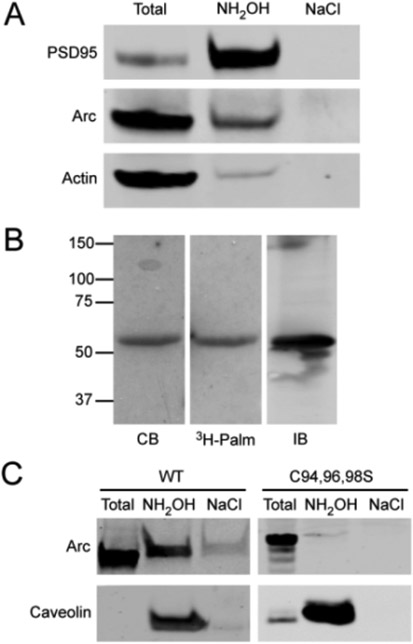
Figure 2.
Palmitoylation of Arc. (A) Palmitoylation of Arc in synaptosomes detected by Acyl-RAC (see Experimental Procedures in the Supporting Information). PSD95 and actin are shown as positive and negative controls. (B) Incorporation of [3H]palmitate into myc-Arc expressed in HeLa cells: lane 1, Coomassie blue (CB)-stained gel of the anti-myc immunoprecipitate; lane 2, corresponding autoradiogram; lane 3, immunoblot of the anti-Arc immunoprecipitate. (C) Palmitoylation of myc-ArcWT or ArcC94,96,98S expressed in HeLa cells determined by Acyl-RAC. Caveolin 1 was the positive control. “Total” represents 1/15 of the input.

