Abstract
目的
构建隐形矫治器远移下颌第一磨牙的有限元模型,探究是否使用微种植体支抗及不同第一磨牙起始位置进行远移时的牙列移动特点。
方法
使用锥形束CT(CBCT)数据构建下颌骨、牙齿、牙周膜及隐形矫治器模型,依据是否辅助微种植体弹性牵引分为无支抗组及微种植体组(第一磨牙与第二磨牙根间),在两组中以第一磨牙起始位置分为工况1:第一磨牙距第二前磨牙0 mm;工况2:第一磨牙距第二前磨牙1 mm,工况3:第一磨牙距第二前磨牙2 mm;工况4:第一磨牙距第二前磨牙3 mm。分析各工况牙列总体位移及各方向位移的数据特点。
结果
无支抗组:除第一磨牙向远中移动外,其余牙均表现为反向移动。微种植体组:除工况1中第二磨牙表现为少量近中移动,其余工况全牙列均表现为远中移动、前牙舌向移动,其中工况4第一磨牙远中位移值最大。随着第一磨牙起始位置向远中改变,第一磨牙向远中、前磨牙向近中及前牙向唇侧移动量增加,而第二磨牙向近中移动量减小。
结论
微种植体能够有效保护前牙支抗,增加磨牙远移的表达率,避免第二磨牙出现往返移动,且第一磨牙移动起始位置与其远移量及其余牙齿移动量有关。
Keywords: 有限元分析, 无托槽隐形矫治器, 磨牙远移, 微种植体支抗, 下颌骨
Abstract
Objective
This study aimed to construct the finite element model of the mandibular first molar with the invisible appliance and explore the dentition movement characteristics of the mandibular first molar when using micro-implant anchorage and different initial positions of the first molar.
Methods
Models of the mandible, tooth, periodontal membrane, and invisible appliance were constructed using cone beam computed tomography (CBCT) data. The two groups were divided into the non-anchorage group and the micro-implant group (between the roots of the first molar and the second molar) based on whether the elastic traction of the micro-implant was assisted or not. The two groups were divided into the following conditions based on the starting position of the first molar: Working condition 1: the distance between the first molar and the second premolar was 0 mm; working condition 2: the distance between the first molar and the second premolar was 1 mm; working condition 3: the distance between the first molar and the second premolar was 2 mm; working condition 4: the distance between the first molar and the second premolar was 3 mm. The data characteristics of total displacement and displacement in each direction of dentition were analyzed.
Results
In the non-anchorage group, all the other teeth showed reverse movement except for the first molar which was moved distally. Meanwhile, in the micro-implant group, except for a small amount of mesial movement of the second molar in working condition 1, the whole dentition in other working conditions presented distal movement and anterior teeth showed lingual movement, among which the distal displacement of the first molar in working condition 4 was the largest. With the change of the initial position of the first molar to the distal, the movement of the first molar to the distal, the premolar to the mesial, and the anterior to the lip increased, while the movement of the second molar to the mesial decreased.
Conclusion
The micro-implant can effectively protect the anterior anchorage, increase the expression rate of molar distancing, and avoid the round-trip movement of the second molar. The initial position of the first molar movement is related to the amount of distancing and the remaining tooth movement.
Keywords: finite element analysis, clear aligner, molar distalization, micro-implant anchorage, mandible
磨牙远移作为一种改善牙列拥挤的非拔牙矫治策略,能够增加牙弓长度,调整咬合关系。在使用传统矫治方法远移下颌磨牙时,被弯制的弓丝及牙面的托槽易造成食物嵌塞并引起不适,且由于舌体的存在,复杂的装置难以就位。随着计算机辅助设计、制造技术的发展,隐形矫治器得到广泛应用,其不仅具有舒适美观的优点,也在磨牙远移方面展现了出色的效率。目前已有临床研究证明,隐形矫治器远移上颌磨牙的效率能够达到86%以上[1]–[2],而对下颌磨牙远移效率仅为76%[3]。有限元分析法作为一种理论力学分析方法,将受力系统划分为有限个相互作用的单元,利用数学方法对真实物理系统进行模拟,使有限数量的未知量逼近真实值。自1980年Takahashi等[4]首次将有限元分析法应用于口腔正畸领域中,其已经成为研究生物力学的重要方法。目前国内外对于使用隐形矫治器远移磨牙的有限元分析,多为模拟第二磨牙远移时进行移动步距、矫治器膜片厚度、牵引方式、支抗设计等相关变量的改变[5]–[14],仍未有对第一磨牙远移的相关研究。因此本研究构建使用隐形矫治器远移下颌第一磨牙的有限元模型,模拟是否设置微种植体支抗牵引以及在不同起始位置进行远移,探究相关生物力学特点。
1. 材料和方法
1.1. 实验研究对象的选择及数据采集
选择一名就诊于吉林大学口腔医院正畸科的成年患者,患者牙列完整,牙齿大小形态正常,牙周状态健康。无外伤史,无系统性疾病史,无正畸治疗史。尖磨牙为安氏Ⅲ类咬合关系,下颌轻度拥挤,拟采用隐形矫治器进行下颌牙列远移治疗。患者行锥形束CT(cone beam computed tomography,CBCT)检测,获取DICOM格式原始文件数据。
1.2. 三维有限元模型的创建
将上述文件导入Mimics Medical 21.0(Materialise公司,比利时)中,通过设置合适的灰度值界限,分离下颌骨及各牙齿,去除孤立单元并填充孔隙,计算生成3D实体后输出为stl文件。将stl文件导入3-Matic 13.0(Materialise公司,比利时)中,使用光滑命令、封装命令、局部推拉命令对牙齿颌骨模型表面进行优化和修复。参照以往研究[15],以牙根外表面扩大0.25 mm创建牙周膜模型。为模拟下颌第一磨牙远移,预先将第二磨牙远中移动3.2 mm,并将第一磨牙以0.2 mm的步距移动至目标位,以牙冠外表面偏置0.75 mm后创建隐形矫治器模型。最终创建包括下颌骨、下颌牙齿、牙周膜及隐形矫治器在内的30个实体模型。组装隐形矫治器-牙齿-牙周膜-下颌骨模型,为实现交界面无缝连接,减少相互干涉,使用拓扑共享方式划分网格。为提高计算精度及适应模型的不规则形状,划分二阶四面体单元。模型共划分为1 164 810个单元,1 425 619个节点,将完成网格划分的模型导入ansys19.2(ANSYS公司,美国)。
1.3. 材料参数赋值及边界条件设定
现阶段大多数学者进行研究时将材料简化为均匀连续、各向同性的线弹性体,实验涉及的材料参数见表1。设定下颌骨髁突及下表面为固定约束,限制其各向自由度。牙根-牙周膜-下颌骨之间为面-面的绑定接触,牙冠外表面与矫治器内表面之间为摩擦接触,摩擦因数为0.2[16]。
表 1. 材料参数.
Tab 1 The parameter of materials
1.4. 模型分组及载荷施加
模型分为对照组(无支抗组)和实验组(微种植体组),微种植体定位于下颌第一磨牙与第二磨牙根间,距离牙槽嵴顶7 mm,向隐形矫治器尖牙牙位唇侧龈缘部位施加指向同侧微种植体1.8 N的牵引力。由于后续分析不包括微种植体的受力情况,因此仅用于施力方向的确定,后续模型将其省略。各组内以下颌第一磨牙起始位置进一步分为工况1~4,工况1:第一磨牙距第二前磨牙0 mm;工况2:第一磨牙距第二前磨牙1 mm;工况3:第一磨牙距第二前磨牙2 mm;工况4:第一磨牙距第二前磨牙3 mm(图1)。使用“两步法”进行载荷施加,第一步:将矫治器与磨牙远移后牙列进行组装,向36、46牙施加返回初始位置的位移约束,计算求得此时隐形矫治器的应力分布。第二步:将第一步求得的矫治器应力作为初始应力施加于矫治前牙列,以模拟隐形矫治器的初戴(图2)。
图 1. 研究分组情况示意图.
Fig 1 Schematic diagram of research groups
图 2. “两步法”示意图.
Fig 2 Diagram of the “two-step” method
1.5. 局部坐标系的设定
为右下颌牙齿建立局部坐标系(图3),坐标原点位于牙齿几何中心(质心),以牙体长轴定义为Z轴,Z轴正方向指向 方,X轴正方向指向近中,Y轴正方形指向舌侧。
图 3. 牙齿局部坐标系.

Fig 3 Local coordinate system of the teeth
2. 结果
由于下颌牙列具有左右两侧对称的特点,因此以右下颌牙齿为例进行分析。观察牙列总位移重叠图(图4)可发现,在无支抗组中,第一磨牙与其余牙齿表现为相反方向的移动趋势,第一磨牙为牙冠向远中移动,自牙冠至牙根其移动量逐渐减小,根尖区表现为近中移动。第二磨牙与前磨牙移动趋势相似,均为牙冠向近中倾斜,牙根移动量可忽略不计。而尖牙和切牙则表现为牙冠向唇侧、向近中的倾斜移动。牙列位移最大值位于第一磨牙牙尖,工况1至工况4分别为9.19e−2、9.32e−2、1.00e−2、1.03e−2 mm。在微种植体组中,第一磨牙仍表现为牙冠向远中、牙根向近中的移动趋势,其移动量较无支抗组均有所增加。而第二磨牙牙冠及牙根移动量均接近0,部分工况中表现为微小远中移动。前磨牙区牙冠向远中、 向倾斜移动,尖牙和切牙则表现为牙冠向远中、舌向倾斜伴有压低的移动趋势。牙列位移最大值仍位于第一磨牙牙尖,工况1至工况4分别为1.27e−2、1.40e−2、1.50e−2、1.54e−2 mm。
图 4. 牙列总位移重叠图.
Fig 4 The overlapping graph of tooth movement
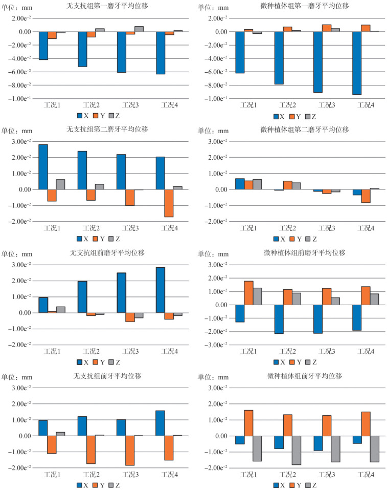
为了进一步量化分析,本研究依据牙齿局部坐标系计算单颗牙齿各方向位移平均值,代表牙齿移动的整体趋势,并绘制下列直方图(图5)。对于第一磨牙,X方向观,两组自工况1至工况4其向远中的移动量依次增加,微种植体支抗显著增加了牙体向远中的整体移动量,增加量自工况1至工况4递增,分别为2.02e−2、2.62e−2、3.00e−2、3.06e−2 mm。Y方向观:无支抗组牙体颊向的移动量依次减小,而微种植体组中表现为舌向移动,并依次增加。Z方向观:两组中各工况未表现出垂直向上变化的明显规律。对于第二磨牙,X方向观:无支抗组其近中移动量依次减小,微种植体组除工况1外,均表现为牙体向远中移动,且远中移动量依次增加。其X方向位移改变量自工况1至工况4分别为2.13e−2、2.44e−2、2.29e−2、2.39e−2 mm。Y方向观,无支抗组各工况及微种植体组中工况3、工况4表现为颊向位移,微种植体组中工况1、工况2为舌向移动。Z方向观,两组中除工况3外,其余工况均为少量 方移动。对于前磨牙:X方向观,无支抗组其近中移动量依次增加,而在微种植体组中,各工况均表现为向远中移动,位移改变量自工况1至工况4分别为2.25e−2、4.12e−2、4.61e−2、4.73e−2 mm。Y方向观:无支抗组除工况1外,均表现为颊向少量移动,而微种植体组中各工况表现为舌向移动。Z方向观:无支抗组工况1及微种植体组各工况表现为伸长移动,而无支抗组工况2至工况4表现为压低移动。对于前牙,无支抗组各工况表现为近中、唇向移动伴有少量伸长移动,而微种植体组中各工况表现为远中、舌侧伴有压低移动。其Y方向位移改变量分别为1.70e−2、1.34e−2、1.80e−2、1.77e−2 mm。
图 5. 牙齿各方向位移直方图.
Fig 5 Histogram of tooth displacement in all directions
3. 讨论
3.1. 下颌磨牙远移的可能性及界限
在临床工作中,能否采用下颌磨牙远移策略进行治疗取决于拥挤量、安氏Ⅲ类错 程度与磨牙后间隙之间的关系。对于成年患者,第二磨牙牙冠远移的界限要从牙冠、牙根两个层面进行观察。牙冠的移动界限通常位于下颌升支前缘,而牙根的移动界限为根尖舌侧的骨皮质,牙冠远中间隙通常大于牙根远中间隙,部分患者甚至存在根尖与下颌舌侧骨皮质接触的现象[23]–[24]。在本例中,下颌第二磨牙的预期远移量为3.2 mm,由于患者下颌轻度拥挤,磨牙关系为开始近中关系,下颌第三磨牙近中阻生,拔除后具有足够的空间进行磨牙远移。研究[25]发现下颌第三磨牙正常萌出及阻生时,其第二磨牙远中间隙均大于无第三磨牙组。对于青少年患者,其下颌体长度的生长发育与下颌升支前缘的改建性骨吸收增加了牙弓的长度。Benjamin Cory[26]追踪了375名8~16岁青少年的侧位片,发现8~14岁期间,下颌第一磨牙远中至下颌升支前缘及下颌升支后缘的间距分别增加了7.7和6.6 mm,而14岁后的生长量仅为2.5和2.3 mm。Richardson[27]在研究中也发现相似规律。近年来,下颌矢状骨面型、垂直生长型、下颌第三磨牙发育程度与磨牙后间隙之间的关系引起了研究者们的关注。安氏Ⅲ类患者的磨牙后间隙通常大于安氏Ⅰ类及安氏Ⅱ类[28],且随着下颌平面角度的增大,磨牙后间隙具有减小的趋势[29],高角型患者通常具有磨牙后间隙不足的特点,容易出现根尖与骨皮质的接触现象。而第三磨牙的发育程度与磨牙后间隙呈正相关[30],磨牙后间隙每增加5 mm,其Nolla分期指数增加1.8[31]。
3.2. 下颌磨牙远移模式及效率
隐形矫治器常规使用序列移动方式,即先远移双侧第二磨牙,待间隙出现后再依次移动第一磨牙,第二、第一前磨牙,最后内收前牙,牙齿移动分步方案呈典型“V”字型。依据同时移动的相邻牙齿数量,分为“密集型”和“疏松型”。前者同时远移的牙齿数量多,移动方案步骤少,但对支抗要求高。而同时移动的牙齿数量越多,对牙齿的控制也越难。后者同时移动的牙齿数量少,远移的实现率更高,但治疗周期长。本研究模拟了单独远移第一磨牙,发现第二磨牙存在不同程度的近中移动趋势,且第一磨牙起始位置越靠近远中,第二磨牙近中移动量越小,前磨牙、前牙近中、唇向移动量越大。这说明其余牙齿受到第一磨牙远移时的反作用力,其余牙齿的移动量与第一磨牙所处位置有关,使用微种植体支抗进行辅助则可以明显减少其余牙齿的不利移动。以第一磨牙远中位移量最大值与设计移动量的比值计算移动效率可以得出:无支抗组中工况一至工况四第一磨牙移动效率分别为45.95%、46.60%、50.00%、50.15%,微种植体组中分别为63.50%、70.00%、75.00%、77.00%。两组第一磨牙远中移动效率自工况1至工况4均表现递增趋势,使用微种植体增加了第一磨牙的远中移动效率。有限元分析模拟的是系统受力后发生形变,使力趋于平衡时的即刻位移,而实际的牙齿移动为牙槽骨受压进行生理性骨改建的过程,本研究计算得出的移动效率可以用于分析不同工况下的变化规律,并不能代表实际的牙齿移动效率。有学者[3]采用无托槽隐形矫治器对下颌轻中度拥挤患者进行推下颌磨牙向远中的治疗,在未使用种植支抗、三类牵引且未设计磨牙压低的情况下,下颌第一、二磨牙冠、根远移有效率分别达到71%、47%和74%、49%,下颌磨牙远移后伴有1.06 mm少量压低,这是隐形矫治器“ 垫效应”的结果,但同时也出现了切牙的唇倾。在临床中应依据患者实际情况实现治疗周期与牙移动表达率的平衡,对于牙冠短小、存在形态缺损且骨质致密的患者,减少同时移动的牙齿数量,设置
 间牵引或微种植体支抗能够减少牙齿间相互作用导致的不利的往返运动。几项研究[32]–[34]发现在使用固定矫治器进行下颌磨牙远移时,
 间Ⅲ类牵引导致咬合平面顺时针旋转,下颌平面角增大,而支抗钉牵引则使咬合平面逆旋,下颌平面角减小,在隐形矫治中是否遵循同样的规律仍有待探讨。需要注意的是,下颌骨颏结节与下颌支前缘间存在一条隆起的骨嵴,即外斜线。相关研究[35]测量发现第一、二磨牙根间颊侧,距离牙槽嵴顶9 mm区域骨皮质厚度为3.23 mm±0.62 mm。下颌外斜线区域牙根颊侧骨皮质以及下颌牙槽骨宽度具有自近中向远中、自
 方向龈方逐渐增大的特点[36]–[37],且颊侧骨皮质平均厚度大于舌侧骨皮质平均厚度[38],在下颌磨牙远移的临床实践中可能出现牙冠舌倾的现象,因此可参照患者CBCT数据预先设计一定的下颌第二磨牙区域扩弓量,以抵消上述不利效应。
3.3. 微种植体支抗的植入部位
微种植体植入后影响其稳定性的因素包括:根间距大小、骨皮质厚度、软组织活动度以及植入角度[39]。临床上进行下颌磨牙远移时,可供选择的微种植体植入部位包括下颌根间区及颊棚区[40]。下颌骨颊侧皮质骨的厚度及密度具有自近中向远中、自牙槽嵴顶至根尖方向逐渐增大的特点[41]。且植入部位越靠近远中,牵引力矢状向分力增大,但口腔黏膜厚度、活动度也增大,容易引起黏膜发炎。本研究中,为尽量增大微种植体支抗弹性牵引的矢状向分力,模拟将微种植体植入于第一磨牙、第二磨牙根间。微种植体组中各工况前牙及前磨牙区支抗得到有效保护,由唇向、近中移动变为舌向、远中移动。第一、第二磨牙远中移动量增加,同时出现少量舌向移动。但研究发现,施加自微种植体相同大小的牵引力后,第一磨牙向远中位移的增加量自工况1至工况4递增,分别为−2.02e−2、−2.62e−2、−3.00e−2、−3.06e−2 mm。但向远中位移的改变量自前磨牙、第一磨牙至第二磨牙递减,如工况2中,微种植体组较无支抗组前磨牙、第一、第二磨牙向远中的位移改变量依次为4.12e−2、2.62e−2、2.44e−2 mm。这说明施加在矫治器尖牙位置的牵引力不能完全传导至第二磨牙,第一磨牙与其邻牙间的矫治器膜片存在形变,这样的形变导致对其远中牙齿的力量衰减。在微种植体组工况1中,第二磨牙仍表现为少量近中移动。多项研究[39],[42]推荐选择第一磨牙与第二磨牙根间作为理想植入部位[43],在第一磨牙与第二磨牙根间距离牙槽嵴顶6~8 mm,以10°~30°的角度植入微种植体能够最大程度避免其与牙根接触。对于磨牙分布远移的隐形矫治患者,可在第二磨牙向远中移动到位后植入支抗钉,既能保持第二磨牙远移效果,又可提供剩余牙远移的支抗力量。下颌颊棚区远离牙根和下颌神经管,不会干扰磨牙远移。颊棚区骨皮质厚度与ANB角呈正相关,与下颌平面角度呈负相关[44]。植入部位越靠近第一磨牙,颊棚区越陡峭,植入时容易滑脱损伤黏膜;而植入部位越靠近第二磨牙,颊棚区越平坦,越容易进行植入[45]。但随着植入支抗钉部位向远中移动,会受到患者张口度、前庭沟深度及颊肌张力等因素的制约,操作难度增大[46]。
Funding Statement
[基金项目] 吉林省自然科学基金(YDZJ202201ZYTS057);吉林省科技厅科技发展计划项目(20210203064SF)
Supported by: Natural Science Foundation of Jilin Province (YDZJ202201ZYTS057); Jilin Scientific and Technological Development Program (20210203064SF).
Footnotes
利益冲突声明:作者声明本文无利益冲突。
References
- 1.任 亚男, 宋 保龙, 封 颖丽, et al. 无托槽隐形矫治器结合微种植体支抗远移上颌磨牙效率的三维重叠研究[J] 中华口腔正畸学杂志. 2018;25(2):92–97. [Google Scholar]; Ren YN, Song BL, Feng YL, et al. Three-dimensional model superimposition study on efficiency of molar distalization by using removable clear appliance combined with micro-implant anchorage[J] Chin J Orthod. 2018;25(2):92–97. [Google Scholar]
- 2.周 洁珉, 白 玉兴, 郝 玮首. 无托槽隐形矫治技术远中移动磨牙效果的三维分析与评价[J] 北京口腔医学. 2011;19(3):157–159. [Google Scholar]; Zhou JM, Bai YX, Hao WS, et al. Three dimensional evaluation of maxillary molar distal movement using invisible bracketless appliance[J] Beijing J Stomatol. 2011;19(3):157–159. [Google Scholar]
- 3.吴 冬雪, 赵 云山, 马 萌, et al. 无托槽隐形矫治器远移下颌磨牙的疗效[J] 中南大学学报: 医学版. 2021;46(10):1114–1121. doi: 10.11817/j.issn.1672-7347.2021.200391. [DOI] [PMC free article] [PubMed] [Google Scholar]; Wu DX, Zhao YS, Ma M, et al. Efficacy of mandibular molar distalization by clear aligner treatment[J] J Cent South Univ: Med Sci. 2021;46(10):1114–1121. doi: 10.11817/j.issn.1672-7347.2021.200391. [DOI] [PMC free article] [PubMed] [Google Scholar]
- 4.Takahashi N, Kitagami T, Komori T. Behaviour of teeth under various loading conditions with finite element method[J] J Oral Rehabil. 1980;7(6):453–461. doi: 10.1111/j.1365-2842.1980.tb00464.x. [DOI] [PubMed] [Google Scholar]
- 5.Jia L, Wang C, Li L, et al. The effects of lingual buttons, precision cuts, and patient-specific attachments during maxillary molar distalization with clear aligners: comparison of finite element analysis[J] Am J Orthod Dentofacial Orthop. 2023;163(1):e1–e12. doi: 10.1016/j.ajodo.2022.10.010. [DOI] [PubMed] [Google Scholar]
- 6.Liu X, Cheng Y, Qin W, et al. Effects of upper-molar distalization using clear aligners in combination with Class Ⅱelastics: a three-dimensional finite element analysis[J] BMC Oral Health. 2022;22(1):546. doi: 10.1186/s12903-022-02526-2. [DOI] [PMC free article] [PubMed] [Google Scholar]
- 7.Fan D, Liu H, Yuan CY, et al. Effectiveness of the attachment position in molar intrusion with clear aligners: a finite element study[J] BMC Oral Health. 2022;22(1):474. doi: 10.1186/s12903-022-02472-z. [DOI] [PMC free article] [PubMed] [Google Scholar]
- 8.Wang Q, Dai D, Wang J, et al. Biomechanical analysis of effective mandibular en-masse retraction using Class Ⅱ elastics with a clear aligner: a finite element study[J] Prog Orthod. 2022;23(1):23. doi: 10.1186/s40510-022-00417-4. [DOI] [PMC free article] [PubMed] [Google Scholar]
- 9.Ji L, Li B, Wu X. Evaluation of biomechanics using different traction devices in distalization of maxillary molar with clear aligners: a finite element study[J] Comput Methods Biomech Biomed Engin. 2023;26(5):559–567. doi: 10.1080/10255842.2022.2073789. [DOI] [PubMed] [Google Scholar]
- 10.Ayidağa C, Kamiloğlu B. Effects of variable composite attachment shapes in controlling upper molar distalization with aligners: a nonlinear finite element study[J] J Healthc Eng. 2021;2021:5557483. doi: 10.1155/2021/5557483. [DOI] [PMC free article] [PubMed] [Google Scholar]
- 11.余 箐, 赵 刚, 叶 之慧, et al. 不同弹性模量的热压膜材料远移上颌磨牙的有限元分析[J] 实用口腔医学杂志. 2022;38(4):450–454. [Google Scholar]; Yu J, Zhao G, Ye ZH, et al. Distalization of maxillary molars by hot pressed membrane materials with different elastic modulus: a finite element analysis[J] J Pract Stomatol. 2022;38(4):450–454. [Google Scholar]
- 12.雷 鹤, 高 冲, 吴 冬雪, et al. 隐形矫治磨牙远移的三维有限元分析[J] 口腔医学. 2021;41(8):692–698. [Google Scholar]; Lei H, Gao C, Wu DX, et al. Three dimensional-finite element analysis of molar distalization with clear aligners[J] Stomatology. 2021;41(8):692–698. [Google Scholar]
- 13.向 彪, 徐 逸晨, 王 梦含, et al. 无托槽隐形矫治矩形附件对上颌磨牙远中移动力学影响的三维有限元分析[J] 临床口腔医学杂志. 2021;37(2):78–82. [Google Scholar]; Xiang B, Xu YC, Wang MH, et al. Finite element analysis on the maxillary molar distal movement with rectangular attachment bonded clear aligner[J] J Clin Stomatol. 2021;37(2):78–82. [Google Scholar]
- 14.迟 敬文, 王 玉俏, 冯 枫, et al. 隐形矫治与固定矫治技术远移上颌磨牙的三维有限元分析[J] 实用口腔医学杂志. 2020;36(1):100–104. [Google Scholar]; Chi JW, Wang YX, Feng F, et al. Three dimensional finite element analysis of maxillary molar distalization by invisible appliance and fixed appliance[J] J Pract Stomatol. 2020;36(1):100–104. [Google Scholar]
- 15.Seo JH, Eghan-Acquah E, Kim MS, et al. Comparative analysis of stress in the periodontal ligament and center of rotation in the tooth after orthodontic treatment depending on clear aligner thickness-finite element analysis study[J] Materials (Basel) 2021;14(2):324. doi: 10.3390/ma14020324. [DOI] [PMC free article] [PubMed] [Google Scholar]
- 16.施 则安, 夏 恺, 罗 良语, et al. 无托槽隐形矫治器联合微种植体内收并压低上前牙的三维有限元分析[J] 华西口腔医学杂志. 2022;40(5):589–596. doi: 10.7518/hxkq.2022.05.013. [DOI] [PMC free article] [PubMed] [Google Scholar]; Shi ZA, Xia K, Luo LY, et al. Three-dimensional finite element analysis of upper anterior teeth retraction and intrusion using clear aligners and mini-implants[J] West China J Stomatol. 2022;40(5):589–596. doi: 10.7518/hxkq.2022.05.013. [DOI] [PMC free article] [PubMed] [Google Scholar]
- 17.Liu L, Song Q, Zhou J, et al. The effects of aligner overtreatment on torque control and intrusion of incisors for anterior retraction with clear aligners: a finite-element study[J] Am J Orthod Dentofacial Orthop. 2022;162(1):33–41. doi: 10.1016/j.ajodo.2021.02.020. [DOI] [PubMed] [Google Scholar]
- 18.Cortona A, Rossini G, Parrini S, et al. Clear aligner orthodontic therapy of rotated mandibular round-shaped teeth: a finite element study[J] Angle Orthod. 2020;90(2):247–254. doi: 10.2319/020719-86.1. [DOI] [PMC free article] [PubMed] [Google Scholar]
- 19.Hong K, Kim WH, Eghan-Acquah E, et al. Efficient design of a clear aligner attachment to induce bodily tooth movement in orthodontic treatment using finite element analysis[J] Materials (Basel) 2021;14(17):4926. doi: 10.3390/ma14174926. [DOI] [PMC free article] [PubMed] [Google Scholar]
- 20.Liu L, Zhan Q, Zhou J, et al. Effectiveness of an anterior mini-screw in achieving incisor intrusion and palatal root torque for anterior retraction with clear aligners[J] Angle Orthod. 2021;91(6):794–803. doi: 10.2319/120420-982.1. [DOI] [PMC free article] [PubMed] [Google Scholar]
- 21.Ma Y, Li S. The optimal orthodontic displacement of clear aligner for mild, moderate and severe periodontal conditions: an in vitro study in a periodontally compromised individual using the finite element model[J] BMC Oral Health. 2021;21(1):109. doi: 10.1186/s12903-021-01474-7. [DOI] [PMC free article] [PubMed] [Google Scholar]
- 22.Jiang T, Wu RY, Wang JK, et al. Clear aligners for maxillary anterior en masse retraction: a 3D finite element study[J] Sci Rep. 2020;10(1):10156. doi: 10.1038/s41598-020-67273-2. [DOI] [PMC free article] [PubMed] [Google Scholar]
- 23.Kim SH, Cha KS, Lee JW, et al. Mandibular skeletal posterior anatomic limit for molar distalization in patients with Class Ⅲ malocclusion with different vertical facial patterns[J] Korean J Orthod. 2021;51(4):250–259. doi: 10.4041/kjod.2021.51.4.250. [DOI] [PMC free article] [PubMed] [Google Scholar]
- 24.Kim SJ, Choi TH, Baik HS, et al. Mandibular posterior anatomic limit for molar distalization[J] Am J Orthod Dentofacial Orthop. 2014;146(2):190–197. doi: 10.1016/j.ajodo.2014.04.021. [DOI] [PubMed] [Google Scholar]
- 25.郭 学强, 刘 新强, 王 铮, et al. 成人下颌磨牙后间隙与第三磨牙关系的三维研究[J] 中华口腔正畸学杂志. 2021;28(2):74–79. [Google Scholar]; Guo XQ, Liu XQ, Wang Z, et al. Association between adult mandibular third molar and retromolar space: a three-dimensional study[J] China J Orthod. 2021;28(2):74–79. [Google Scholar]
- 26.Benjamin Cory L. A study of the mandibular third molar area[J] Am J Orthod. 1953;39(5):366–373. [Google Scholar]
- 27.Richardson ME. Development of the lower third molar from 10 to 15 years[J] Angle Orthod. 1973;43(2):191–193. doi: 10.1043/0003-3219(1973)043<0191:DOTLTM>2.0.CO;2. [DOI] [PubMed] [Google Scholar]
- 28.Fan Z, Zhang Q, Jiang Y, et al. Mandibular retromolar space in adults with different sagittal skeletal patterns[J] Angle Orthod. 2022;92(5):606–612. doi: 10.2319/112021-854.1. [DOI] [PMC free article] [PubMed] [Google Scholar]
- 29.Zhao Z, Wang Q, Yi P, et al. Quantitative evaluation of retromolar space in adults with different vertical facial types[J] Angle Orthod. 2020;90(6):857–865. doi: 10.2319/121219-787.1. [DOI] [PMC free article] [PubMed] [Google Scholar]
- 30.Marchiori DF, Packota GV, Boughner JC. Third-molar mineralization as a function of available retromolar space[J] Acta Odontol Scand. 2016;74(7):509–517. doi: 10.1080/00016357.2016.1209240. [DOI] [PubMed] [Google Scholar]
- 31.Ghougassian SS, Ghafari JG. Association between mandibular third molar formation and retromolar space[J] Angle Orthod. 2014;84(6):946–950. doi: 10.2319/120113-883.1. [DOI] [PMC free article] [PubMed] [Google Scholar]
- 32.Nakamura M, Kawanabe N, Kataoka T, et al. Comparative evaluation of treatment outcomes between temporary anchorage devices and Class Ⅲ elastics in Class Ⅲ malocclusions[J] Am J Orthod Dentofacial Orthop. 2017;151(6):1116–1124. doi: 10.1016/j.ajodo.2016.10.040. [DOI] [PubMed] [Google Scholar]
- 33.He S, Gao J, Wamalwa P, et al. Camouflage treatment of skeletal Class Ⅲ malocclusion with multiloop edgewise arch wire and modified Class Ⅲ elastics by maxillary mini-implant anchorage[J] Angle Orthod. 2013;83(4):630–640. doi: 10.2319/091312-730.1. [DOI] [PMC free article] [PubMed] [Google Scholar]
- 34.Roberts WE, Viecilli RF, Chang C, et al. Biology of biomechanics: finite element analysis of a statically determinate system to rotate the occlusal plane for correction of a skeletal Class Ⅲ open-bite malocclusion[J] Am J Orthod Dentofacial Orthop. 2015;148(6):943–955. doi: 10.1016/j.ajodo.2015.10.002. [DOI] [PubMed] [Google Scholar]
- 35.穆 荣荣, 张 丹, 张 扬. 下颌后牙区颊侧骨皮质厚度及牙槽宽度的CBCT测量分析[J] 口腔医学. 2014;34(8):604–607. [Google Scholar]; Mu RR, Zhang D, Zhang Y. Cortical bone thickness in the buccal posterior region and bone depth of mandible: a CBCT analysis[J] Stomatology. 2014;34(8):604–607. [Google Scholar]
- 36.朱 恬尔. 不同块状自体骨上置法骨增量术在口腔种植修复中的应用[D] 杭州: 浙江大学; 2020. [Google Scholar]; Zhu TE. Application of different onlay block autogenous bone graftingin oral implant prosthesis[D] Hangzhou: Zhejiang University; 2020. [Google Scholar]
- 37.吴 晓雪. 下颌骨外斜线区微种植钉远移下牙列的研究[D] 西安: 空军军医大学; 2018. [Google Scholar]; Wu XX. A study on distalization of the lower dentition by using miniscrews in the external oblique area[D] Xi'an: Air Force Medical University; 2018. [Google Scholar]
- 38.韩 敏. 不同生长型人群下颌磨牙区牙槽骨形态的CBCT研究[D] 济南: 山东大学; 2013. [Google Scholar]; Han M. Association between mandibular posterior alveolar morphology and growth pattern: a CBCT study[D] Jinan: Shandong University; 2013. [DOI] [PMC free article] [PubMed] [Google Scholar]
- 39.Nucera R, Lo Giudice A, Bellocchio AM, et al. Bone and cortical bone thickness of mandibular buccal shelf for mini-screw insertion in adults[J] Angle Orthod. 2017;87(5):745–751. doi: 10.2319/011117-34.1. [DOI] [PMC free article] [PubMed] [Google Scholar]
- 40.范 星星, 赵 桂芝, 柯 杰. 下颌第二磨牙远中移动的研究进展[J] 2014;22(2):119–120. [Google Scholar]; Fan XX, Zhao GZ, Ke J. Progress in the study of distal movement of mandibular second molars[J] Beijing J Stomatol. 2014;22(2):119–120. [Google Scholar]
- 41.Liu H, Wu X, Tan J, et al. Safe regions of miniscrew implantation for distalization of mandibular dentition with CBCT[J] Prog Orthod. 2019;20(1):45. doi: 10.1186/s40510-019-0297-6. [DOI] [PMC free article] [PubMed] [Google Scholar]
- 42.Farnsworth D, Rossouw PE, Ceen RF, et al. Cortical bone thickness at common miniscrew implant placement sites[J] Am J Orthod Dentofacial Orthop. 2011;139(4):495–503. doi: 10.1016/j.ajodo.2009.03.057. [DOI] [PubMed] [Google Scholar]
- 43.Wang Y, Sun J, Shi Y, et al. Buccal bone thickness of posterior mandible for microscrews implantation in molar distalization[J] Ann Anat. 2022;244:151993. doi: 10.1016/j.aanat.2022.151993. [DOI] [PubMed] [Google Scholar]
- 44.吴 瑶, 徐 依山, 李 真真, et al. 不同骨面型下颌颊棚区骨量的特征分析[J] 口腔医学研究. 2022;38(4):362–366. [Google Scholar]; Wu Y, Xu YS, Li ZZ, et al. Characteristics analysis of bone quantity of mandibular buccal shelf with different skeletal pattern[J] J Oral Sci Res. 2022;38(4):362–366. [Google Scholar]
- 45.张 瑞, 陈 星, 黄 晓峰. 200例口腔患者下颌颊棚区骨斜面坡度的锥形束CT研究[J] 中华口腔正畸学杂志. 2020;27(3):121–124. [Google Scholar]; Zhang R, Chen X, Huang XF. The Cone-Beam CT study on the slope of oblique plane at the mandibular buccal shelf area in 200 cases[J] Chin J Orthod. 2020;27(3):121–124. [Google Scholar]
-
46.李 俊. 下颌磨牙根颊侧微种植体支抗钉在骨性Ⅲ类错
 畸形患者全牙列远移中的应用效果[J] 中国民康医学. 2021;33(24):75–77. [Google Scholar]; Li J. Application effects of buccal micro-implant anchorage nails of mandibular molar roots in whole dentition distalization of patients with skeletal class Ⅲ malocclusion[J] Med J Chin People's Health. 2021;33(24):75–77. [Google Scholar]


