Abstract
Background
Venous leg ulcers (VLUs) are a serious manifestation of chronic venous disease affecting up to 3% of the adult population. This typically recalcitrant and recurring condition significantly impairs quality of life, and its treatment places a heavy financial burden upon healthcare systems. The longstanding mainstay treatment for VLUs is compression therapy. Surgical removal of incompetent veins reduces the risk of ulcer recurrence. However, open surgery is an unpopular option amongst people with VLU, and many people are unsuitable for it. The efficacy of the newer, minimally‐invasive endovenous techniques has been established in uncomplicated superficial venous disease, and these techniques can also be used in the management of VLU. When used with compression, endovenous ablation aims to further reduce pressure in the veins of the leg, which may impact ulcer healing.
Objectives
To determine the effects of superficial endovenous ablation on the healing and recurrence of venous leg ulcers and the quality of life of people with venous ulcer disease.
Search methods
In April 2022 we searched the Cochrane Wounds Specialised Register; the Cochrane Central Register of Controlled Trials (CENTRAL); Ovid MEDLINE (including In‐Process & Other Non‐Indexed Citations); Ovid Embase and EBSCO CINAHL Plus. We also searched clinical trials registries for ongoing and unpublished studies, and scrutinised reference lists of relevant included studies as well as reviews, meta‐analyses and health technology reports to identify additional studies. There were no restrictions on the language of publication, but there was a restriction on publication year from 1998 to April 2022 as superficial endovenous ablation is a comparatively new technology.
Selection criteria
Randomised controlled trials (RCTs) comparing endovenous ablative techniques with compression versus compression therapy alone for the treatment of VLU were eligible for inclusion. Studies needed to have assessed at least one of the following primary review outcomes related to objective measures of ulcer healing such as: proportion of ulcers healed at a given time point; time to complete healing; change in ulcer size; proportion of ulcers recurring over a given time period or at a specific point; or ulcer‐free days. Secondary outcomes of interest were patient‐reported quality of life, economic data and adverse events.
Data collection and analysis
Two reviewers independently assessed studies for eligibility, extracted data, carried out risk of bias assessment using the Cochrane RoB 1 tool, and assessed GRADE certainty of evidence.
Main results
The previous version of this review found no RCTs meeting the inclusion criteria. In this update, we identified two eligible RCTs and included them in a meta‐analysis. There was a total of 506 participants with an active VLU, with mean durations of 3.1 months ± 1.1 months in the EVRA trial and 60.5 months ± 96.4 months in the VUERT trial. Both trials randomised participants to endovenous treatment and compression or compression alone, however the compression alone group in the EVRA trial received deferred endovenous treatment (after ulcer healing or from six months).
There is high‐certainty evidence that combined endovenous ablation and compression compared with compression therapy alone, or compression with deferred endovenous treatment, improves time to complete ulcer healing (pooled hazard ratio (HR) 1.41, 95% CI 1.36 to 1.47; I2 = 0%; 2 studies, 466 participants). There is moderate‐certainty evidence that the proportion of ulcers healed at 90 days is probably higher with combined endovenous ablation and compression compared with compression therapy alone or compression with deferred endovenous treatment (risk ratio (RR) 1.14, 95% CI 1.00 to 1.30; I2 = 0%; 2 studies, 466 participants). There is low‐certainty evidence showing an unclear effect on ulcer recurrence at one year in people with healed ulcers with combined endovenous treatment and compression when compared with compression alone or compression with deferred endovenous treatment (RR 0.29, 95% CI 0.03 to 2.48; I2 = 78%; 2 studies, 460 participants). There is also low‐certainty evidence that the median number of ulcer‐free days at one year may not differ (306 (interquartile range (IQR) 240 to 328) days versus 278 (IQR 175 to 324) days) following combined endovenous treatment and compression when compared with compression and deferred endovenous treatment; (1 study, 450 participants).
There is low‐certainty evidence of an unclear effect in rates of thromboembolism between groups (RR 2.02, 95% CI 0.51 to 7.97; I2 = 78%, 2 studies, 506 participants). The addition of endovenous ablation to compression is probably cost‐effective at one year (99% probability at GBP 20,000/QALY; 1 study; moderate‐certainty evidence).
Authors' conclusions
Endovenous ablation of superficial venous incompetence in combination with compression improves leg ulcer healing when compared with compression alone. This conclusion is based on high‐certainty evidence. There is moderate‐certainty evidence to suggest that it is probably cost‐effective at one year and low certainty evidence of unclear effects on recurrence and complications. Further research is needed to explore the additional benefit of endovenous ablation in ulcers of greater than six months duration and the optimal modality of endovenous ablation.
Keywords: Adult; Humans; Leg Ulcer; Neoplasm Recurrence, Local; Varicose Ulcer; Varicose Ulcer/surgery; Veins; Wound Healing
Plain language summary
What are the benefits and risks of endovenous ablation (using heat or medication to permanently close veins) for treating venous leg ulcers
Key messages
Compared with compression treatment alone, or compression with endovenous treatment (but delayed until after an ulcer has healed):
‐ endovenous ablation and compression together reduce time to complete ulcer healing;
‐ endovenous treatment and compression together probably increase the proportion of ulcers healed at 90 days;
‐ combined endovenous treatment and compression may not change ulcer recurrence or the number of ulcer‐free days at one year.
Further research is needed into:
‐ the effects of endovenous ablation for chronic (long‐term) ulcers;
‐ how effective different endovenous ablation techniques are.
What are venous leg ulcers?
Venous leg ulcers (VLUs) are open sores below the knee that are caused by chronic venous disease.
Veins in the calf normally pump blood back towards the heart. Valves in these veins prevent the blood from flowing back down the leg. If these valves fail, pressure can increase in the leg, which can lead to inflammation, skin breakdown and ulcer formation.
Venous leg ulcers take a long time to heal and often reoccur. This significantly impairs quality of life, and treating VLUs places a heavy financial burden upon healthcare systems.
How are venous leg ulcers treated?
Venous leg ulcers are usually treated with compression therapy. This involves wearing socks or stockings that help to increase blood flow in the legs.
Problematic veins can also be surgically removed. However, this is not a popular option amongst patients, and many people are not able to have this kind of surgery due to other medical problems.
Endovenous ablation is a newer, minimally‐invasive technique that treats venous disease by blocking certain veins. Medication is injected into the vein, or a device is inserted into the vein and then heated up to permanently close it.
Endovenous ablation combined with compression therapy aims to further reduce pressure in the veins of the leg, which may impact ulcer healing.
What did we want to find out?
We wanted to find out if endovenous ablation combined with compression therapy:
‐ helps to heal VLUs;
‐ prevents VLUs from reoccurring;
‐ improves quality of life;
‐ is cost‐effective;
‐ was associated with any unwanted effects.
What did we do? We searched for studies that looked at endovenous ablation with compression therapy versus compression therapy alone for treating VLUs. There were no restrictions on the publication language.
We compared and summarised the studies’ results and rated our confidence in the evidence, based on factors such as study methods and sizes.
What did we find?
In our original review we did not find any eligible studies. In this update we found two new relevant studies. We found one UK study with 450 participants and one Brazilian study with 56 participants.
Compared with compression therapy alone, or compression with deferred endovenous treatment:
‐ combined endovenous ablation and compression improves time to complete ulcer healing;
‐ combined endovenous treatment and compression probably improves the proportion of ulcers healed at 90 days;
‐ combined endovenous treatment and compression may not have an effect on ulcer recurrence at one year or the number of ulcer‐free days at one year;
‐ combined endovenous treatment and compression is probably cost‐effective at one year;
‐ combined endovenous treatment and compression may not have an effect on blood clots (venous thromboembolism).
What are the limitations of the evidence?
The majority of the participants had an ulcer for less than six months. Although the results were similar in those with longer‐term ulcers, further research is required to confirm this.
This review did not find any studies which assessed whether some techniques of endovenous ablation are more effective than others for people with venous leg ulcers.
How up to date is this evidence?
This review updates our previous review. The evidence is up to date to April 2022.
Summary of findings
Summary of findings 1. Summary of findings: endovenous ablation and compression vs compression alone for venous leg ulcers.
| Endovenous ablation of superficial venous incompetence and compression compared with compression alone for venous leg ulcers | ||||||
|
Patient or population: adults with venous leg ulcers. Settings: outpatient hospital care and/or in community care Intervention: endovenous ablation of superficial venous incompetence with standard wound care that includes compression Comparison: standard wound care that includes compression | ||||||
| Outcomes | Illustrative comparative risk* (95% CI) | Relative effect (95% CI) | No of Participants (studies) | Quality of the evidence (GRADE) | Comments | |
| Risk with standard wound care |
Risk with endovenous ablation and standard wound care |
|||||
|
Ulcer healing time (measured as days from randomisation and followed‐up to 1 year) |
78 days | 56 days (47.95 to 64.05) |
HR 1.41 (1.36 to 1.47) | 466 (2 RCTs) |
⊕⊕⊕⊕ High | Risk of performance bias in both included studies was high as the nature of the interventions in the treatment groups meant that neither participants nor treating clinicians were blinded at the time treatments were performed. Blinding was not possible in these studies. The outcome was measured by blinded assessors. The outcomes in the control arms were equal to or better than those reported in the independent literature. This increases our confidence that the true effect lies close to that of the estimate of the effect, so we did not downgrade the certainty of evidence. |
|
Proportion of ulcers healed at 90 days (90 days follow‐up) |
616 per 1000 | 705 per 1000 | RR 1.14 (1.00 to 1.30) | 466 (2 RCTs) |
⊕⊕⊕⊝ Moderatea | |
| Proportion of ulcers healed at one year (52 week follow up) | 270 per 1000 | 215 per 1000 | RR 1.08 (1.02 to 1.14 ) | 505 (2 RCTs) | ⊕⊕⊕⊝ Moderatea | |
|
Ulcer recurrence (1 year follow‐up) |
37 per 1000 | 448 per 1000 | RR 0.29 (0.03 to 2.48) |
460 (2 RCTs) |
⊕⊕⊕⊝ Lowb | |
|
Ulcer‐free time (measured as days from healing of index ulcer to recurrence) |
278 (175 to 324) |
306 (240 to 328) |
450 (1 RCT) |
⊕⊕⊕⊝ Lowc | Ulcer‐free time was only reported by the EVRA study; it was not possible to calculate an effect estimate. We downgraded the certainty of evidence by two levels due to serious indirectness as the intervention was given to both groups. | |
| *The basis for the assumed risk (e.g. the median control group risk across studies) is provided in footnotes. The corresponding risk (and its 95% confidence interval) is based on the assumed risk in the comparison group and the relative effect of the intervention (and its 95% CI). CI: confidence interval; HR: hazard ratio; RR: risk ratio | ||||||
| GRADE Working Group grades of evidence High quality: further research is very unlikely to change our confidence in the estimate of effect. Moderate quality: further research is likely to have an important impact on our confidence in the estimate of effect and may change the estimate. Low quality: further research is very likely to have an important impact on our confidence in the estimate of effect and is likely to change the estimate. Very low quality: we are very uncertain about the estimate. | ||||||
aDowngraded one level: for serious imprecision due to wide confidence intervals. bDowngraded two levels: one for serious imprecision due to low sample size and one for risk of bias. cDowngraded two levels: two for very serious indirectness due to intervention being given to both groups in the EVRA trial.
Background
For a glossary of terms see Appendix 1.
Description of the condition
Venous leg ulcers (VLU) are thought to have a prevalence of approximately 1% to 3% in the adult population (Fletcher 2003; Gallenkemper 2008; Graham 2003; Grey 2006), and are more common in older people and women (Baker 1991; Callam 1985; Iglesias 2004; Lees 1992; London 2000; Margolis 2002). Prevalence of active ulceration is 0.5% and healed ulcers affect up to 2.4% of people over the age of 70 (Gallenkemper 2008; Gloviczki 2009). Overall incidence rates are 15 to 30 per 100,000 person years in the western world (Gallenkemper 2008; Heit 2001).
Active ulceration has a profoundly detrimental effect upon quality of life, leading to significant pain and restriction in mobility, which significantly impacts people's physical and social roles (Carradice 2011b; Green 2014; Hareendran 2005; Herber 2007; Iglesias 2005; Michaels 2009; Phillips 2018). Healing times are often protracted, sometimes taking many years, with some ulcers failing to heal (Moffatt 1995; Ruckley 1998). One large trial found that even with treatment and close monitoring, only 65% of ulcers healed within 24 weeks, and it can be three or more years before 90% are healed (Barwell 2004). Real world data are less optimistic, with healing rates of 50% at one year (Guest 2018). Once healed, VLU are subject to cyclical recurrence, with recurrence rates of between 26% and 70% within one year after healing (Barwell 2004; Franks 1995; Ghauri 2000; Grey 2006; Lees 1992; Monk 1982).
The aetiology (cause) of VLU is poorly understood. The underlying pathology is the presence of relative venous hypertension, brought on by reflux (reverse flow) or obstruction/occlusion of the veins. These can be seen in combination, but the majority are due to reflux alone. Normally the veins in the calf are compressed during muscle contraction (walking), causing a flow of blood, against gravity, towards the heart. Valves in the veins prevent reverse flow back down the leg. Failure of these valves causes reflux, and this reduction in the ability to clear blood from the veins in the leg increases the pressure. This leads to back‐pressure on venules and capillaries within the soft tissues, causing inflammation and interruption of gas exchange between cells and the capillaries. These effects culminate in skin breakdown and ulcer formation after minor, innocuous trauma, or even spontaneously. Furthermore, healing is prolonged, or even arrested, due to the hostile environment created by these processes.
There are two systems of veins in the leg: the deep system and the superficial system. These communicate with each other at two main junctions, the saphenofemoral junction in the groin and the saphenopopliteal junction behind the knee. Among people with ulcers, 51% to 53% have isolated reflux in the superficial system, 32% to 44% have reflux in both the deep and superficial systems, and 5% to 15% have reflux confined to the deep system alone (Barwell 2004).
A number of incompetent perforating veins may also be present, though they are highly variable, and a systematic review failed to confirm optimal timing or treatment technique for incompetent perforators (Gloviczki 2011). Discussion of the role of perforators is beyond the scope of this review. Venous ulcer disease constitutes the most severe end of the spectrum of chronic venous insufficiency, as categorised by the CEAP (Clinical severity, Etiology, Anatomy, and Pathophysiology) classification system (Eklof 2004; Khilnani 2010). Clinical severity is scored from C0 (no disease) to C5 (healed ulcer) and C6 (active ulceration) (Eklof 2004; Khilnani 2010).
Description of the intervention
Existing treatment
The longstanding mainstay of VLU treatment is compression therapy, which has been shown to clearly improve ulcer healing and recurrence rates (Nelson EA 2014; Partsch 2008; Shi 2021). Compression is thought to work by increasing pressure in the tissues and facilitating venous blood return to the heart, hence reducing venous hypertension in the limb. There are many different variations of compression systems, but multi‐component systems (including several layers of different materials) appear to be more effective than single‐component systems (O'Meara 2009). Significant costs are associated with the treatment of venous ulceration because of the chronic and relapsing nature of the condition, and the need for high levels of nursing, and, in some cases, social care. Western healthcare systems spend around 1% to 3% of their budget in this area (Bosanquet 1992; Ellison 2002; Gallenkemper 2008; Gloviczki 2009; Guest 2018; Kurz 1999; Nelzen 2000; Purwins 2010; Ragnarson Tennvall 2005; Ruckley 1997; Van den Oever 1998). In the USA alone, the cost of treating venous leg ulcer patients has been estimated at USD 14.9 billion per year (Bradford Rice 2014). Compression treatment only offers a benefit during active treatment and it can be bulky and uncomfortable to wear, which affects compliance. The impact on the quality of life of patients and their relatives, alongside the significant costs of treatment, means there is an urgent need for better therapies to improve the outcomes of VLU healing.
A randomised clinical trial (RCT) from the UK has shown that conventional superficial venous ligation and stripping, in addition to compression bandaging, significantly reduced the recurrence of venous ulceration after healing, although the overall time to achieve healing was unaffected (Barwell 2004). It is unclear whether this negative result was due to the challenges of delivering a general anaesthetic procedure with associated morbidity in this patient population, or to technical issues with the trial conduct. People with venous ulcer disease are typically elderly, and many have significant comorbidities. Consequently, a significant proportion are judged to be unsuitable for conventional surgery under a general anaesthetic. Previous work also suggests that around 25% of patients refuse conventional surgery when it is offered (Ghauri 1998). These factors have limited the potential benefits of surgery in the management of venous ulcer disease.
Endovenous ablation
Catheter‐directed endovenous ablative techniques have increased in popularity since 1998 (Carradice 2008; HES online 2019‐20), replacing surgery as the suggested first‐line treatment for chronic venous insufficiency (Marsden 2013). These minimally invasive techniques work by permanently occluding superficial incompetent veins, allowing venous return to continue via the remaining healthy veins and thereby abolishing the cycle of venous reflux, hypertension and inflammation that leads to skin ulceration. There are several available endovenous treatment options, which can be grouped by their underlying treatment mechanism into thermal or non‐thermal methods.
Thermal ablation
The two most popular methods of thermal energy delivery are endovenous laser ablation (EVLA) and radiofrequency ablation (RFA). With both techniques, intraluminal (inside the vein) heat energy causes collagen to contract and endothelium to be denuded. Occlusion of the target vein is caused by thickening of the vein wall, contraction of the lumen and fibrosis of the vein in the long term. Whilst thermal methods of ablation are to date typically associated with the highest rates of successful occlusion, they must be used in conjunction with tumescent anaesthesia. This involves the ultrasound‐guided injection of local anaesthetic solution all around the vein's length. It stops the patient feeling the pain of the procedure, protects surrounding structures, such as nerves and the skin, from injury and collapses the vein, which increases the efficiency of energy transfer. The injection of this fluid is typically associated with some discomfort.
Non‐thermal ablation
Sclerotherapy is the most established non‐surgical method of treating chronic venous insufficiency. It can be performed using a variety of chemicals; however, detergent agents such as sodium tetradecyl sulphate are frequently used. Most commonly injected as a foam, these detergent agents displace blood and form micelles that disrupt and denature (destroy) proteins in the cell wall resulting in intimal cell death. This leads to vein thrombosis in the short term, followed by an inflammatory process leading to fibrosis and vein occlusion in the long term.
Sclerotherapy offers some advantages over other techniques in that it is quick and can be applied to veins in difficult areas, such as underlying the ulcer; it is relatively inexpensive and avoids the need for tumescent anaesthesia. However, long‐term success rates in large trials of chronic venous insufficiency do not match those seen with thermal ablation (Brittenden 2014; Brittenden 2019).
More recently, catheter‐directed non‐thermal techniques have found increasing popularity. These include mechanochemical ablation (MOCA) and cyanoacrylate glue. The MOCA catheter incorporates a rapidly revolving tip which causes a physical denudation of the vessel intima. This is used in combination with a liquid sclerotherapy agent with the aim of increasing the efficacy of sclerotherapy. There is evidence to suggest that it achieves an increase in efficacy, but may still fail to reach that of thermal ablation (Mohamed 2020; Nugroho 2020), and the purported benefits in terms of reduced pain have not been supported.
Cyanoacrylate glue is a relatively novel non‐surgical method for endovenous ablation which, like foam sclerotherapy, is non‐thermal and does not require tumescent local anaesthesia, and unlike foam sclerotherapy does not require the use of sclerosant. Instead, cyanoacrylate glue relies on rapid obliteration of the lumen and a 'foreign body' response leading to fibrosis of the vein and occlusion. Recent evidence suggests some potential advantages over the established endothermal ablative techniques (Dimech 2020), and similar long term success rates (Morrison 2020).
How the intervention might work
As venous hypertension drives the underlying inflammation causing venous ulceration, it is hoped that interventions aimed at resolving the hypertension itself will result in healing and a reduction in ulcer recurrence. The four‐year follow‐up of the ESCHAR study highlighted the value of surgical removal of superficial venous incompetence in addition to compression therapy in terms of reduced recurrence alone (Gohel 2007). Endovenous treatment of the incompetent superficial venous system may demonstrate similar benefits.
The 'walk‐in, walk‐out' local anaesthetic techniques may be more acceptable to people with venous ulcers, avoiding the difficulties associated with general anaesthesia and minimising morbidity, recovery time and early recurrence following intervention (Carradice 2011c; Carradice 2011d; Darwood 2008; Disselhoff 2008; Mekako 2006; Subramonia 2010). As these techniques can be performed in an 'office‐based' environment rather than traditional surgical facilities, it may be feasible to use them in less‐wealthy health economies as well as saving considerable costs in advanced healthcare systems. In pandemics such as COVID‐19, this would also facilitate these interventions being performed in a 'green' pathway, reducing the risk of infection for patients.
Why it is important to do this review
Venous ulceration is a protracted and recurring condition that is particularly challenging to treat. It has a profound impact on patients, in terms of pain, physical disability and social isolation. It also places a heavy burden on healthcare systems and this is projected to increase as population age increases (Guest 2018). Endovenous techniques have a proven advantage over surgery in uncomplicated venous disease (Carradice 2011c; Carradice 2011d; Wallace 2018), and their use is growing in the management of venous ulcers. Therefore, a systematic review and appraisal of the existing evidence for the effects of endovenous ablation on venous ulcer healing and recurrence will assess whether evidence‐based recommendations can be made to guide decisions regarding future implementation of the technique and the need for further research.
Objectives
To determine the effects of superficial endovenous ablation on the healing and recurrence of venous leg ulcers and the quality of life of people with venous ulcer disease.
Methods
Criteria for considering studies for this review
Types of studies
For changes to this section since the protocol (Carradice 2011a) and previous version of the review (Samuel 2013), please see Differences between protocol and review.
Included studies were published randomised clinical trials using endovenous ablative techniques in conjunction with standard compression therapy versus local standard of care, which must include compression. We excluded cluster‐randomised trials and quasi‐randomised trials where, for example, treatment allocation was made through alternation or by date of birth. Please note Differences between protocol and review.
Types of participants
Included studies recruited participants of any age undergoing treatment for venous ulcer disease (levels C5 (healed venous ulcer) and C6 (active venous ulcer); Eklof 2004; Khilnani 2010), in which venous reflux was demonstrated in the superficial venous system preoperatively using duplex ultrasound. Excluded studies included those where ulceration was thought to be of a mixed or non‐venous aetiology (e.g. arterial, vasculitis), as well as those involving participants with an ankle brachial pressure index (ABPI) of less than 0.8 or who required interventions for peripheral arterial disease.
Types of interventions
We included any endovenous ablative technique such as RFA, EVLA, foam sclerotherapy, MOCA, or cyanoacrylate adhesive occlusion in conjunction with compression therapy versus compression alone.
Types of outcome measures
To be eligible for inclusion, trials had to assess at least one of the following primary outcomes of this review (below). Please note Differences between protocol and review.
Primary outcomes
Objective measures of healing such as the following:
time to complete healing measured in days or weeks;
proportion of ulcers healed at a given time point measured at preset follow‐up points, i.e. at 90 days or one year;
change in ulcer size (measured objectively in centimetres squared);
proportion of ulcers recurring over a given time period, i.e. at one year, or time to recurrence from healing measured in days or weeks;
ulcer‐free days over a given time period measured in days or weeks.
The methods used to assess the outcomes are described in Measures of treatment effect.
Secondary outcomes
Secondary outcomes:
quality of life (patient‐reported using standardised quality of life questionnaires);
economic data and cost‐effectiveness analysis;
adverse events.
Search methods for identification of studies
Electronic searches
We searched the following electronic databases to identify reports of relevant RCTs:
the Cochrane Wounds Specialised Register (searched 19 April 2022);
the Cochrane Central Register of Controlled Trials (CENTRAL; 2022, Issue 3) in the Cochrane Library (searched 19 April 2022);
Ovid MEDLINE including In‐Process & Other Non‐Indexed Citations (1946 to 19 April 2022);
Ovid Embase (1974 to 19 April 2022);
EBSCO CINAHL Plus (Cumulative Index to Nursing and Allied Health Literature; 1937 to 19 April 2022).
The search strategies for the Cochrane Wounds Specialised Register, CENTRAL, Ovid MEDLINE, Ovid Embase and EBSCO CINAHL Plus can be found in Appendix 2. We combined the Ovid MEDLINE search with the Cochrane Highly Sensitive Search Strategy for identifying randomised trials in MEDLINE: sensitivity‐ and precision‐maximising version (2008 revision) (Lefebvre 2022). We combined the Embase search with the Ovid Embase filter developed by the UK Cochrane Centre (Lefebvre 2022). We combined the CINAHL Plus search with the trial filter developed by Glanville 2019. There were no restrictions with respect to language.
We also searched the following clinical trials registries:
ClinicalTrials.gov (www.clinicaltrials.gov) (searched 19 April 2022);
World Health Organization (WHO) International Clinical Trials Registry Platform (www.who.int/clinical-trials-registry-platform). This database was last searched 19 April 2022.
In addition to the above, we carried out a separate, more sensitive CENTRAL search, designed specifically to identify trial registry records.
Cochrane Central Register of Controlled Trials (CENTRAL; 2022, Issue 3) in the Cochrane Library via Cochrane Register of Studies (searched 19 April 2022).
Search strategies for clinical trial registries can be found in Appendix 2.
Details of the search strategies used for the previous version of the review are given in Samuel 2013.
Searching other resources
We checked the citation lists of papers identified by the above strategies for further reports of eligible studies. Where key information was missing or unclear, we contacted the corresponding authors of identified studies.
Data collection and analysis
The data collection and analysis was performed according to the methods stated in the published protocol (Carradice 2011a). Changes from the protocol or previous published versions of the review are documented in Differences between protocol and review.
Two authors (ICC, DC) of the previous version and this version of this review were co‐authors/collaborators in one of the trials included in the review. To prevent any form of bias, they were not involved in extracting data or assessing risk of bias for any of the studies in which they were co‐authors/collaborators.
The review authors were not blinded in the selection of studies, the assessment of bias or the extraction of data.
Selection of studies
Two review authors (PC, LH) independently assessed the titles and available abstracts of all studies identified by the updated searches and excluded any clearly irrelevant studies. They retrieved full‐length articles for all potentially relevant studies and used these to identify studies that met the inclusion criteria. Disagreements about inclusion were resolved by consensus, and, if this failed, by the arbitration of a third review author (AM).
We created a flow diagram using the PRISMA template (Liberati 2009). The flowchart is shown in Figure 1 and includes the number of:
1.
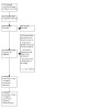
Study flow diagram (PRISMA) for all search results.
records identified by the database and other searches;
records after removal of duplicates;
records excluded after preliminary screening (i.e. of titles and abstracts);
records retrieved in full text;
records or studies excluded after assessment of the full text with brief reasons;
studies included in qualitative synthesis and quantitative syntheses (meta‐analysis).
Data extraction and management
Two review authors (PC, LH) independently extracted the following data using a standardised form:
methods (number of participants eligible and randomised, adequacy of randomisation, allocation concealment, blinding, completeness of follow‐up);
participant characteristics and exclusions;
type of endovenous intervention;
study dates;
number of participants per group;
prospective registration in a clinical trials registry;
information about ethics approval, consent, and conflict of interest;
source of funding;
outcomes.
Any discrepancies were resolved through discussion.
We extracted data for outcomes of interest (means and standard deviations for continuous outcomes, number of events for dichotomous outcomes, and hazard ratio and 95% confidence intervals for time‐to‐event data) from the published reports. Where necessary, we contacted study authors were contacted for clarification, further information, and/or raw data.
Assessment of risk of bias in included studies
Two review authors (PC, LH) independently assessed the eligible trials for risk of bias (RoB) using the Cochrane RoB 1 tool (Higgins 2017). This tool addresses six specific domains, namely sequence generation, allocation concealment, blinding, incomplete outcome data, selective outcome reporting, and other potential sources of bias (see Appendix 3 for details of the criteria on which the judgements were based). We assessed blinding and completeness of outcome data for each outcome separately, and completed a risk of bias table for each eligible study. Any disagreements between review authors were resolved by consensus. We contacted study authors to resolve any ambiguities. We have presented our assessments as a risk of bias summary figure, which shows all the judgements in a cross‐tabulation of study by entry.
Measures of treatment effect
The results of each included study were charted on forest plots as point estimates, i.e. risk ratios (RR) with corresponding 95% confidence intervals (CI) for dichotomous outcomes, mean difference (MD) with 95% CI for continuous outcomes, and hazard ratio (HR) with 95% CI for time‐to‐event outcomes (e.g. time to healing). The estimated log hazards ratio was calculating using the (O‐E/V) method (Higgins 2022a). Results that could not be shown in this way are reported in the text of the review.
Unit of analysis issues
If included studies randomised at the participant level and measured outcomes at the ulcer level, the participant was considered as the unit of analysis when the number of ulcers assessed appeared equal to the number of participants (e.g. one ulcer per person). Where studies randomised ulcers or legs as opposed to individuals and there were multiple ulcers per participant, and we were unable to obtain further information from study authors, we did not include them in the meta‐analysis. See Differences between protocol and review.
Dealing with missing data
Where data or information were missing from the trial reports, we contacted study authors to provide this, as per review protocol Carradice 2011a. Where it was not possible to ascertain the healed status of participants, we conducted a completed case analysis only. No imputations for missing data were made. See Differences between protocol and review.
Assessment of heterogeneity
Prior to meta‐analysis, we assessed clinical homogeneity with respect to participant demographics, types of therapy, comparator treatment and the nature of the outcomes reported. Where studies were clinically heterogeneous, we have described their data separately. Where studies were clinically homogeneous, we assessed statistical heterogeneity using the Q test (Chi² and the I² measure). A Chi² test resulting in a P value less than 0.10 was interpreted as indicating significant statistical heterogeneity. In order to assess and quantify the possible magnitude of heterogeneity across studies, we used the I² measure, interpreted as follows:
0% to 40% would be viewed as indicative of low levels of heterogeneity that may not be important;
30% to 60% may represent moderate heterogeneity;
50% to 90% would represent substantial heterogeneity;
75% to 100% would represent considerable heterogeneity and unsuitability for meta‐analysis (Deeks 2022).
Assessment of reporting biases
Two review authors (PC, LH) independently assessed any evidence of reporting bias and reported this as recommended (Higgins 2022b). Where there was doubt, we contacted the study authors for clarification, to acquire unpublished data and provide an English language version of the original trial protocol in order to assess the risk of bias.
Data synthesis
For clinically homogeneous studies with similar participants, comparators and outcome measures, we pooled data in a meta‐analysis using a fixed‐effect model. For studies differing in participants and comparators but estimating similar intervention effects, we used a random‐effects model. For time‐to‐event data, we converted estimates of hazard ratio (HR) and 95% CI into the log rank observed minus expected events and variance of the log rank, and we pooled these estimates using a random‐effect model (Deeks 2022). Where statistical synthesis of data from more than one study was not possible or considered inappropriate, we conducted a narrative review of eligible studies.
Subgroup analysis and investigation of heterogeneity
We planned subgroup analyses to determine whether effect size was influenced by the following factors:
severity of ulcers at baseline, determined by size (more than 5 cm2 versus 5 cm2 or less) or ulcer duration (more than six months versus six months or less) at baseline;
different endovenous thermal techniques;
different forms of compression;
the presence of infection (determined by clinical features and positive culture);
aetiology of the ulcer (occlusive or reflux).
However, the small number of studies and sample size in studies precluded subgrouping of the data.
Sensitivity analysis
We had planned to undertake sensitivity analysis to investigate the robustness of the treatment effect by removing studies classified as high in risk of bias. This, however, was not possible as only two studies were included in the meta analysis.
Summary of findings and assessment of the certainty of the evidence
The main outcomes of the review are presented in a summary of findings table. This table presents key information concerning the certainty of the evidence, the magnitude of the effects of the interventions examined, and the sum of available data for the main outcomes (Schünemann 2022a). The summary of findings table also includes an overall grading of the evidence related to each of the primary outcomes, using the GRADE approach. The GRADE approach defines the certainty of a body of evidence as the extent to which one can be confident that an estimate of effect or association is close to the true quantity of specific interest. The certainty of a body of evidence involves consideration of within‐trial risk of bias, directness of evidence, heterogeneity, precision of effect estimates, and risk of publication bias (Schünemann 2022b). The following outcomes are presented in the summary of findings table for the comparison of endovenous ablation and standard care with standard care alone:
time to complete healing;
proportion of ulcers healed at a given time point;
proportion of ulcers recurring over a given time period;
ulcer‐free days over a given time period.
For other outcomes, we conducted a GRADE assessment and presented it in a narrative format within the Results section, but not in the summary of findings table. For the GRADE assessment of cost‐effectiveness evidence, we assessed the RCT evidence on which the evaluation was based.
Results
Description of studies
See: Characteristics of included studies, Characteristics of excluded studies, Characteristics of ongoing studies.
Results of the search
For the update of this review, we identified a total of 1142 records from electronic searches. After removal of duplicates there were 810 records, of which 802 could be excluded on the basis of their titles and abstracts. We reviewed eight full‐text articles, of which four met the inclusion criteria. Two articles referred to the same trial at different lengths of follow‐up (Gohel 2018; Gohel 2020), and one article was an economic outcomes paper (Epstein 2019). We excluded Gohel 2020 and Epstein 2019 as there was no other study with a comparable length of follow‐up or economic data. One other study was ongoing with no available results and therefore not included in the analysis (see Characteristics of ongoing studies) (De Oliveira). There were no studies awaiting classification. Two trials were eligible for inclusion in the qualitative and quantitative analysis (meta analysis) (Gohel 2018; Puggina 2020). The results of all searches to date are summarised in Figure 1.
Included studies
Two RCTs met the eligibility criteria.
An RCT of early endovenous ablation in venous ulceration (EVRA); Gohel 2018.
An RCT of the effects of saphenous and perforating veins radiofrequency ablation in venous ulcer healing (VUERT); Puggina 2020.
We excluded Gohel 2020 from quantitative synthesis (meta analysis) as it pertained to longer term follow‐up data of Gohel 2018, with no matched duration data from another study. We conducted a GRADE assessment of the outcomes and presented these assessments in a narrative format (Appendix 4).
Types of participants
We included two studies in this update, Gohel 2018 and Puggina 2020, which enrolled a total of 506 participants. Both studies enrolled participants with active VLU. Gohel 2018 specified the minimum duration of an eligible VLU to be six weeks and the maximum to be six months, while Puggina 2020 set a minimum duration of only four weeks. The mean ulcer duration in the Gohel 2018 trial was 3.1 months ± 1.1 months, and in the Puggina 2020 trial it was 60.5 months ± 96.4 months.
The mean (standard deviation) ages of participant in years was 67 (± 15) and 69 (± 14) in Gohel 2018 and 53 (± 13) and 54 (± 12) in Puggina 2020.
Gohel 2018 was conducted in the UK and Puggina 2020 in Brazil.
Types of interventions
Both included studies used endovenous ablative techniques in conjunction with standard wound care which included compression.
Puggina 2020 used RFA exclusively with a standardised technique for all participants, followed by two‐layer compression bandaging.
As a multicentre study, Gohel 2018 used a variety of endovenous ablative techniques and compression therapies as part of standard care, as the local clinical team determined the method and strategy of endovenous treatment and compression use: 49.6% of the participants received foam sclerotherapy, 31.7% received endothermal ablation, 12.1% received endothermal ablation and foam sclerotherapy, 2.2% received MOCA, and 1.3% received MOCA with foam sclerotherapy, with the remaining participants not receiving any endovenous treatment. Types of compression used were not recorded in detail per participant, but multilayer elastic compression (two to four layers), short‐stretch compression, and compression hosiery were all deemed to be acceptable.
Comparators were standard wound care, which included compression in the same fashion as the intervention group.
Following ulcer healing or at six months, Gohel 2018 allowed participants in the control arm to cross over and undergo endovenous ablation. This was justified by the extrapolation of previous data (Barwell 2004), suggesting that surgical treatment following ulcer healing reduced recurrence rates and was considered by the authors to constitute best practice. Conversely, Puggina 2020 aimed to keep participants in their allocated groups. This difference significantly limits the comparability of the studies after six months for the outcome of recurrence and limits the differential treatment effect with regard to both recurrence and ulcer‐free days.
Excluded studies
The previous version of this review excluded a total of four studies: three were case studies (Pannier 2007; Sharif 2007; Teo 2010), and Viarengo 2007 was a quasi‐randomised trial.
For this update, we identified and excluded three new published studies (Dillavou 2010; Nieves 2015; Zhou 2016). We excluded Dillavou 2010 as the study was abandoned due to difficulties in recruiting; neither the treatment nor the control group in Nieves 2015 received compression therapy; and both the treatment and control group in Zhou 2016 received endovenous laser ablation.
This brings the total number of excluded studies to seven, described in Characteristics of excluded studies.
Ongoing studies
No ongoing studies were identified in the previous version of this review.
For this update, we did not identify any ongoing studies through published trial protocols, but we identified one ongoing study from trial registry records (De Oliveira). See Characteristics of ongoing studies.
Risk of bias in included studies
See Figure 2 and Figure 3 for the risk of bias summary; details of the risk of bias judgements for each domain and their rationales for each study are given in Characteristics of included studies.
2.
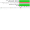
Risk of bias graph: review authors' judgements about each risk of bias item presented as percentages across all included studies.
3.
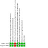
Risk of bias summary: review authors' judgements about each risk of bias item for each included study.
Allocation
Selection bias risk was low in both included studies (Gohel 2018; Puggina 2020). Both randomised participants via computer‐generated numbers or sequences. Both studies were also at low risk of bias with regards to allocation: the VUERT trial used sealed, opaque, sequentially numbered envelopes to conceal allocations (Puggina 2020), whereas in EVRA, allocation was performed centrally via computer independently to the locally recruiting team (Gohel 2018).
Blinding
Risk of performance bias in both included studies was high as the nature of the interventions in the treatment groups meant that neither participants nor treating clinicians were blinded at the time treatments were performed. It should be noted that it is not reasonably possible to blind treating clinicians or participants in trials of this nature in an ethical context. Outcome assessments in both studies, however, were carried out by independent blinded assessors, therefore we deemed the risk of detection bias to be low. In EVRA photography was used to support the objective assessment of wound healing. It is important to note that participants were not recruited into either study unless the treating clinicians were at equipoise, meaning they were in a state of genuine uncertainty regarding the comparative therapeutic merits of each arm in the trial.
Incomplete outcome data
Risk of attrition bias was low in both included studies. Dropout rates were less than 10%. There was no evidence to suggest differential dropout from the different study arms.
Selective reporting
Risk of reporting bias was low in both included studies. The manuscripts published outlined all the results that the protocols set out to measure.
Other potential sources of bias
No other potential sources of bias were identified.
Effects of interventions
See: Table 1
Please see Table 1.
Comparison of endovenous ablation with compression versus compression alone in the management of venous leg ulcers
We included two studies, with a total number of 506 participants (Gohel 2018; Puggina 2020).
Primary outcomes
Time to complete ulcer healing
Both included studies reported the time from randomisation to complete ulcer healing. A total of 466 participants were included in the meta analysis for this outcome. Combined endovenous treatment with compression reduced time to ulcer healing when compared with compression alone or compression with deferred endovenous treatment (HR 1.41, 95% CI 1.36 to 1.47; I2 = 0%; Analysis 1.1; Figure 4). The median ulcer healing time in EVRA trial was 56 days (95% CI 49 days to 66 days) in the intervention group and 82 days in the control group (95% CI 69 days to 92 days). In the VUERT trial the median ulcer healing time was 68 days in the intervention group (95% CI 57 days to 147 days) and 77 days in the control group (95% CI 72 days to 198 days). This is high‐certainty evidence (Table 1).
1.1. Analysis.
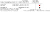
Comparison 1: Endovenous ablation and compression vs compression alone for venous leg ulcers, Outcome 1: Ulcer healing
4.
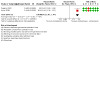
Figure 4: Forest plot of time to ulcer healing
Proportion of ulcers healed at 90 days
Both studies reported ulcer healing rates at three months. A total of 466 participants were included in this analysis. Ulcer healing rates were probably better with combined endovenous treatment and compression when compared with compression alone or compression with deferred endovenous treatment (RR 1.14, 95% CI 1.00 to 1.30; I2 = 0%; Analysis 1.2; Figure 5). We downgraded this evidence to moderate certainty due to imprecision (Table 1).
1.2. Analysis.
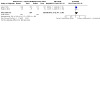
Comparison 1: Endovenous ablation and compression vs compression alone for venous leg ulcers, Outcome 2: Proportion of ulcers healed at 90 days
5.
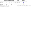
Figure 5: Forest plot of proportion of ulcers healed at 90 days
Proportion of ulcers healed at one year
Both studies reported ulcer healing rates at 12 months. A total of 505 participants were included in this analysis. There is probably no difference in ulcer healing rates with combined endovenous treatment and compression when compared with compression alone or compression with deferred endovenous treatment (RR 1.08, 95% CI 1.02 to 1.14; I2 = 80%; Analysis 1.3; Figure 6). This is likely to have been influenced by cross‐over in the EVRA trial. We downgraded this evidence to moderate certainty due to imprecision (Table 1).
1.3. Analysis.
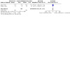
Comparison 1: Endovenous ablation and compression vs compression alone for venous leg ulcers, Outcome 3: Proportion of ulcers healed at one year
6.
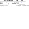
Figure 6: Forest plot of proportion of ulcers healed at one year (NB participants in the compression alone group in Gohel 2018 were offered ablation from 6 months)
Proportion of ulcers recurring at one year in participants with healed ulcers
Both studies reported the number of ulcers recurring at one year in participants with healed ulcers. A total of 460 participants were included in this analysis. There is low‐certainty evidence that the effect of endovenous treatment on the proportion of ulcers recurring at one year is unclear when compared with compression alone or compression with deferred endovenous treatment (RR 0.29, 95% CI 0.03 to 2.48; I2 = 78%; Analysis 1.4; Figure 7). The recurrence rate at one year in the Puggina 2020 trial was 3.7% (1/27) in the intervention group compared with 44.8% (13/29) in the compression group. The proportion of ulcers that recurred in Gohel 2018 at one year was 11.4% (24/210) in the intervention group compared with 16.5% (32/194) in the control group. We downgraded this evidence to low certainty for imprecision and risk of bias (Table 1). This is likely to have been influenced by cross‐over in the EVRA trial.
1.4. Analysis.
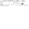
Comparison 1: Endovenous ablation and compression vs compression alone for venous leg ulcers, Outcome 4: Ulcer recurrence
7.
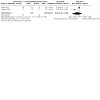
Figure 7: forest plot of ulcer recurrence (NB participants in the compression alone group of Gohel 2018 were offered ablation following ulcer healing)
Change in ulcer size
Though both studies reported ulcer sizes in their participant groups at baseline, neither of the trials reported change in ulcer size during the follow‐up period.
Ulcer‐free time during the first year postrandomisation
Only the EVRA trial reported this outcome. The effect of endovenous ablation on ulcer‐free days is unclear. Median and interquartile range (IQR) ulcer‐free time at one year follow‐up was 306 days (240 days to 328 days) in the early intervention and compression group compared with 278 days (175 days to 324 days) with compression and deferred endovenous treatment. Again, it is relevant that the methodology of the EVRA trial may have reduced the effect size from what would have been seen if the control arm had not received the intervention upon healing or at six months. We downgraded this evidence to low certainty due to indirectness (Table 1).
Secondary outcomes
Complications
The most common complication in the endovenous treatment arms was venous thromboembolism (VTE). The EVRA trial reported 2.7% cases (6/224) of calf vein deep vein thrombosis (DVT) in the early intervention group. In the deferred intervention group calf vein thrombosis occurred in 1.3% of participants (3/226). In all cases this occurred after endovenous treatment. In the VUERT trial no episodes of DVT or pulmonary embolism were detected. The effect of endovenous ablation on thromboembolism is unclear (RR 2.02, 95% CI 0.51 to 7.97; 2 studies, 506 participants; Analysis 1.5). This is low‐certainty evidence, downgraded twice due to imprecision.
1.5. Analysis.
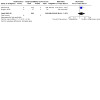
Comparison 1: Endovenous ablation and compression vs compression alone for venous leg ulcers, Outcome 5: Thromboembolism (complications)
Quality of life outcomes
Quality of life outcomes are only available at present in the EVRA trial (Gohel 2018). VUERT did record quality of life outcomes, but these are not yet available (Puggina 2020). The EVRA trial Gohel 2018 reported quality of life using the Aberdeen Varicose Vein Questionnaire (AVVQ), EQ‐5D‐5L, and SF‐36 measures at six weeks, six months or 12 months. There were no clear differences between the groups at 12 months (AVVQ: MD ‐1.8 points, 95% CI ‐4.0 to 0.3; EQ‐5D‐3L: 1.3, 95% CI ‐2.1 to 4.8; SF‐36 physical component summary: 0.3, 95% CI ‐1.7 to 2.3; SF‐36 mental component summary ‐0.7, 95% CI ‐2.7 to 1.4). This is low‐certainty evidence, downgraded twice for indirectness as the intervention was given to both groups in the EVRA trial (Gohel 2018).
Economic outcomes
The EVRA trial (Gohel 2018) reported a separate cost‐utility analysis at one year in Epstein 2019. This outcome was not included in the summary of findings table, but we nonetheless rated this using the GRADE assessment of the certainty of evidence and presented these assessments in a narrative format (Appendix 4). Epstein 2019 performed a within‐trial cost‐utility analysis with a time horizon of one year, from the perspective of the UK National Health Service and Personal Social Services. The primary outcome was quality adjusted life years (QALYs), collected using EQ‐5D‐5L and mapped onto health utility values (Dolan 1997). Data were available for 76.4% of participants. The mean cost difference was GBP 163 and the mean difference in QALY was 0.041. The incremental cost‐effectiveness ratio (ICER) was GBP 3976/QALY. The probability of endovenous treatment being more cost‐effective is 89% at a threshold of GBP 20,000. When imputed data were included, the mean difference in cost was GBP ‐72 and the mean difference in QALY was 0.058. The probability of endovenous therapy being cost‐effective was 99% at a threshold of GBP 20,000. We downgraded this outcome to moderate‐certainty evidence for imprecision related to missing data.
Economic data were not available from the VUERT trial.
Discussion
Summary of main results
This Cochrane Review synthesises RCT evidence on the effects of endovenous ablation of superficial venous incompetence in the healing of VLU. We identified two eligible RCTs with a total of 506 participants, both of which we included in the meta‐analysis (Gohel 2018; Puggina 2020). With the addition of these studies to the previous version of the review, there now is high‐certainty evidence that endovenous ablation in combination with compression therapy is associated with reduced ulcer healing time compared with compression therapy alone, or compression with deferred endovenous treatment. Both included RCTs demonstrated that high‐quality compression treatment alone, when performed by experts in the context of a clinical trial, is highly efficacious, achieving much higher healing rates than are reported in the literature. Even the most favourable ulcer healing rates described in the literature were achieved in the included control arms in half the time reported previously in the literature (Barwell 2004; Guest 2018; Heyer 2017), demonstrating the high quality of treatment in the control arm. Despite this, the addition of endovenous ablation resulted in a significant improvement in healing time. In association with this, there is also moderate‐certainty evidence suggesting that, when compared with compression alone, or compression with deferred endovenous treatment, endovenous ablation in combination with compression is probably associated with a higher number of ulcers healed at 90 days. There is also low‐certainty evidence of unclear effects of endovenous ablation on recurrence rates and on ulcer‐free time. An economic analysis based upon one study found moderate‐certainty evidence demonstrating that endovenous ablation is probably a cost‐effective treatment for venous leg ulcer at one year (99% probability at a threshold of GBP 20,000/QALY).
Complications postintervention were well reported in both studies, but this is low‐certainty evidence due to imprecision. DVT (above the calf) post‐endovenous treatment is a rare complication reported in around 0.3 to 0.5% of participants (Healy 2018; Jia 2007; Marsh 2010). There is some evidence that it is more common in people with ulcers. Calf vein DVT is associated with far fewer complications than femoropopliteal or iliofemoral DVT (Kirkilesis 2020; Robert‐Ebadi 2017). Conventional clinical compression duplex used to detect DVT in most clinical DVT services does not assess for calf vein DVT. It is a marker of the quality of the EVRA study that subtle complications such as this were detected and managed to prevent significant complications.
Lastly, there is a place for further research into whether endovenous ablation in combination with compression impacts quality of life. These outcomes were only reported by Gohel 2018, and the certainty of evidence is low.
Overall completeness and applicability of evidence
These studies have been designed and executed to a high standard. The included participants allow generalisability of the findings to those developing venous leg ulcers. The studies may be clinically skewed in favour of the control arm, as the healing rates in the control groups were much higher than those previously reported in real world data (Guest 2018), but this has the benefit of adding considerable confidence to the validity of the conclusions.
The limitations of the review include the exclusion of participants with chronic ulceration (more than six months) from the EVRA trial (Gohel 2018). The mean duration of ulceration in the VUERT trial (Puggina 2020), however, was much longer (mean 60.5 months ± 96.4 months). Despite this marked difference, the outcomes were broadly similar. The quality of care prior to trial participation is unknown, and therefore it is difficult to say with confidence whether chronic ulcers in the trial healthcare systems are similar to those in other systems. In practice, chronic ulcers are associated with good healing rates in the hands of experts, but further research is needed to address this.
The approach of the EVRA trial was not whether to perform ablation in addition to compression, but rather when to do it, with one group receiving it within two weeks and the other waiting until after ulcer healing. The high‐quality compression treatment in the deferred group ensured high healing rates, but also high intervention rates posthealing. If the research question is not when, but rather whether to perform ablation, then this study is also likely to have under‐represented the benefits of ablation due to this cross over. This is an explanation for the difference in recurrence rate estimates between the trials in Figure 7, and may have resulted in an underestimate in the increase in ulcer‐free days associated with the intervention. Treatment of superficial venous reflux (albeit with open surgery) has previously been shown to be associated with a reduction in recurrence (Gohel 2007), and is the reason why the EVRA authors thought it unethical not to offer delayed intervention to the control arm. The lack of association with recurrence in this review should therefore be treated with caution.
The included studies were not designed or powered to evaluate the relative efficacy of the different methods of endovenous ablation in the healing of leg ulcers. Further research would be needed to draw any conclusion on the effectiveness of these different modalities or treatment strategies.
Quality of the evidence
The certainty of evidence is high for the primary outcome of time to ulcer healing. The certainty of evidence is moderate for the primary outcome of proportion of ulcers healed at 90 days and the secondary outcome of cost‐effectiveness. We downgraded the evidence for these outcomes due to imprecision arising from wide confidence intervals and missing data, respectively. The certainty of evidence is low for the primary outcomes ulcer recurrence at one year and ulcer‐free time, and for the secondary outcomes complications and quality of life. We downgraded the evidence for these outcomes due to imprecision, risk of bias or indirectness.
Potential biases in the review process
The reviewers were not blinded regarding the source of the studies under evaluation. Some of the reviewers were co‐authors/collaborators on the EVRA trial and as such were not involved in the critical appraisal of this study as part of the review.
Agreements and disagreements with other studies or reviews
The previous version of this review did not identify any suitable studies, and we have not identified any other reviews on this issue.
Authors' conclusions
Implications for practice.
Endovenous ablation of superficial venous incompetence in combination with compression improves leg ulcer healing when compared with compression alone. This conclusion is based on high‐certainty evidence. There is moderate‐certainty evidence to suggest that it is probably cost‐effective at one year, but there is low‐certainty evidence of unclear effects of the treatment on recurrence rates and complications.
Implications for research.
Further research is needed to explore the additional benefit of endovenous ablation in ulcers of greater than six months duration and the optimal modality of endovenous ablation.
What's new
| Date | Event | Description |
|---|---|---|
| 27 July 2023 | New search has been performed | First update, new search. Title changed to include all types of endovenous ablation. Two studies now included. |
| 27 July 2023 | New citation required and conclusions have changed | Updated. Conclusions changed. |
History
Protocol first published: Issue 12, 2011 Review first published: Issue 10, 2013
Acknowledgements
The Academic Vascular Surgery Unit Hull: Bankole Akomolafe, Gemma Boam, Hayley Crane, Anna Firth, Josie Hatfield, Brian Johnson, Rakesh Kapur, Judith Long, Robert Lonsdale, Anthony Mekako, Joanne Palmer, Sean Pymer, Paul Renwick, Tracey Roe, Amber Stafford and Junaid Sultan.
The authors would like to thank Rebeca Illescas for her help with translating the records written in Spanish and liaising on our behalf with the authors.
The authors would also like to thank Nehemiah Samuel and Tom Wallace for their previous work on this review, peer reviewers: Sarah Rhodes and En Lin Goh for providing comments on this review update; peer reviewers: David Margolis, Andrea Nelson, Caroline Main, Gill Worthy, Fausto Biancari, Iain McCallum and David Voegli for providing feedback on the previous version of the review; and copy editors: Jenny Bellorini and Elizabeth Royle for the previous version of this review.
Appendices
Appendix 1. Glossary of terms
Capillaries: small blood vessels, that connect arteries and veins, they carry oxygen and nutrients and take away carbon dioxide and waste products to and from individual cells throughout the body
Catheter: a tube that delivers something into or out of the body
Denude: to strip something away
Duplex ultrasound: technology that involves the reflection of ultrasound waves fired into the body to create a real‐time picture of the veins inside the body, and to show the speed and direction of blood flow within them
Endothelium: inner‐most layer of a vein
'Green' pathway: this is a patient pathway designed for patients in which Covid‐19 infection has been excluded and avoids them coming into contact with other patients, staff or facilities who may be infected or contaminated with Covid‐19.
Inflammatory process/inflammation: the body's natural response to tissue damage that can be dysfunctional and leave lasting tissue damage.
Intima: the innermost layer of a blood vessel
Lumen: the central cavity of a tubular or other hollow structure
Micelle: an aggregate of molecules in a colloidal solution, such as those formed by detergents
MOCA: mechanochemical ablation. A method of endovenous ablation using both mechanical damage with a rotating wire and the injection of sclerotherapy
Perforating veins: small veins connecting the deep to the superficial venous system
Tumescent: the practice of injecting a very dilute solution of local anaesthetic into tissue until it becomes firm and tense (tumescent).
Venous hypertension: high blood pressure within veins.
Venous incompetence/insufficiency/reflux: the presence of abnormal retrograde (reverse) flow within the veins caused by valvular dysfunction.
Venules: smaller size veins
Appendix 2. Search strategies
Cochrane Wounds Specialised Register
1 MESH DESCRIPTOR Ablation Techniques EXPLODE ALL AND INREGISTER
2 ablation or ablative AND INREGISTER
3 (endovenous near3 laser*) AND INREGISTER
4 (EVL or EVLA or EVLO or EVLT or EVTA or ELA) AND INREGISTER
5 (VenaCure) AND INREGISTER
6 (radiofrequency near2 ablati*) AND INREGISTER
7 (RFA or RFO) AND INREGISTER
8 (VeneFit) AND INREGISTER
9 (VNUS or ClosureFAST or VCF or RFITT) AND INREGISTER
10 (electrocoagulation or cauter* or cryosurge* or cryoablat*) AND INREGISTER
11 MESH DESCRIPTOR Steam EXPLODE ALL AND INREGISTER
12 ((endovenous or vein*) near2 steam*) AND INREGISTER
13 (EVSA) AND INREGISTER
14 MESH DESCRIPTOR Sclerotherapy EXPLODE ALL AND INREGISTER
15 MESH DESCRIPTOR Sclerosing Solutions EXPLODE ALL AND INREGISTER
16 MESH DESCRIPTOR Sodium Tetradecyl Sulfate EXPLODE ALL AND INREGISTER
17 MESH DESCRIPTOR Cyanoacrylates EXPLODE ALL AND INREGISTER
18 sclerotherap* AND INREGISTER
19 UGFS AND INREGISTER
20 sodium tetradecyl sul* AND INREGISTER
21 (MOCA or Clarivein) AND INREGISTER
22 ((endovenous or occlusion) near2 (glue* or adhesive* or superglu*)) AND INREGISTER
23 (cyanoacrylate* or acrylate*) AND INREGISTER
24 #1 OR #2 OR #3 OR #4 OR #5 OR #6 OR #7 OR #8 OR #9 OR #10 OR #11 OR #12 OR #13 OR #14 OR #15 OR #16 OR #17 OR #18 OR #19 OR #20 OR #21 OR #22 OR #23
25 MESH DESCRIPTOR Leg Ulcer EXPLODE ALL AND INREGISTER
26 ((varicose next ulcer*) or (venous next ulcer*) or (leg next ulcer*) or (stasis next ulcer*) or (crural next ulcer*) or (ulcus cruris) or (ulcer* next cruris)) AND INREGISTER
27 MESH DESCRIPTOR Venous Insufficiency EXPLODE ALL AND INREGISTER
28 ("chronic venous insufficiency" or CVI) AND INREGISTER
29 #25 OR #27 OR #26 OR #28
30 #24 AND #29
The Cochrane Central Register of Controlled Clinical Trials (CENTRAL) Trial Registry specific search combined with above search via the Cochrane Specialised Register
31 (NCT0* or ACTRN* or ChiCTR* or DRKS* or EUCTR* or eudract* or IRCT* or ISRCTN* or JapicCTI* or JPRN* or NTR0* or NTR1* or NTR2* or NTR3* or NTR4* or NTR5* or NTR6* or NTR7* or NTR8* or NTR9* or SRCTN* or UMIN0*):AU AND CENTRAL:TARGET
32 http*:SO AND CENTRAL:TARGET
33 #31 OR #32 AND CENTRAL:TARGET
The Cochrane Central Register of Controlled Clinical Trials (CENTRAL)
#1 MeSH descriptor: [Ablation Techniques] explode all trees
#2 (ablation* or ablative*):ti,ab,kw
#3 (endovenous near/3 laser*):ti,ab,kw
#4 (EVL or EVLA or EVLO or EVLT or EVTA or ELA):ti,ab,kw
#5 (VenaCure):ti,ab,kw
#6 (radiofrequency near/2 ablati*):ti,ab,kw
#7 (RFA or RFO):ti,ab,kw
#8 (VeneFit):ti,ab,kw
#9 (VNUS or ClosureFAST or VCF or RFITT):ti,ab,kw
#10 (electrocoagulation or cauter* or cryosurge* or cryoablat*):ti,ab,kw
#11 MeSH descriptor: [Steam] explode all trees
#12 ((endovenous or vein*) near/2 steam*):ti,ab,kw
#13 (EVSA):ti,ab,kw
#14 MeSH descriptor: [Sclerotherapy] explode all trees
#15 MeSH descriptor: [Sclerosing Solutions] explode all trees
#16 MeSH descriptor: [Sodium Tetradecyl Sulfate] explode all trees
#17 MeSH descriptor: [Cyanoacrylates] explode all trees
#18 (sclerotherap*):ti,ab,kw
#19 (UGFS):ti,ab,kw
#20 (sodium tetradecyl sul*):ti,ab,kw
#21 (MOCA or Clarivein):ti,ab,kw
#22 ((endovenous or occlusion) near/2 (glue* or adhesive* or superglu*)):ti,ab,kw
#23 (cyanoacrylate* or acrylate*):ti,ab,kw
#24 {or #1‐#23}
#25 MeSH descriptor: [Leg Ulcer] explode all trees
#26 ((varicose next ulcer*) or (venous next ulcer*) or (leg next ulcer*) or (stasis next ulcer*) or (crural next ulcer*) or (ulcus cruris) or (ulcer* next cruris)):ti,ab,kw
#27 MeSH descriptor: [Venous Insufficiency] explode all trees
#28 ("chronic venous insufficiency" or CVI):ti,ab,kw
#29 {or #25‐#28}
#30 {and #24, #29}
Ovid MEDLINE
1 exp Ablation Techniques/
2 (ablation* or ablative*).tw.
3 (endovenous adj3 laser*).tw.
4 (EVL or EVLA or EVLO or EVLT or EVTA or ELA).tw.
5 VenaCure.tw.
6 (radiofrequency adj3 ablati*).tw.
7 (RFA or RFO).tw.
8 VeneFit.tw.
9 (VNUS or ClosureFAST or VCF or RFITT).tw.
10 (electrocoagulation or cauter* or cryosurge* or cryoablat*).tw.
11 Steam/
12 ((endovenous or vein*) adj2 steam*).tw.
13 EVSA.tw.
14 Sclerotherapy/
15 Sclerosing solutions/
16 Sodium Tetradecyl Sulfate/
17 Cyanoacrylates/
18 sclerotherap*.tw.
19 UGFS.tw.
20 sodium tetradecyl sul*.tw.
21 (MOCA or Clarivein).tw.
22 ((endovenous or occlusion) adj2 (glue* or adhesive* or superglu*)).tw.
23 (cyanoacrylate* or acrylate*).tw.
24 or/1‐23
25 exp Leg Ulcer/
26 (varicose ulcer* or venous ulcer* or leg ulcer* or stasis ulcer* or crural ulcer* or ulcus cruris).tw.
27 exp Venous Insufficiency/
28 (chronic venous insufficiency or CVI).tw.
29 or/25‐28
30 and/24,29
31 randomized controlled trial.pt.
32 controlled clinical trial.pt.
33 randomi?ed.ab.
34 placebo.ab.
35 clinical trials as topic.sh.
36 randomly.ab.
37 trial.ti.
38 or/31‐37
39 exp animals/ not humans.sh.
40 38 not 39
41 30 and 40
42 limit 41 to yr="1998 ‐ 2020"
Ovid Embase
1 ablation therapy/
2 (ablation* or ablative*).tw.
3 exp laser surgery/
4 (endovenous adj3 laser*).tw.
5 (EVL or EVLA or EVLO or EVLT or ELA).tw.
6 VenaCure.tw.
7 catheter ablation/
8 (radiofrequency adj3 ablati*).tw.
9 (RFA or RFO).tw.
10 VeneFit.tw.
11 (VNUS or ClosureFAST or VCF or RFITT).tw.
12 (electrocoagulation or cauter* or cryosurge* or cryoablat*).tw.
13 water vapor/
14 ((endovenous or vein*) adj2 steam*).tw.
15 EVSA.tw.
16 exp sclerotherapy/
17 exp sclerosing agent/
18 tetradecyl sulfate sodium/
19 Cyanoacrylates/
20 sclerotherap*.tw.
21 UGFS.tw.
22 sodium tetradecyl sul*.tw.
23 (MOCA or Clarivein).tw.
24 ((endovenous or occlusion) adj2 (glue* or adhesive* or superglu*)).tw.
25 (cyanoacrylate* or acrylate*).tw.
26 or/1‐25
27 exp leg ulcer/
28 (varicose ulcer* or venous ulcer* or leg ulcer* or stasis ulcer* or crural ulcer* or ulcus cruris).tw.
29 exp vein insufficiency/
30 (chronic venous insufficiency or CVI).tw.
31 or/27‐30
32 and/26,31
33 Randomized controlled trials/
34 Single‐Blind Method/
35 Double‐Blind Method/
36 Crossover Procedure/
37 (random* or factorial* or crossover* or cross over* or cross‐over* or placebo* or assign* or allocat* or volunteer*).ti,ab.
38 (doubl* adj blind*).ti,ab.
39 (singl* adj blind*).ti,ab.
40 or/33‐39
41 exp animals/ or exp invertebrate/ or animal experiment/ or animal model/ or animal tissue/ or animal cell/ or nonhuman/
42 human/ or human cell/
43 and/41‐42
44 41 not 43
45 40 not 44
46 32 and 45
47 limit 46 to yr="1998 ‐ 2020"
EBSCO CINAHL Plus
S54 S52 AND S53
S53 PY 1998‐2020
S52 S28 AND S51
S51 S50 NOT S49
S50 S29 OR S30 OR S31 OR S32 OR S33 OR S34 OR S35 OR S36 OR S37 OR S38 OR S39 OR S40 OR S41 OR S42 OR S43
S49 S47 NOT S48
S48 MH (human)
S47 S44 OR S45 OR S46
S46 TI (animal model*)
S45 MH (animal studies)
S44 MH animals+
S43 AB (cluster W3 RCT)
S42 MH (crossover design) OR MH (comparative studies)
S41 AB (control W5 group)
S40 PT (randomized controlled trial)
S39 MH (placebos)
S38 MH (sample size) AND AB (assigned OR allocated OR control)
S37 TI (trial)
S36 AB (random*)
S35 TI (randomised OR randomized)
S34 MH cluster sample
S33 MH pretest‐posttest design
S32 MH random assignment
S31 MH single‐blind studies
S30 MH double‐blind studies
S29 MH randomized controlled trials
S28 S22 AND S27
S27 S23 OR S24 OR S25 OR S26
S26 AB ( (chronic venous insufficiency or CVI) ) OR TI ( (chronic venous insufficiency or CVI) )
S25 (MH "Venous Insufficiency+")
S24 TI ( (varicose ulcer* or venous ulcer* or leg ulcer* or stasis ulcer* or crural ulcer* or ulcus cruris) ) OR AB ( (varicose ulcer* or venous ulcer* or leg ulcer* or stasis ulcer* or crural ulcer* or ulcus cruris) )
S23 (MH "Leg Ulcer+")
S22 S1 OR S2 OR S3 OR S4 OR S5 OR S6 OR S7 OR S8 OR S9 OR S10 OR S11 OR S12 OR S13 OR S14 OR S15 OR S16 OR S17 OR S18 OR S19 OR S20 OR S21
S21 TI ( (cyanoacrylate* or acrylate*) ) OR AB ( (cyanoacrylate* or acrylate*) )
S20 TI ( ((endovenous or occlusion) N2 (glue* or adhesive* or superglu*)) ) OR AB ( ((endovenous or occlusion) N2 (glue* or adhesive* or superglu*)) )
S19 TI ( (MOCA or Clarivein) ) OR AB ( (MOCA or Clarivein) )
S18 TI sodium tetradecyl sul* OR AB sodium tetradecyl sul*
S17 TI UGFS OR AB UGFS
S16 TI sclerotherap* OR AB sclerotherap*
S15 (MH "Sclerosing Solutions")
S14 (MH "Sclerotherapy")
S13 TI EVSA OR AB EVSA
S12 TI ( ((endovenous or vein*) N2 steam*) ) OR AB ( ((endovenous or vein*) N2 steam*) )
S11 (MH "Steam")
S10 TI ( (electrocoagulation or cauter* or cryosurge* or cryoablat*) ) OR AB ( (electrocoagulation or cauter* or cryosurge* or cryoablat*) )
S9 TI ( (VNUS or ClosureFAST or VCF or RFITT) ) OR AB ( (VNUS or ClosureFAST or VCF or RFITT) )
S8 TI venefit OR AB venefit
S7 TI ( RFA or RFO ) OR AB ( RFA or RFO )
S6 TI (radiofrequency N3 ablati*) OR AB (radiofrequency N3 ablati*)
S5 TI VenaCure OR AB VenaCure
S4 TI ( (EVL or EVLA or EVLO or EVLT or EVTA or ELA) ) OR AB ( (EVL or EVLA or EVLO or EVLT or EVTA or ELA) )
S3 TI endovenous N3 laser* OR AB endovenous N3 laser*
S2 TI ( ablation or ablative* ) OR AB ( ablation or ablative* )
S1 (MH "Ablation Techniques+")
US National Institutes of Health Ongoing Trials Register (ClinicalTrials.gov)
endovenous OR ablation OR ablative OR thermal OR RFA OR steam OR heat OR sclerotherapy OR glue OR cyanoacrylate OR EVLT OR EVLA OR EVSA OR radiofrequency | Venous Leg Ulcer
endovenous OR ablation OR ablative OR thermal OR RFA OR steam OR heat OR sclerotherapy OR glue OR cyanoacrylate OR EVLT OR EVLA OR EVSA OR radiofrequency | Ulcer Venous
endovenous OR ablation OR ablative OR thermal OR RFA OR steam OR heat OR sclerotherapy OR glue OR cyanoacrylate OR EVLT OR EVLA OR EVSA OR radiofrequency | varicose ulcer
endovenous OR ablation OR ablative OR thermal OR RFA OR steam OR heat OR sclerotherapy OR glue OR cyanoacrylate OR EVLT OR EVLA OR EVSA OR radiofrequency | Venous Stasis Ulcer
endovenous OR ablation OR ablative OR thermal OR RFA OR steam OR heat OR sclerotherapy OR glue OR cyanoacrylate OR EVLT OR EVLA OR EVSA OR radiofrequency | Venous Ulceration
World Health Organization International Clinical Trials Registry Platform
endovenous OR ablation OR ablative OR thermal OR RFA OR steam OR heat OR sclerotherapy OR glue OR cyanoacrylate OR EVLT OR EVLA OR EVSA OR radiofrequency Venous Leg Ulcer [Title]
endovenous OR ablation OR ablative OR thermal OR RFA OR steam OR heat OR sclerotherapy OR glue OR cyanoacrylate OR EVLT OR EVLA OR EVSA OR radiofrequency | Venous Leg Ulcer [Condition]
endovenous OR ablation OR ablative OR thermal OR RFA OR steam OR heat OR sclerotherapy OR glue OR cyanoacrylate OR EVLT OR EVLA OR EVSA OR radiofrequency I Ulcer venous [Title]
endovenous OR ablation OR ablative OR thermal OR RFA OR steam OR heat OR sclerotherapy OR glue OR cyanoacrylate OR EVLT OR EVLA OR EVSA OR radiofrequency I Ulcer venous [Condition]
endovenous OR ablation OR ablative OR thermal OR RFA OR steam OR heat OR sclerotherapy OR glue OR cyanoacrylate OR EVLT OR EVLA OR EVSA OR radiofrequency I Varicose Ulcer [Title]
endovenous OR ablation OR ablative OR thermal OR RFA OR steam OR heat OR sclerotherapy OR glue OR cyanoacrylate OR EVLT OR EVLA OR EVSA OR radiofrequency I Varicose Ulcer [Condition]
endovenous OR ablation OR ablative OR thermal OR RFA OR steam OR heat OR sclerotherapy OR glue OR cyanoacrylate OR EVLT OR EVLA OR EVSA OR radiofrequency I venous stasis ulcer [Title]
endovenous OR ablation OR ablative OR thermal OR RFA OR steam OR heat OR sclerotherapy OR glue OR cyanoacrylate OR EVLT OR EVLA OR EVSA OR radiofrequency I venous stasis ulcer [Condition]
endovenous OR ablation OR ablative OR thermal OR RFA OR steam OR heat OR sclerotherapy OR glue OR cyanoacrylate OR EVLT OR EVLA OR EVSA OR radiofrequency I venous ulceration [Title]
endovenous OR ablation OR ablative OR thermal OR RFA OR steam OR heat OR sclerotherapy OR glue OR cyanoacrylate OR EVLT OR EVLA OR EVSA OR radiofrequency I venous ulceration [Condition]
Appendix 3. Risk of bias criteria
1. Was the allocation sequence randomly generated?
Low risk of bias
The investigators describe a random component in the sequence generation process such as: referring to a random number table; using a computer random number generator; coin tossing; shuffling cards or envelopes; throwing dice; drawing of lots.
High risk of bias
The investigators describe a non‐random component in the sequence generation process. Usually, the description would involve some systematic, non‐random approach, for example: sequence generated by odd or even date of birth; sequence generated by some rule based on date (or day) of admission; sequence generated by some rule based on hospital or clinic record number.
Unclear
Insufficient information about the sequence generation process to permit a judgement of low or high risk of bias.
2. Was the treatment allocation adequately concealed?
Low risk of bias
Participants and investigators enrolling participants could not foresee assignment because one of the following, or an equivalent method, was used to conceal allocation: central allocation (including telephone, web‐based and pharmacy‐controlled randomisation); sequentially‐numbered drug containers of identical appearance; sequentially‐numbered, opaque, sealed envelopes.
High risk of bias
Participants or investigators enrolling participants could possibly foresee assignments and thus introduce selection bias, such as allocation based on: using an open random allocation schedule (e.g. a list of random numbers); assignment envelopes used without appropriate safeguards (e.g. if envelopes were unsealed or non‐opaque or not sequentially numbered); alternation or rotation; date of birth; case record number; any other explicitly unconcealed procedure.
Unclear
Insufficient information available to permit a judgement of low or high risk of bias. This is usually the case if the method of concealment is not described or not described in sufficient detail to allow a definite judgement to be made, for example if the use of assignment envelopes is described, but it remains unclear whether envelopes were sequentially numbered, opaque and sealed.
3. Blinding: was knowledge of the allocated interventions adequately prevented during the study?
Low risk of bias
Any one of the following.
No blinding, but the review authors judge that the outcome and the outcome measurement are not likely to be influenced by lack of blinding.
Blinding of participants and key study personnel ensured, and unlikely that the blinding could have been broken.
Either participants or some key study personnel were not blinded, but outcome assessment was blinded and the non‐blinding of others was unlikely to introduce bias.
High risk of bias
Any one of the following.
No blinding or incomplete blinding, and the outcome or outcome measurement is likely to be influenced by lack of blinding.
Blinding of key study participants and personnel attempted, but likely that the blinding could have been broken.
Either participants or some key study personnel were not blinded, and the non‐blinding of others was likely to introduce bias.
Unclear
Either of the following.
Insufficient information available to permit a judgement of low or high risk of bias.
The study did not address this outcome.
4. Were incomplete outcome data adequately addressed?
Low risk of bias
Any one of the following.
No missing outcome data.
Reasons for missing outcome data unlikely to be related to true outcome (for survival data, censoring unlikely to be introducing bias).
Missing outcome data balanced in numbers across intervention groups, with similar reasons for missing data across groups.
For dichotomous outcome data, the proportion of missing outcomes compared with observed event risk not enough to have a clinically relevant impact on the intervention effect estimate.
For continuous outcome data, plausible effect size (difference in means or standardised difference in means) among missing outcomes not enough to have a clinically relevant impact on observed effect size.
Missing data have been imputed using appropriate methods.
High risk of bias
Any one of the following.
Reason for missing outcome data likely to be related to true outcome, with either imbalance in numbers or reasons for missing data across intervention groups.
For dichotomous outcome data, the proportion of missing outcomes compared with observed event risk was enough to induce clinically relevant bias in intervention effect estimate.
For continuous outcome data, plausible effect size (difference in means or standardised difference in means) among missing outcomes was enough to induce clinically relevant bias in observed effect size.
‘As‐treated’ analysis done with substantial departure in the intervention received from that assigned at randomisation.
Potentially inappropriate application of simple imputation.
Unclear
Either of the following.
Insufficient reporting of attrition/exclusions to permit judgement of low or high risk of bias (e.g. number randomised not stated, no reasons for missing data provided).
The study did not address this outcome.
5. Are reports of the study free of suggestion of selective outcome reporting?
Low risk of bias
Either of the following.
The study protocol is available and all of the study’s pre‐specified (primary and secondary) outcomes that are of interest in the review have been reported in the pre‐specified way.
The study protocol is not available but it is clear that the published reports include all expected outcomes, including those that were pre‐specified (convincing text of this nature may be uncommon)
High risk of bias
Any one of the following.
Not all of the study’s pre‐specified primary outcomes have been reported.
One or more primary outcomes were reported using measurements, analysis methods or subsets of the data (e.g. subscales) that were not pre‐specified.
One or more reported primary outcomes were not pre‐specified (unless clear justification for their reporting is provided, such as an unexpected adverse effect).
One or more outcomes of interest in the review are reported incompletely so that they cannot be entered in a meta‐analysis.
The study report fails to include results for a key outcome that would be expected to have been reported for such a study.
Unclear
Insufficient information available to permit judgement of low or high risk of bias. It is likely that the majority of studies will fall into this category.
6. Other sources of potential bias
Low risk of bias
The study appears to be free of other sources of bias.
High risk of bias
There is at least one important risk of bias. For example, the study:
had a potential source of bias related to the specific study design used; or
stopped early due to some data‐dependent process (including a formal‐stopping rule); or
had extreme baseline imbalance; or
has been claimed to have been fraudulent; or
had some other problem.
Unclear
There may be a risk of bias, but there is either:
insufficient information to assess whether an important risk of bias exists; or
insufficient rationale or evidence that an identified problem will introduce bias.
Appendix 4. Results of studies not included in pooled analyses
This appendix contains the results of studies which reported specified outcomes but could not be included in the pooled analyses we conducted.
Gohel 2020
Types of participants
Gohel 2018, and therefore Gohel 2020, enrolled participants with active venous leg ulcers (VLU) and specified the minimum age of an eligible VLU to be six weeks and the maximum age to be six months. The study contacted 420 participants for the long term follow‐up.
Gohel 2020 was conducted in the UK.
Types of interventions
Gohel 2018, and therefore Gohel 2020, used endovenous ablative techniques in conjunction with standard wound care which included compression.
As a multicentre study, Gohel 2018, and therefore Gohel 2020, used a variety of endovenous ablative techniques and compression therapies as part of standard care, as the local clinical team determined the method and strategy of endovenous treatment and compression used. 49.6% of the participants in the early‐intervention arm received foam sclerotherapy, 31.7% received endothermal ablation only, 12.1% received endothermal ablation and foam sclerotherapy, 2.2% received mechanochemical ablation (MOCA), and 1.3% received MOCA with foam sclerotherapy. Types of compression used were not recorded in detail per participant but multilayer elastic compression (two to four layers), short‐stretch compression, and compression hosiery were all deemed to be acceptable.
Comparators were standard wound care which included compression in the same fashion as the intervention group.
Risk of bias in study
See Figure 2 and Figure 3 for the risk of bias summary; details of the risk of bias judgements for each domain and their rationales for each study are given in Characteristics of included studies.
Allocation (selection bias)
Selection bias risk was low in Gohel 2020. The study randomised patients via computer‐generated numbers or sequences. Risk of bias with regards to allocation was also low as the study allocation was performed centrally via computer independently to the locally recruiting team (Gohel 2020).
Blinding (performance bias and detection bias)
Risk of performance bias in this study was high as the nature of the interventions in the treatment groups meant that neither participants nor treating clinicians were blinded at the time treatments were performed. Blinding of outcome assessment (detection bias) was also high in this study as it was not recorded if assessors were blinded to group allocation for the long term follow‐ups. However, a standardised questionnaire was used for telephone call follow‐ups. It is also unclear how medical record review was conducted.
Incomplete outcome data (attrition bias)
Risk of attrition bias was high in this study due to variation in follow‐up modality (combination of telephone call follow‐up, medical records review or both).
Selective reporting (reporting bias)
Risk of reporting bias was in this study. The manuscripts published outline all the results that the protocol set out to measure.
Other potential sources of bias
No other potential sources of bias were identified.
Effects of interventions
Primary outcomes
Time to first ulcer recurrence from healing of index ulcer
Time to first recurrence from ulcer healing was similar in the early‐intervention group and the deferred‐intervention group (hazard ratio (HR) 0.82, 95% CI 0.57 to 1.17; P = 0.28). We downgraded this evidence to low certainty due to serious risk of bias and serious indirectness (as both groups received intervention) (Characteristics of included studies; Figure 3).
Time to first ulcer recurrence from randomisation
Time to ulcer recurrence from randomisation was similar in the early‐intervention group and the deferred‐intervention group (HR 0.86, 95% CI 0.60 to 1.24; P = 0.43). We downgraded this evidence to low certainty due to serious risk of bias and serious indirectness (Characteristics of included studies; Figure 3).
Secondary outcomes
Time to ulcer healing of index ulcer and recurrent ulcers
Time to ulcer healing of the primary ulcer was shorter in the early‐intervention group compared with the deferred‐intervention group (HR 1.36, 95% CI 1.12 to 1.64; P = 0.002), but there was no difference in the healing of recurrent ulcers between the two groups (HR 1.10, 95% CI 0.79 to 1.54; P = 0.58). We downgraded this evidence to low certainty due to serious risk of bias and serious indirectness (Characteristics of included studies; Figure 3).
Ulcer‐free time
The median ulcer‐free time was 1137 days (interquartile range (IQR) 860 days to 1411 days) in the early‐intervention group and 1090 days (IQR 625 days to 1364 days) in the deferred‐intervention group. There was no difference between the groups (HR 0.84, 95% CI 0.69 to 1.02; P = 0.07). We downgraded this evidence to low certainty due to serious risk of bias and serious indirectness (Characteristics of included studies; Figure 3).
Patient reported quality of life
The differences in quality of life outcomes did not reach statistical significance. We downgraded this evidence to low certainty due to serious risk of bias and serious indirectness (Characteristics of included studies; Figure 3).
Epstein 2019
Epstein 2019 evaluated the cost‐effectiveness of early versus late endovenous ablation for superficial venous reflux using results from the EVRA trial (Gohel 2018). They performed a within‐trial cost‐utility analysis with a one‐year time horizon. The perspective of the evaluation was from the UK National Health Service and Personal Social Services. The primary outcome, quality adjusted life years (QALYs), was determined using EQ‐5D‐5L. To determine health utility, responses were converted to the three‐level scale and mapped to the health states developed by Dolan 1997. They also performed a sensitivity analysis using the EQ‐5D‐3L data with the Devlin 2018 health utility values. The paper focused on ulcer‐related resource use and collected data from monthly telephone calls to participants. This method increases the risk of recall bias. Costs were determined by the published data, national unit costs for the UK and manufacturers' list prices in GBP, converted to euros. No discounting or inflation was used as the time horizon was one year. For missing participants, they imputed data at each time point.
A cost‐effectiveness analysis (CEA) using regression methods and Monte Carlo resamples with replacement was performed. They reported that the mean cost per participant over one year was similar between groups (GBP 2514 (early) versus GBP 2516 (late)). There was similar resource use between groups. Overall, 23.6% of data were missing for ED‐5D‐5L or cost data at one year – therefore only 76.4% of participants were included in the CEA. The mean difference in cost per participant was GBP 163 and the difference in QALYs was 0.041 at one year. The incremental cost‐effectiveness ratio (ICER) was GBP 3976/QALY. Based on this, the estimated probability of early endovenous therapy being cost‐effective at a threshold of GBP 20,000/QALY was 89%. When imputed missing data were included, the mean difference in cost per participant decreased to ‐GBP 72 and the mean difference in QALYs increased to 0.058. The estimated cost‐effectiveness probably increased to 99%, at the threshold of GBP 20 000/QALY.
It is therefore likely that early endovenous therapy is cost‐effective at one year. This is likely to be due to faster ulcer healing. The impact of ulcer recurrence is inconclusive as there were too few events in the trial. The paper also reported that improved quality of life in the early group, with little impact on cost, further supported the likelihood of endovenous therapy’s cost‐effectiveness. We downgraded this evidence to moderate certainty due to imprecision.
Data and analyses
Comparison 1. Endovenous ablation and compression vs compression alone for venous leg ulcers.
| Outcome or subgroup title | No. of studies | No. of participants | Statistical method | Effect size |
|---|---|---|---|---|
| 1.1 Ulcer healing | 2 | Hazard Ratio (IV, Fixed, 95% CI) | 1.41 [1.36, 1.47] | |
| 1.2 Proportion of ulcers healed at 90 days | 2 | 466 | Risk Ratio (M‐H, Fixed, 95% CI) | 1.14 [1.00, 1.30] |
| 1.3 Proportion of ulcers healed at one year | 2 | 505 | Risk Ratio (M‐H, Fixed, 95% CI) | 1.08 [1.02, 1.14] |
| 1.4 Ulcer recurrence | 2 | 460 | Risk Ratio (M‐H, Random, 95% CI) | 0.29 [0.03, 2.48] |
| 1.5 Thromboembolism (complications) | 2 | 506 | Risk Ratio (M‐H, Fixed, 95% CI) | 2.02 [0.51, 7.97] |
Characteristics of studies
Characteristics of included studies [ordered by study ID]
Gohel 2018.
| Study characteristics | ||
| Methods |
Study design: prospective, randomised multicentre study Study grouping: parallel Ethics and informed consent: yes Follow‐up period: 1 year Sample size estimate: yes ITT analysis: yes, number randomised: 450, number analysed: 450 Funding: National Institute for Health Research Health Technology Assessment Program. Preregistration: yes |
|
| Participants |
Location: UK Intervention group: n = 224, control group: n = 226 Mean age (SD): early intervention group 67 (± 15); deferred intervention group 69 (± 14) Inclusion criteria: participants older than 18 years of age with an open venous leg ulcer present for between 6 weeks and 6 months; an ankle–brachial index of 0.8 or higher; primary or recurrent superficial venous reflux. Exclusion criteria: pregnancy; person unable to adhere to compression therapy; person with deep venous occlusive disease or any other condition precluding superficial venous ablation; non‐venous leg ulcers; leg ulcers requiring skin grafting. |
|
| Interventions |
Aim/s: to evaluate the role of early endovenous treatment of superficial venous reflux as an adjunct to compression therapy in participants with venous leg ulcers. Group 1 (early intervention) intervention: combined early endovenous treatment (within 2 weeks of randomisation) and compression therapy. Group 2 (deferred intervention) intervention: compression therapy with deferred endovenous treatment (after healing of ulceration or at 6 months if ulceration remains). Study date/s: October 2013 to September 2016 |
|
| Outcomes |
Validity of measure/s: superficial venous reflux pre‐defined, ulcer healing predefined. Time points: 6 weeks, 6 months, 12 months |
|
| Notes | ||
| Risk of bias | ||
| Bias | Authors' judgement | Support for judgement |
| Random sequence generation (selection bias) | Low risk | Online 1:1 randomisation stratified by treatment centre to ensure equality across centres. |
| Allocation concealment (selection bias) | Low risk | Participants were recruited locally at each centre and were assigned their randomisation centrally. |
| Blinding of participants and personnel (performance bias) All outcomes | High risk | As blinding was not possible, performance bias may have occurred. It should be noted that it is not reasonably possible to blind treating clinicians or participants in trials of this nature in an ethical context. |
| Blinding of outcome assessment (detection bias) All outcomes | Low risk | Primary outcome assessment was performed by two blinded and independent observers with conflict resolution by a third. |
| Incomplete outcome data (attrition bias) All outcomes | Low risk | Loss to follow‐up rate was < 10%. |
| Selective reporting (reporting bias) | Low risk | Prespecified protocol, power calculation and subgroup analysis. |
| Other bias | Low risk | No other biases detected. |
Puggina 2020.
| Study characteristics | ||
| Methods |
Study design: prospective, randomised, single study Study grouping: parallel Ethics and informed consent: yes Follow‐up period: 1 year Sample size estimate: yes ITT analysis: yes, number randomised: 56, number analysed: 56 Funding: government research funding agency (São Paulo Research Foundation – FAPESP) Preregistration: yes |
|
| Participants |
Location: Brazil Intervention group: n = 27, control group: n = 29 Mean age (SD): RFA group 53.48 (± 12.73); Control group 54.06 (± 11.70) Inclusion criteria: participants between the ages of 18 and 80 with an active venous leg ulcer presented for more than 4 weeks, ABPI > 0.8 and either long or short or both saphenous vein incompetence. Exclusion criteria: people with evidence of significant arterial obstruction (ABPI < 0.8); previous history or current evidence of occlusion/obstruction and previous deep venous thrombosis in the index leg; evidence of previous superficial thrombophlebitis in the target saphenous vein; pregnancy; contraindications for surgery even under local anaesthetic; other causes of ulceration in legs; inability to walk or severe ankle ankylosis; no evidence of insufficiency of at least 1 saphenous vein and 1 perforating vein in the index leg; saphenous vein diameter > 12 mm. |
|
| Interventions |
Aim/s: to investigate whether RFA of insufficient saphenous and perforating veins could prevent VLU recurrence and increase healing rates. Group 1 (RFA group) intervention: RFA of insufficient saphenous and perforating veins followed by compression using a two‐layer compressive system changed weekly. Group 2 (control group) intervention: compression alone treatment using a two‐layer compressive system changed weekly. Study date/s: January 2015 to August 2017 |
|
| Outcomes |
Validity of measure/s: superficial venous reflux predefined, ulcer healing predefined. Time points: 6 weeks, 12, weeks, 24 weeks, 52 weeks (1 year) |
|
| Notes | ||
| Risk of bias | ||
| Bias | Authors' judgement | Support for judgement |
| Random sequence generation (selection bias) | Low risk | An independent remote computerised randomisation service. |
| Allocation concealment (selection bias) | Low risk | Use of sealed, opaque, sequentially numbered envelopes. |
| Blinding of participants and personnel (performance bias) All outcomes | High risk | As blinding was not possible, performance bias may have occurred. It should be noted that it is not reasonably possible to blind treating clinicians or participants in trials of this nature in an ethical context. |
| Blinding of outcome assessment (detection bias) All outcomes | Low risk | The ultrasound evaluation before and after treatment was performed by an independent physician who had no information about participants' treatment and outcome. |
| Incomplete outcome data (attrition bias) All outcomes | Low risk | Only one dropout in control group. |
| Selective reporting (reporting bias) | Low risk | Prespecified protocol and subgroup analysis. |
| Other bias | Low risk | No other biases detected. |
ABPI: ankle brachial pressure index; RFA: radiofrequency ablation; VLU: venous leg ulcer
Characteristics of excluded studies [ordered by study ID]
| Study | Reason for exclusion |
|---|---|
| Dillavou 2010 | Trial abandoned due to difficulties in recruiting. |
| Nieves 2015 | Compression therapy was not given to the treatment group or the control group. |
| Pannier 2007 | Case series. |
| Sharif 2007 | Case series. |
| Teo 2010 | Case series. |
| Viarengo 2007 | Quasi‐randomisation: allocation of treatment without clear description of sequence generation in the publication. However, personal communication from the authors stated that the first case was decided by a draw held by the participant through red and blue cards, in which the red card allocated compression therapy and the blue card combined compression and EVLA treatment. From this initial treatment chosen by draw, subsequent participants were allocated alternately to compression or compression plus EVLA treatment. |
| Zhou 2016 | Endovenous laser treatment was given to both the treatment and the control group. Study investigated the effects of ozone therapy rather than the effects of endovenous ablation. |
EVLA: endovenous laser ablation
Characteristics of ongoing studies [ordered by study ID]
De Oliveira.
| Study name | Comparison of the efficacy and safety of endovenous radiofrequency ablation, endovenous laser ablation, foam sclerotherapy, and elastic compression for the treatment of severe chronic venous insufficiency (CEAP 6) |
| Methods | Randomised, open‐label, controlled trial. |
| Participants | People with active venous ulcers. |
| Interventions | Four groups of participants. Group A participants received compression therapy, Group B participants received radiofrequency ablation, Group C participants received endovenous laser ablation and Group D received foam sclerotherapy. |
| Outcomes |
Preliminary results published as abstract. Awaiting completion of follow‐up (estimated by the authors around the end of 2020) for publication. |
| Starting date | |
| Contact information | mlsobreira@gmail.com |
| Notes |
Differences between protocol and review
The protocol (Carradice 2011a) and original review (Samuel 2013) only included endothermal ablation techniques, such as radiofrequency and laser ablation. This updated review includes all currently practised endovenous ablation techniques with the provision of this provided in the protocol. The search strategies were updated by adding synonyms and acronyms for endovenous ablation.
The protocol did not specify whether the primary outcomes of this review had to have been assessed for a study to be included (Carradice 2011a). This updated review clarifies that at least one of the stated primary outcomes of this review had to be investigated for a study to be included.
The protocol did not specify that cluster‐randomised trials and quasi‐randomised trials were to be excluded (Carradice 2011a). This updated review has clarified this, and those studies were excluded.
The re‐run of the searches identified a number of studies on the management of perforator disease. At the time the protocol was written (Carradice 2011a), it was not anticipated that RCTs of perforator management were likely to be forthcoming. The specific management of perforator disease is out of the scope of the review.
The protocol stated that if missing data were not obtained from the trial authors, missing data would be reported based on assumptions of both healing/no recurrence and non‐healing/recurrence, performing a sensitivity analysis on the effect of these extreme assumptions (Carradice 2011a). This review included only complete‐case analysis.
The protocol did not specify that if included studies had randomised at the participant level and measured outcomes at the ulcer level, the participant was considered as the unit of analysis when the number of ulcers assessed appeared equal to the number of participants (e.g. one ulcer per person) (Carradice 2011a). Where studies randomised ulcers or legs as opposed to individuals and there were multiple ulcers per participant, and we were unable to obtain further information from study authors, we did not include them in the meta‐analysis. This is now specified in this updated review.
Given the wealth of evidence on the use of compression as part of standard care in the management of VLU (Nelson EA 2014; Partsch 2008; Shi 2021), the control/comparator group as well as the intervention group had to be offered compression in order to be eligible for inclusion.
Contributions of authors
Paris L Cai: designed the review update; co‐ordinated the review update; extracted data; checked quality of data extraction; analysed or interpreted data; undertook quality assessment; produced the first draft of the review update; contributed to writing or editing the review update; wrote to study authors/experts/companies; approved the final review update prior to submission.
Louise H Hitchman: designed the review update; extracted data; checked quality of data extraction; analysed or interpreted data; undertook quality assessment; performed statistical analysis; contributed to writing or editing the review update; wrote to study authors/experts/companies; approved the final review update prior to submission.
Abduraheem H Mohamed: designed the review update; extracted data; checked quality of data extraction; analysed or interpreted data; undertook quality assessment; contributed to writing or editing the review update; approved the final review update prior to submission.
George E Smith: conceived the review; designed the review update; analysed or interpreted data; checked quality assessment; checked quality of statistical analysis; contributed to writing or editing the review update; advised on the review update; secured funding; performed previous work that was the foundation of the current review update; approved the final review update prior to submission.
Ian Chetter: conceived the review; designed the review update; analysed or interpreted data; checked quality assessment; checked quality of statistical analysis; contributed to writing or editing the review update; advised on the review update; secured funding; performed previous work that was the foundation of the current review update; approved the final review update prior to submission.
Daniel Carradice: conceived the review; designed the review update; analysed or interpreted data; checked quality assessment; checked quality of statistical analysis; contributed to writing or editing the review update; advised on the review update; secured funding; performed previous work that was the foundation of the current review update; approved the final review update prior to submission; is guarantor of the review update.
Contributions of editorial base
Nicky Cullum (Joint Co‐ordinating Editor): edited the protocol and the previous version of this review; advised on methodology, interpretation and content. Approved the final protocol and review prior to publication.
Gill Norman: edited this update; advised on methodology, interpretation and content.
Gill Rizzello (Managing Editor): co‐ordinated the editorial process. Advised on interpretation and content. Edited the update.
Sophie Bishop (Information Specialist): ran the search and edited the search methods section.
Tom Patterson (Editorial Assistant): wrote the Plain Language Summary and edited the references.
Sources of support
Internal sources
-
No internal source of support, UK
The authors received no internal sources of support
External sources
-
National Institute for Health Research (NIHR), UK
This project was supported by the National Institute for Health Research via Cochrane Infrastructure funding to Cochrane Wounds. The views expressed are those of the authors and not necessarily those of the NIHR or the Department of Health and Social Care.
Declarations of interest
Paris L Cai: I work as a health professional.
Louise H Hitchman: I work as a health professional.
Abduraheem H Mohamed: I work as a health professional.
George E Smith: I have received consultancy fees and payments/honoraria for lectures from BSN Medical Inc. (now part of Essity Medical Solutions).
Ian Chetter: I work as a health professional. I have worked as a consultant and developed training materials for the technology companies Angiodynamics and Medtronic, and I have also been a collaborator/co‐author on one of the included trials. I was not involved in extracting data or assessing risk of bias for this trial.
Daniel Carradice: I work as a health professional. I have worked as a consultant and developed training materials for the technology companies Angiodynamics and Medtronic, and I have also been a collaborator/co‐author on one of the included trials. I was not involved in extracting data or assessing risk of bias for this trial.
New search for studies and content updated (conclusions changed)
References
References to studies included in this review
Gohel 2018 {published data only}
- Epstein DM, Gohel MS, Heatley F, Liu X, Bradbury A, Bulbulia R, et al. Cost-effectiveness analysis of a randomized clinical trial of early versus deferred endovenous ablation of superficial venous reflux in patients with venous ulceration. British Journal of Surgery 2019;106(5):555-62. [DOI: 10.1002/bjs.11082] [DOI] [PubMed] [Google Scholar]
- Gohel MS, Heatley F, Liu X, Bradbury A, Bulbulia R, Cullum N, et al. A randomized trial of early endovenous ablation in venous ulceration. New England Journal of Medicine 2018;378(22):2105-14. [PMID: ] [DOI] [PubMed] [Google Scholar]
- Gohel MS, Mora J, Szigeti M, Epstein DM, Heatley F, Bradbury A, et al. Long-term clinical and cost-effectiveness of early endovenous ablation in venous ulceration: a randomized clinical trial. JAMA Surgery 2020;155(12):1113-21. [DOI: 10.1001/jamasurg.2020.3845] [DOI] [PMC free article] [PubMed] [Google Scholar]
Puggina 2020 {published data only}
References to studies excluded from this review
Dillavou 2010 {published data only}
- Dillavou ED. Perforator vein injection for symptomatic venous disease (Dillavou). clinicaltrials.gov/ct2/show/nct03363633.
Nieves 2015 {published data only}
- Nieves E, Ramírez M, Fajardo E, Camacho E, Giraldo Ó. Venous ulcer treatment with sclerofoam versus a conventional treatment: randomized prospective clinical trial [Tratamiento de la úlcera venosa con escleroespuma frente a un método convencional. Ensayo clínico, prospectivo y aleatorizado]. Angiología 2015;67:32-7. [DOI: 10.1016/j.angio.2014.08.002] [DOI] [Google Scholar]
Pannier 2007 {published data only}
- Pannier F, Rabe E. Endovenous laser treatment with the 980 nm diode laser in patients with venous leg ulcers [Endovenöse Lasertherapie mit dem 980-nm-Diodenlaser bei Ulcus cruris venosum]. Phlebologie 2007;36:179-85. [DOI: 10.1055/s-0037-1622181] [DOI] [Google Scholar]
Sharif 2007 {published data only}
- Sharif MA, Lau LL, Lee B, Hannon RJ, Soong CV. Role of endovenous laser treatment in the management of chronic venous insufficiency. Annals of Vascular Surgery 2007;21:551-5. [PMID: ] [DOI] [PubMed] [Google Scholar]
Teo 2010 {published data only}
- Teo TK, Tay KH, Lin SE, Tan SG, Lo RH, Taneja M, et al. Endovenous laser therapy in the treatment of lower-limb venous ulcers. Journal of Vascular and Interventional Radiology 2010;21:657-62. [PMID: ] [DOI] [PubMed] [Google Scholar]
Viarengo 2007 {published data only}
- Viarengo LM, Potério-Filho J, Potério GM, Menezes FH, Meirelles GV. Endovenous laser treatment for varicose veins in patients with active ulcers: measurement of intravenous and perivenous temperatures during the procedure. Dermatology Surgery 2007;33(10):1234-42. [DOI: 10.1111/j.1524-4725.2007.33259.x] [DOI] [PubMed] [Google Scholar]
Zhou 2016 {published data only}
- Zhou YT, Zhao XD, Jiang JW, Li XS, Wu ZH. Ozone gas bath combined with endovenous laser therapy for lower limb venous ulcers: a randomized clinical trial. Journal of Investigative Surgery 2016;29:5:254-9. [DOI: 10.3109/08941939.2016.1149637] [DOI] [PubMed] [Google Scholar]
References to ongoing studies
De Oliveira {published data only}
- De Oliveira AC, Sobreira ML, Bertanha M, Jaldin RG, Rosa FD, Angeleli P, et al. Comparison of the efficacy and safety of endovenous radiofrequency ablation, endovenous laser ablation, foam sclerotherapy, and elastic compression for the treatment of severe chronic venous insufficiency (CEAP 6). Vascular and Endovascular Surgery 2018;52(8S):S44-5. [DOI: 10.1177/1538574418797220] [DOI] [Google Scholar]
Additional references
Baker 1991
- Baker SR, Stacey MC, Jopp-McKay AG, Hoskin SE, Thompson PJ. Epidemiology of chronic venous ulcers. British Journal of Surgery 1991;78(7):864-7. [DOI: 10.1002/bjs.1800780729] [DOI] [PubMed] [Google Scholar]
Barwell 2004
- Barwell JR, Davies CE, Deacon J, Harvey K, Minor J, Sassano A, et al. Comparison of surgery and compression with compression alone in chronic venous ulceration (ESCHAR study): randomised controlled trial. Lancet 2004;363(9424):1854-9. [DOI: 10.1016/S0140-6736(04)16353-8] [DOI] [PubMed] [Google Scholar]
Bosanquet 1992
- Bosanquet N. Costs of venous ulcers - from maintenance therapy to investment programs. Phlebology 1992;7:44-6. [Google Scholar]
Bradford Rice 2014
- Bradford Rice J, Desai U, Cummings AK, Birnbaum HG, Skornicki M, Parsons N. Burden of venous leg ulcers in the United States. Journal of Medical Economics 2014;17(5):347-56. [DOI: 10.3111/13696998.2014.903258] [DOI] [PubMed] [Google Scholar]
Brittenden 2014
- Brittenden J, Cotton SC, Elders A, Ramsay CR, Norrie J, Burr J, et al. A randomised trial comparing treatments for varicose veins. New England Journal of Medicine 2014;371(13):1218-27. [DOI: 10.1056/NEJMoa1400781] [DOI] [PubMed] [Google Scholar]
Brittenden 2019
- Brittenden J, Cooper D, Dimitrova M, Scotland G, Cotton SC, Elders A, et al. Five-year outcomes of a randomized trial of treatments for varicose veins. New England Journal of Medicine 2019;381(10):912-22. [DOI: 10.1056/NEJMoa1805186] [DOI] [PubMed] [Google Scholar]
Callam 1985
- Callam MJ, Ruckley CV, Harper DR, Dale JJ. Chronic ulceration of the leg: extent of the problem and provision of care. British Medical Journal (Clinical research ed.) 1985;290(6485):1855-6. [DOI: 10.1136/bmj.290.6485.1855] [DOI] [PMC free article] [PubMed] [Google Scholar]
Carradice 2008
- Carradice D, Chetter I. Endovenous laser ablation in the management of varicose veins. In: Greenhalgh RM, editors(s). Vascular and Endovascular Consensus Update. London (UK): BIBA, 2008:581-4. [Google Scholar]
Carradice 2011b
- Carradice D, Mazari FA, Samuel N, Allgar V, Hatfield J, Chetter IC. Modelling the effect of venous disease on quality of life. British Journal of Surgery 2011;98(8):1089-98. [DOI: 10.1002/bjs.7500] [DOI] [PubMed] [Google Scholar]
Carradice 2011c
- Carradice D, Mekako AI, Mazari FA, Samuel N, Hatfield J, Chetter IC. Randomized clinical trial of endovenous laser ablation compared with conventional surgery for great saphenous varicose veins. British Journal of Surgery 2011;98(4):501-10. [DOI: 10.1002/bjs.7394] [DOI] [PubMed] [Google Scholar]
Carradice 2011d
- Carradice D, Mekako AI, Mazari FA, Samuel N, Hatfield J, Chetter IC. Clinical and technical outcomes from a randomized clinical trial of endovenous laser ablation compared with conventional surgery for great saphenous varicose veins. British Journal of Surgery 2011;98(8):1117-23. [DOI: 10.1002/bjs.7615] [DOI] [PubMed] [Google Scholar]
Darwood 2008
- Darwood RJ, Theivacumar N, Dellagrammaticas D, Mavor AI, Gough MJ. Randomized clinical trial comparing endovenous laser ablation with surgery for the treatment of primary great saphenous varicose veins. British Journal of Surgery 2008;95(3):294-301. [DOI: 10.1002/bjs.6101] [DOI] [PubMed] [Google Scholar]
Deeks 2022
- Deeks JJ, Higgins JP, Altman DG, editor(s). Chapter 10: Analysing data and undertaking meta-analyses. In: Higgins JP, Thomas J, Chandler J, Cumpston M, Li T, Page MJ, Welch VA, editor(s). Cochrane Handbook for Systematic Reviews of Interventions version 6.3 (updated February 2022). Cochrane, 2022. Available from www.training.cochrane.org/handbook.
Devlin 2018
- Devlin NJ, Shah KK, Feng Y, Mulhern B, Van Hout B. Valuing health-related quality of life: an EQ-5D-5L value set for England. Health Economics 2018;27(1):7-22. [DOI: 10.1002/hec.3564] [DOI] [PMC free article] [PubMed] [Google Scholar]
Dimech 2020
- Dimech AP, Cassar K. Efficacy of cyanoacrylate glue ablation of primary truncal varicose veins compared to existing endovenous techniques: a systematic review of the literature. Surgery Journal 2020;6(2):e77-86. [DOI: 10.1055/s-0040-1708866] [DOI] [PMC free article] [PubMed] [Google Scholar]
Disselhoff 2008
- Disselhoff BC, Der Kinderen DJ, Kelder JC, Moll FL. Randomized clinical trial comparing endovenous laser with cryostripping for great saphenous varicose veins. British Journal of Surgery 2008;95(10):1232-8. [DOI: 10.1002/bjs.6351] [DOI] [PubMed] [Google Scholar]
Dolan 1997
- Dolan P. Modeling valuations for EuroQol health states. Medical Care 1997;35(11):1095-108. [DOI: 10.1097/00005650-199711000-00002] [DOI] [PubMed] [Google Scholar]
Eklof 2004
- Eklof B, Rutherford RB, Bergan JJ, Carpentier PH, Gloviczki P, Kistner RL, et al. Revision of the CEAP classification for chronic venous disorders: consensus statement. Journal of Vascular Surgery 2004;40(6):1248-52. [DOI: 10.1016/j.jvs.2004.09.027] [DOI] [PubMed] [Google Scholar]
Ellison 2002
- Ellison DA, Hayes L, Lane C, Tracey A, McCollum CN. Evaluating the cost and efficacy of leg ulcer care provided in two large UK health authorities. Journal of Wound Care 2002;11(2):47-51. [DOI: 10.12968/jowc.2002.11.2.26366] [DOI] [PubMed] [Google Scholar]
Epstein 2019
- Epstein DM, Gohel MS, Heatley F, Liu X, Bradbury A, Bulbulia R, et al. Cost-effectiveness analysis of a randomized clinical trial of early versus deferred endovenous ablation of superficial venous reflux in patients with venous ulceration. British Journal of Surgery 2019;106(5):555-62. [DOI: 10.1002/bjs.11082] [DOI] [PubMed] [Google Scholar]
Fletcher 2003
- Fletcher J. Measuring the prevalence and incidence of chronic wounds. Professional Nurse 2003;18(7):384-8. [PMID: ] [PubMed] [Google Scholar]
Franks 1995
- Franks PJ, Oldroyd MI, Dickson D, Sharp EJ, Moffatt CJ. Risk factors for leg ulcer recurrence: a randomized trial of two types of compression stocking. Age and Ageing 1995;24(6):490-4. [DOI: 10.1093/ageing/24.6.490] [DOI] [PubMed] [Google Scholar]
Gallenkemper 2008
- Gallenkemper G. Guidelines for diagnosis and therapy of venous ulcers (version 8 2008) ICD10: 183.0 (without inflammation) and 183.2 (with inflammation). Phlebologie 2008;37(6):308-29. [Google Scholar]
Ghauri 1998
- Ghauri AS, Nyamekye I, Grabs AJ, Farndon JR, Whyman MR, Poskitt KR. Influence of a specialised leg ulcer service and venous surgery on the outcome of venous leg ulcers. European Journal of Vascular and Endovascular Surgery 1998;16(3):238-44. [DOI: 10.1016/s1078-5884(98)80226-8] [DOI] [PubMed] [Google Scholar]
Ghauri 2000
- Ghauri AS, Taylor MC, Deacon JE, Whyman MR, Earnshaw JJ, Heather BP, et al. Influence of a specialized leg ulcer service on management and outcome. British Journal of Surgery 2000;87(8):1048-56. [DOI: 10.1046/j.1365-2168.2000.01491.x] [DOI] [PubMed] [Google Scholar]
Glanville 2019
- Glanville J, Dooley G, Wisniewski S, Foxlee R, Noel-Storr A. Development of a search filter to identify reports of controlled clinical trials within CINAHL Plus. Health Information and Libraries Journal 2019;36(1):73-90. [DOI] [PubMed] [Google Scholar]
Gloviczki 2009
- Gloviczki P, Gloviczki ML. Evidence on efficacy of treatments of venous ulcers and on prevention of ulcer recurrence. Perspectives in Vascular Surgery and Endovascular Therapy 2009;21(4):259-68. [DOI: 10.1177/1531003510373660] [DOI] [PubMed] [Google Scholar]
Gloviczki 2011
- Gloviczki P, Comerota AJ, Dalsing MC, Eklof BG, Gillespie DL, Gloviczki ML, et al. The care of patients with varicose veins and associated chronic venous diseases: clinical practice guidelines of the Society for Vascular Surgery and the American Venous Forum. Journal of Vascular Surgery 2011;53(5 Suppl):2-48S. [DOI: 10.1016/j.jvs.2011.01.079] [DOI] [PubMed] [Google Scholar]
Gohel 2007
- Gohel MS, Barwell JR, Taylor M, Chant T, Foy C, Earnshaw JJ, et al. Long term results of compression therapy alone versus compression plus surgery in chronic venous ulceration (ESCHAR): randomised controlled trial. BMJ 2007;335:83-7. [DOI: 10.1136/bmj.39216.542442.BE] [DOI] [PMC free article] [PubMed] [Google Scholar]
Gohel 2020
- Gohel MS, Mora J, Szigeti M, Epstein DM, Heatley F, Bradbury A, et al. Long-term clinical and cost-effectiveness of early endovenous ablation in venous ulceration: a randomized clinical trial. JAMA Surgery 2020;155(12):1113-21. [DOI: 10.1001/jamasurg.2020.3845] [DOI] [PMC free article] [PubMed] [Google Scholar]
Graham 2003
- Graham ID, Harrison MB, Nelson EA, Lorimer K, Fisher A. Prevalence of lower-limb ulceration: a systematic review of prevalence studies. Advances in Skin and Wound Care 2003;16(6):305-16. [DOI: 10.1097/00129334-200311000-00013.] [DOI] [PubMed] [Google Scholar]
Green 2014
- Green J, Jester R, McKinley R, Pooler A. The impact of chronic venous leg ulcers: a systematic review. Journal of Wound Care 2014;23:601-12. [PMID: ] [DOI] [PubMed] [Google Scholar]
Grey 2006
- Grey JE, Harding KG, Enoch S. Venous and arterial leg ulcers. BMJ 2006;332(7537):347-50. [DOI: 10.1136/bmj.332.7537.347] [DOI] [PMC free article] [PubMed] [Google Scholar]
Guest 2018
- Guest JF, Fuller GW, Vowden P. Venous leg ulcer management in clinical practice in the UK: costs and outcomes. International Wound Journal 2018;15(1):29-37. [DOI: 10.1111/iwj.12814] [DOI] [PMC free article] [PubMed] [Google Scholar]
Hareendran 2005
- Hareendran A, Bradbury A, Budd J, Geroulakos G, Hobbs R, Kenkre J, et al. Measuring the impact of venous leg ulcers on quality of life. Journal of Wound Care 2005;14(2):53-7. [DOI: 10.12968/jowc.2005.14.2.26732] [DOI] [PubMed] [Google Scholar]
Healy 2018
- Healy D, Kimura S, Power D, Elhaj A, Abdeldaim Y, Cross KS, et al. A systematic review and meta-analysis of thrombotic events following endovenous thermal ablation of the great saphenous vein. European Journal of Vascular and Endovascular Surgery 2018;56(3):410-24. [DOI: 10.1016/j.ejvs.2018.05.008] [DOI] [PubMed] [Google Scholar]
Heit 2001
- Heit JA, Rooke TW, Silverstein MD, Mohr DN, Lohse CM, Petterson TM, et al. Trends in the incidence of venous stasis syndrome and venous ulcer: a 25-year population-based study. Journal of Vascular Surgery 2001;33(5):1022-7. [DOI: 10.1067/mva.2001.113308] [DOI] [PubMed] [Google Scholar]
Herber 2007
- Herber OR, Schnepp W, Rieger MA. A systematic review on the impact of leg ulceration on patients' quality of life. Health Quality of Life Outcomes 2007;Jul 25(5):44. [DOI: 10.1186/1477-7525-5-44] [DOI] [PMC free article] [PubMed] [Google Scholar]
HES online 2019‐20
- HES online: hospital episode statistics. digital.nhs.uk/data-and-information/data-tools-and-services/data-services/hospital-episode-statistics (accessed 03 January 2021).
Heyer 2017
- Heyer K, Protz K, Glaeske G, Augustin M. Epidemiology and use of compression treatment in venous leg ulcers: nationwide claims data analysis in Germany. International Wound Journal 2017;14(2):338-43. [DOI: 10.1111/iwj.12605] [DOI] [PMC free article] [PubMed] [Google Scholar]
Higgins 2017
- Higgins JP, Altman DG, Sterne JA, editor(s). Chapter 8: Assessing risk of bias in included studies. In: Higgins JP, Churchill R, Chandler J, Cumpston MS, editor(s). Cochrane Handbook for Systematic Reviews of Interventions Version 5.2.0 (updated June 2017). Cochrane, 2017. Available from training.cochrane.org/handbook/archive/v5.2.
Higgins 2022a
- Higgins JP, Li T, Deeks JJ, editor(s). Chapter 6: Choosing effect measures and computing estimates of effect. In: Higgins JP, Thomas J, Chandler J, Cumpston M, Li T, Page MJ, Welch VA, editor(s). Cochrane Handbook for Systematic Reviews of Interventions version 6.3 (updated February 2022). Cochrane, 2022. Available from www.training.cochrane.org/handbook.
Higgins 2022b
- Boutron I, Page MJ, Higgins JPT, Altman DG, Lundh A, Hróbjartsson A. Chapter 7: Considering bias and conflicts of interest among the included studies. In: Higgins JPT, Thomas J, Chandler J, Cumpston M, Li T, Page MJ, Welch VA, editor(s). Cochrane Handbook for Systematic Reviews of Interventions version 6.3 (updated February 2022). Cochrane, 2022. Available from www.training.cochrane.org/handbook.
Iglesias 2004
- Iglesias C, Nelson EA, Cullum NA, Torgerson DJ. VenUS I: a randomised controlled trial of two types of bandage for treating venous leg ulcers. Health Technology Assessment 2004;8(29):1-105. [DOI: 10.3310/hta8290] [DOI] [PubMed] [Google Scholar]
Iglesias 2005
- Iglesias CP, Birks Y, Nelson EA, Scanlon E, Cullum NA. Quality of life of people with venous leg ulcers: a comparison of the discriminative and responsive characteristics of two generic and a disease specific instruments. Quality of Life Research 2005;14(7):1705-18. [DOI: 10.1007/s11136-005-2751-9] [DOI] [PubMed] [Google Scholar]
Jia 2007
- Jia X, Mowatt G, Burr JM, Cassar K, Cook J, Fraser C. Systematic review of foam sclerotherapy for varicose veins. British Journal of Surgery 2007;94(8):925-36. [DOI: 10.1002/bjs.5891] [DOI] [PubMed] [Google Scholar]
Khilnani 2010
- Khilnani NM, Grassi CJ, Kundu S, D’Agostino HR, Khan AA, McGraw JK, et al. Multi-society consensus quality improvement guidelines for the treatment of lower-extremity superficial venous insufficiency with endovenous thermal ablation from the Society of Interventional Radiology, Cardiovascular Interventional Radiological Society of Europe, American College of Phlebology, and Canadian Interventional Radiology Association. Journal of Vascular and Interventional Radiology 2010;1:14-31. [PMID: ] [DOI] [PubMed] [Google Scholar]
Kirkilesis 2020
- Kirkilesis G, Kakkos SK, Bicknell C, Salim S, Kakavia K. Treatment of distal deep vein thrombosis. Cochrane Database of Systematic Reviews 2020, Issue 4. Art. No: CD013422. [DOI: 10.1002/14651858.CD013422.pub2] [DOI] [PMC free article] [PubMed] [Google Scholar]
Kurz 1999
- Kurz X, Kahn SR, Abenhaim L, Clement D, Norgren L, Baccaglini U, et al. Chronic venous disorders of the leg: epidemiology, outcomes, diagnosis and management. Summary of an evidence-based report of the VEINES task force. Venous Insufficiency Epidemiologic and Economic Studies. International Angiology 1999;18(2):83-102. [PMID: ] [PubMed] [Google Scholar]
Lees 1992
- Lees TA, Lambert D. Prevalence of lower limb ulceration in an urban health district. British Journal of Surgery 1992;79(10):1032-4. [DOI: 10.1002/bjs.1800791015] [DOI] [PubMed] [Google Scholar]
Lefebvre 2022
- Lefebvre C, Glanville J, Briscoe S, Featherstone R, Littlewood A, Marshall C, et al. Chapter 4: Searching for and selecting studies. In: Higgins JP, Thomas J, Chandler J, Cumpston M, Li T, Page MJ, Welch VA (editors). Cochrane Handbook for Systematic Reviews of Interventions version 6.3 (updated February 2022). Cochrane, 2022. Available from www.training.cochrane.org/handbook.
Liberati 2009
- Liberati A, Altman DG, TetzlaF J, Mulrow C, Gøtzsche PC, Ioannidis JP, et al. The PRISMA statement for reporting systematic reviews and meta-analyses of studies that evaluate health care interventions: explanation and elaboration. Annals of Internal Medicine 2009;151:W65-94. [DOI: 10.7326/0003-4819-151-4-200908180-00136] [DOI] [PubMed] [Google Scholar]
London 2000
- London NJ, Donnelly R. ABC of arterial and venous disease. Ulcerated lower limb. BMJ 2000;320(7249):1589-91. [DOI: 10.1136/bmj.320.7249.1589] [DOI] [PMC free article] [PubMed] [Google Scholar]
Margolis 2002
- Margolis DJ, Bilker W, Santanna J, Baumgarten M. Venous leg ulcer: incidence and prevalence in the elderly. Journal of the American Academy of Dermatology 2002;46(3):381-6. [DOI: 10.1067/mjd.2002.121739] [DOI] [PubMed] [Google Scholar]
Marsden 2013
- Marsden G, Perry M, Kelley K, Davies AH. Diagnosis and management of varicose veins in the legs: summary of NICE guidance. BMJ 2013;347:f4279. [DOI: 10.1136/bmj.f4279] [DOI] [PubMed] [Google Scholar]
Marsh 2010
- Marsh P, Price, BA, Holdstock, J, Harrison C, Whiteley MS. Deep Vein Thrombosis (DVT) after venous thermoablation techniques: rates of Endovenous Heat-Induced Thrombosis (EHIT) and classical DVT after radiofrequency and endovenous laser ablation in a single centre. European Journal of Vascular and Endovascular Surgery 2010;40(4):521-27. [DOI: 10.1016/j.ejvs.2010.05.011] [DOI] [PubMed] [Google Scholar]
Mekako 2006
- Mekako AI, Hatfield J, Bryce J, Lee D, McCollum PT, Chetter I. A non randomised controlled trial of endovenous laser therapy and surgery in the treatment of varicose veins. Annals of Vascular Surgery 2006;20(4):451-7. [DOI: 10.1007/s10016-006-9095-y] [DOI] [PubMed] [Google Scholar]
Michaels 2009
- Michaels JA, Campbell WB, King BM, Macintyre J, Palfreyman SJ, Shackley P, et al. A prospective randomised controlled trial and economic modelling of antimicrobial silver dressings versus non-adherent control dressings for venous leg ulcers: the VULCAN trial. Health Technology Assessment 2009;13(56):1-114. [DOI: 10.3310/hta13560] [DOI] [PubMed] [Google Scholar]
Moffatt 1995
- Moffatt CJ, Dorman MC. Recurrence of leg ulcers within a community ulcer service. Journal of Wound Care 1995;4(2):57-61. [DOI: 10.12968/jowc.1995.4.2.57] [DOI] [PubMed] [Google Scholar]
Mohamed 2020
- Mohamed AH, Leung C, Wallace T, Smith G, Carradice D, Chetter I. A randomized controlled trial of endovenous laser ablation versus mechanochemical ablation with clarivein in the management of superficial venous incompetence (LAMA Trial). Annals of Surgery 2020 Jan 21 [Epub ahead of print];273:e188-95. [DOI: 10.1097/SLA.0000000000003749] [DOI] [PubMed] [Google Scholar]
Monk 1982
- Monk BE, Sarkany I. Outcome of treatment of venous stasis ulcers. Clinical and Experimental Dermatology 1982;7(4):397-400. [DOI: 10.1111/j.1365-2230.1982.tb02447.x] [DOI] [PubMed] [Google Scholar]
Morrison 2020
- Morrison N, Gibson K, Vasquez M, Weiss R, Jones A. Five-year extension study of patients from a randomized clinical trial (VeClose) comparing cyanoacrylate closure versus radiofrequency ablation for the treatment of incompetent great saphenous veins. Journal of Vascular Surgery: Venous and lymphatic disorders 2020;8(6):978-89. [DOI: 10.1016/j.jvsv.2019.12.080] [DOI] [PubMed] [Google Scholar]
Nelson EA 2014
- Nelson EA, Bell-Sayer SE. Compression for preventing recurrence of venous ulcers. Cochrane Database of Systematic Reviews 2014, Issue 9. Art. No: CD002303. [DOI: 10.1002/14651858.CD002303.pub3] [DOI] [PMC free article] [PubMed] [Google Scholar]
Nelzen 2000
- Nelzen O. Leg ulcers: economic aspects. Phlebology 2000;15:110-4. [DOI: 10.1177/026835550001500305] [DOI] [Google Scholar]
Nugroho 2020
- Nugroho J, Wardhana A, Ghea C. Mechanical Occlusion Chemically Assisted Ablation (MOCA) for saphenous vein insufficiency: a meta-analysis of a randomized trial. International Journal of Vascular Medicine 2020 Jan 29 [Epub ahead of print]. [DOI: 10.1155/2020/8758905] [DOI] [PMC free article] [PubMed]
O'Meara 2009
- O'Meara S, Cullum NA, Nelson EA. Compression for venous leg ulcers. Cochrane Database of Systematic Reviews 2012, Issue 11. Art. No: CD000265. [DOI: 10.1002/14651858.CD000265.pub3] [DOI] [PubMed] [Google Scholar]
Partsch 2008
- Partsch H, Flour M, Smith PC. Indications for compression therapy in venous and lymphatic disease consensus based on experimental data and scientific evidence. Under the auspices of the IUP. International Angiology 2008;27(3):193-219. [PMID: ] [PubMed] [Google Scholar]
Phillips 2018
- Phillips P, Lumley E, Duncan R, Aber A, Woods HB, Jones GL, et al. A systematic review of qualitative research into people's experiences of living with venous leg ulcers. Journal of Advanced Nursing 2018;74:550-63. [PMID: ] [DOI] [PubMed] [Google Scholar]
Purwins 2010
- Purwins S, Herberger K, Debus ES, Rustenbach SJ, Pelzer P, Rabe E, et al. Cost-of-illness of chronic leg ulcers in Germany. International Wound Journal 2010;7(2):97-102. [DOI: 10.1111/j.1742-481X.2010.00660.x] [DOI] [PMC free article] [PubMed] [Google Scholar]
Ragnarson Tennvall 2005
- Ragnarson Tennvall G, Hjelmgren J. Annual costs of treatment for venous leg ulcers in Sweden and the United Kingdom. Wound Repair and Regeneration 2005;13(1):13-8. [DOI: 10.1111/j.1067-1927.2005.130103.x] [DOI] [PubMed] [Google Scholar]
Robert‐Ebadi 2017
- Robert-Ebadi H, Righini M. Management of distal deep vein thrombosis. Thrombosis Research 2017;149:48-55. [DOI: 10.1016/j.thromres.2016.11.009] [DOI] [PubMed] [Google Scholar]
Ruckley 1997
- Ruckley CV. Socioeconomic impact of chronic venous insufficiency and leg ulcers. Angiology 1997;48(1):67-9. [DOI: 10.1177/000331979704800111] [DOI] [PubMed] [Google Scholar]
Ruckley 1998
- Ruckley CV. Caring for patients with chronic leg ulcer. BMJ 1998;316:407. [DOI: 10.1136/bmj.316.7129.407] [DOI] [PMC free article] [PubMed] [Google Scholar]
Schünemann 2022a
- Schünemann HJ, Vist GE, Higgins JP, Santesso N, Deeks JJ, Glasziou P, et al. Chapter 15: Interpreting results and drawing conclusions. In: Higgins JP, Thomas J, Chandler J, Cumpston M, Li T, Page MJ, Welch VA (editors). Cochrane Handbook for Systematic Reviews of Interventions version 6.3 (updated February 2022). Cochrane, 2022. Available from www.training.cochrane.org/handbook.
Schünemann 2022b
- Schünemann HJ, Higgins JP, Vist GE, Glasziou P, Akl EA, Skoetz N, et al. Chapter 14: Completing ‘Summary of findings’ tables and grading the certainty of the evidence. In: Higgins JP, Thomas J, Chandler J, Cumpston M, Li T, Page MJ, Welch VA (editors). Cochrane Handbook for Systematic Reviews of Interventions version 6.3 (updated February 2022). Cochrane, 2022. Available from www.training.cochrane.org/handbook.
Shi 2021
- Shi C, Dumville JC, Cullum N, Connaughton E, Norman G. Compression bandages or stockings versus no compression for treating venous leg ulcers. Cochrane Database of Systematic Reviews 2021, Issue 7. Art. No: CD013397. [DOI: 10.1002/14651858.CD013397.pub2] [DOI] [PMC free article] [PubMed] [Google Scholar]
Subramonia 2010
- Subramonia S, Lees T. Randomized clinical trial of radiofrequency ablation or conventional high ligation and stripping for great saphenous varicose veins. British Journal of Surgery 2010;97(3):328-36. [DOI: 10.1002/bjs.6867] [DOI] [PubMed] [Google Scholar]
Van den Oever 1998
- Van den Oever R, Hepp B, Debbaut B, Simon I. Socio-economic impact of chronic venous insufficiency. An underestimated public health problem. International Angiology 1998;17(3):161-7. [PMID: ] [PubMed] [Google Scholar]
Wallace 2018
- Wallace T, El-Sheikha J, Nandhra S, Leung C, Mohamed A, Harwood A, et al. Long-term outcomes of endovenous laser ablation and conventional surgery for great saphenous varicose veins. British Journal of Surgery 2018;105(13):1759-67. [DOI: 10.1002/bjs.10961] [DOI] [PubMed] [Google Scholar]
References to other published versions of this review
Carradice 2011a
- Carradice D, Samuel N, Smith GE, Wallace T, Chetter IC. Endovenous thermal ablation for venous ulcer disease. Cochrane Database of Systematic Reviews 2011, Issue 12. Art. No: CD009494. [DOI: 10.1002/14651858.CD009494] [DOI] [PMC free article] [PubMed] [Google Scholar]
Samuel 2013
- Samuel N, Carradice D, Wallace T, Smith GE, Chetter IC. Endovenous thermal ablation for healing venous ulcers and preventing recurrence. Cochrane Database of Systematic Reviews 2013, Issue 10. Art. No: CD009494. [DOI: 10.1002/14651858.CD009494.pub2] [DOI] [PMC free article] [PubMed] [Google Scholar]


