Abstract
The genome of bacteriophage P1 harbors a gene coding for a 162-amino-acid protein which shows 66% amino acid sequence identity to the Escherichia coli single-stranded DNA-binding protein (SSB). The expression of the P1 gene is tightly regulated by P1 immunity proteins. It is completely repressed during lysogenic growth and only weakly expressed during lytic growth, as assayed by an ssb-P1/lacZ fusion construct. When cloned on an intermediate-copy-number plasmid, the P1 gene is able to suppress the temperature-sensitive defect of an E. coli ssb mutant, indicating that the two proteins are functionally interchangeable. Many bacteriophages and conjugative plasmids do not rely on the SSB protein provided by their host organism but code for their own SSB proteins. However, the close relationship between SSB-P1 and the SSB protein of the P1 host, E. coli, raises questions about the functional significance of the phage protein.
Bacteriophage P1 infects several enterobacterial species, including Escherichia coli (60). The ability to mediate generalized transduction of chromosomal markers between different strains (35) has gained P1 tremendous practical importance in the construction of new laboratory strains and in the fine mapping of the E. coli chromosome (2). Despite its widespread use in many laboratories around the world, surprisingly little is known about other aspects of the virulent life cycle of bacteriophage P1. Only approximately 60% of the complete nucleotide sequence of the P1 genome is currently accessible in databases. As a consequence, many P1 genes which have been mapped genetically (54, 55, 59) have not yet been identified and characterized physically. One of these genes was described as early as 1982, when Johnson (28) reported that some mutants of bacteriophage P1 were able to suppress a temperature-sensitive defect in the E. coli single-stranded DNA-binding (SSB) protein. E. coli SSB plays an essential role in three fundamental cellular processes, namely, DNA replication, recombination, and repair (for reviews of E. coli SSB, see Chase [5], Lohmann and Ferrari [36], and Meyer and Laine [37]). Also in the 1980s, many bacteriophages and conjugative plasmids were shown to code for their own SSB proteins, and the nucleotide sequences of most of the respective genes have been determined (reference 15 and references therein). For bacteriophage P1, it was found that mutations in the auxiliary repressor protein Lxc (53) led to the expression of SSB-P1 during lysogenic growth (47). However, the P1 ssb gene remained elusive, despite major efforts to localize it (47).
In this study we report the nucleotide sequence of the P1 ssb gene, show that the expression of ssb-P1 is regulated by the P1 proteins C1 (12, 19) and Lxc (53), and demonstrate that SSB-P1 is sufficient to complement a temperature-sensitive ssb mutant of E. coli. A multiple sequence alignment, including SSB proteins encoded by bacteria, plasmids, and bacteriophages, was constructed. It showed that SSB-P1 has a high degree of sequence similarity to its bacterial counterparts. A possible role of SSB-P1 in the lytic growth cycle of the bacteriophage is discussed.
MATERIALS AND METHODS
Standard procedures and DNA sequencing.
Standard DNA techniques, liquid media, and agar plates were used as described by Sambrook et al. (44). Antibiotics were added as appropriate at concentrations of 100 μg/ml for ampicillin, 25 μg/ml for kanamycin, and 25 μg/ml for chloramphenicol. DNA-sequencing reactions were performed as described by Sanger et al. (45), using a Thermo Sequenase-based sequencing kit (Amersham).
Bacterial strains.
The E. coli K-12 strains used were UT580 [F′ Tetr traΔ36 lacIqΔ(lacZ)M15 proA+B+/supD thi Δ(lac-proAB)] (24), KLC438 (F− mel thy rha), and KLC436 (F− ssb-1 mel thy rha) (51). The ssb-1 allele specifies a temperature-sensitive protein carrying a His55Tyr substitution (37).
Bacteriophages.
The bacteriophages used in this study were P1-15::Tn2680 (40), P1Cm (25), P1Cmclr.100 (25, 43), and P1Cm lxc-2 (42). The lxc* gene of P1Cm lxc-2 contains an uncharacterized mutation affecting the function of the auxiliary repressor protein Lxc. The c1(Ts) genes of P1-15::Tn2680 and P1Cmclr.100 contain uncharacterized mutations rendering the C1 protein temperature sensitive. Lysogenic derivatives of different E. coli strains were constructed according to the procedure of Rosner (43). Phage DNA was isolated as described by Iida and Arber (26).
Vectors and plasmids.
The vectors pUC19 (58), pBR322 (3), and pACYC184 (4) and the lacZ fusion vector pNM481 (39) were used to clone different P1 restriction fragments. The plasmid pAM1 carries a ColD replication origin and a kanamycin resistance marker (22). The plasmids pAM2b and pAM8 are derivatives of pAM1, carrying in addition the P1 c1 gene and both the P1 c1 and lxc genes, respectively (20, 22). The pAM plasmids were used to analyze the effect of P1 repressor proteins on the expression of ssb-P1.
Plasmids constructed in this work.
Total P1 DNA was cleaved with the restriction enzymes EcoRI or BamHI, and the P1 restriction fragments EcoRI-4 (pHAL245 and pBR322), EcoRI-10 (pHAL246 and pUC19), and BamHI-6 (pHAL247 and pUC19) were cloned into the indicated vectors cleaved with the corresponding restriction enzymes. These plasmids and several subclones served as templates in sequencing reactions. In order to clone ssb-P1 separately from any other P1 function, we used the plasmid pHAL245 as a template in a PCR, including the two oligonucleotide primers (DNA Technology A/S, Aarhus, Denmark) SSB1 (5′GGG AAT TCG ATC CCT TTA GAA GAC ACA GGA T3′) and SSB3 (5′GGG GAT CCG CGC GTG CCA TTG CCA ACT TTG GCG TT3′). The 699-bp product of the PCR was cleaved with the restriction enzyme EcoRI and cloned into the EcoRI/EcoRV site of the cloning vector pACYC184, resulting in plasmid pHAL251. A 327-bp fragment was cleaved out of pHAL251 with the restriction enzymes XmnI and EcoRI and was then cloned into the EcoRI/SmaI site of the lacZ fusion vector pNM481. In the resulting indicator plasmid construct, pHAL252, an SSB-P1–LacZ fusion protein was expressed under the control of the ssb-P1 promoter.
Detection of ssb-P1 promoter activity.
Cultures of the strain UT580 carrying the indicator plasmid pHAL252 and of derivatives of this strain (carrying in addition one of the following plasmids or P1 prophages: pAM1, pAM2b, pAM8, P1-15::Tn2680, P1Cmclr.100, P1Cm, or P1Cm lxc-2) were grown into exponential growth phase up to an optical density at 600 nm of 0.6. The cultures were then assayed for LacZ activity according to the method of Miller (38). A qualitative indication of promoter activity was obtained by spreading the above-mentioned strains on agar plates containing the lactose analog 5-bromo-4-chloro-3-indolyl-β-d-galactopyranoside (X-Gal). Duplicates of the plates were incubated overnight at 30 and 42°C. Blue colonies indicated the expression of the SSB-P1–LacZ fusion protein from pHAL252.
Computer analysis.
For nucleotide sequence comparison and handling, the Wisconsin package, version 9.1, of the Genetics Computer Group (10) was used. Database searches were done with the programs Advanced BLAST and PSI-BLAST at the NCBI web server (1, 40a).
Nucleotide sequence accession number.
The new nucleotide sequence reported in this paper has been submitted to the GenBank Nucleotide Sequence Data Library and has the accession no. AF125376.
RESULTS
Determination of the nucleotide sequence of ssb-P1.
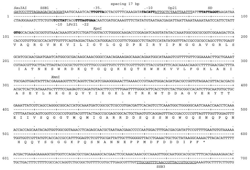
Intrigued by the fact that the location of ssb-P1 had not been determined previously, we decided to investigate an uncharacterized segment of the P1 chromosome located between map positions 15 and 24 (for a circular map of bacteriophage P1, see Yarmolinsky and Lobocka [59]). We cloned the restriction fragments EcoRI-4 and EcoRI-10 of the P1 isolate P1-15::Tn2680, determined the nucleotide sequence of these two fragments, and aligned them with respect to each other. The resulting 7,885-bp sequence includes the gene lysA, coding for the P1 lysozyme (46), and lies adjacent to the recently published sequence of the P1 dar operon (27). Figure 1 shows a physical map of the sequence, indicating the presence of five open reading frames. Two of them, darB′ and lysA, are reading in a counterclockwise orientation, while three are oriented clockwise. Two of the latter, orf17 and orf23 (numbered according to their respective map positions on the P1 chromosome [59]), show no significant homology to other known sequences in the databases. The third open reading frame was found to code for a small, 162-amino-acid protein which showed 66% amino acid sequence identity to the E. coli SSB protein, and it was therefore called ssb-P1. Figure 2 shows the nucleotide sequence of ssb-P1, which was further corroborated by determining the corresponding sequence of an independent P1 isolate, P1Cm. The P1 ssb gene starts with a GTG codon and is preceded by a weak E. coli consensus promoter (17). Immediately downstream of the −10 region of the E. coli promoter, a 17-bp asymmetric consensus binding site for the major repressor protein C1 (13, 52) of bacteriophage P1 was found. This C1 binding site, Op21, was identified previously by Citron et al. (6) on a short DNA fragment excluding ssb-P1. A P1-specific late promoter sequence (32, 33), LPr21, was located immediately upstream of the ssb promoter, reading in the opposite direction, expressing the lysA gene (46).
FIG. 1.
Physical map of a segment of the P1 chromosome flanking ssb-P1. Only the cleavage sites of the restriction enzymes EcoRI, BamHI, KpnI, and PstI are shown. The open boxes represent open reading frames, and the hatched box shows the location of the resident IS1 element. A prime indicates that only part of the gene or genetic element is shown.
FIG. 2.
Nucleotide sequence of the P1 ssb gene and its promoter region. The recognition sequences of the restriction enzymes Sau3AI, KpnI, and XmnI are indicated in italic. The sequences of the two oligonucleotide primers SSB1 and SSB3, used to PCR amplify ssb-P1, are underlined. Promoter elements, like the −35, −22, and −10 sequences, and a potential Shine-Dalgarno sequence for ssb-P1 are shown in boldface. The 17-bp binding site of the major P1 repressor protein C1, Op21, is underlined. The open reading frame of ssb-P1, starting with a GTG codon, shown in boldface, is translated into the single-letter amino acid code below the sequence. The nucleotide sequence shown is part of a larger sequence deposited in the GenBank Nucleotide Sequence Data Library.
Regulation of ssb-P1 expression.
The arrangement of promoter elements shown in Fig. 2 indicated that ssb-P1 is expressed from a weak E. coli consensus promoter, regulated by the repressor proteins C1 (12, 19) and Lxc (53). The major P1 repressor protein C1 binds to 17-bp asymmetrical sequences with the consensus ATT GCT CTA ATA AAT TT and reduces the activity of promoter sequences located in the vicinity (21). The auxiliary repressor protein Lxc does not bind DNA on its own but interacts with DNA-bound C1 and in such a ternary complex usually increases the level of repression exerted by C1 (53). However, as Lxc also lowers the concentration of C1 protein in the cell, its effect on different c1-regulated promoter sequences can vary significantly (21, 50). The 17-bp sequence found in the promoter of ssb-P1, ATT GCT CTA ATT AAT TT, shows only a single mismatch (shown in boldface) with the consensus C1 binding site. To experimentally confirm the idea that ssb-P1 is regulated from the promoter shown in Fig. 2, we constructed a fusion of ssb-P1 to lacZ (see Materials and Methods). The resulting indicator plasmid, pHAL252, was then assayed in the presence or absence of different P1 functions. In Table 1, the 685 Miller activity units expressed from pHAL252 in the absence of any P1 functions was set to 100%. If the major repressor protein C1 was expressed from plasmid pAM2b, expression of the SSB-P1–LacZ fusion protein from pHAL252 was reduced to less than 50%, demonstrating that the ssb-P1 promoter is indeed regulated by C1. If the corepressor protein Lxc was expressed, in addition to C1, from plasmid pAM8, expression from pHAL252 was further reduced. In the presence of a P1 lysogen, either P1-15::Tn2680, P1Cmclr.100, or P1Cm, expression from pHAL252 was reduced to background levels, showing that SSB-P1 is not expressed during lysogenic growth. These results agree with the finding of Johnson (28) that wild-type P1 does not suppress an E. coli ssb(Ts) mutation. These in vivo results also confirm the in vitro data of Citron et al. (6) showing that Op21 is a functional binding site for the C1 repressor protein.
TABLE 1.
Regulation of ssb-P1 expression
| Strain | Relevant P1 functionsa | Miller LacZ unitsb | % Activityc | Phenotype of colonies on X-Gal plates at:
|
|
|---|---|---|---|---|---|
| 30°C | 42°C | ||||
| UT580(pNM481) | 51 ± 11 | 7 | White | White | |
| UT580(pHAL252) | 685 ± 69 | 100 | Blue | Blue | |
| UT580(pHAL252, pAM1) | 671 ± 74 | 98 | Blue | Blue | |
| UT580(pHAL252, pAM2b) | c1′ | 309 ± 39 | 45 | Blue | Blue |
| UT580(pHAL252, pAM8) | c1′; lxc′ | 223 ± 36 | 33 | Blue | Blue |
| UT580(pHAL252, P1-15::Tn2680) | c1(Ts); lxc | 67 ± 12 | 10 | White | Blue |
| UT580(pHAL252, P1Cmclr.100) | c1(Ts); lxc | 74 ± 12 | 11 | White | Blue |
| UT580(pHAL252, P1Cm) | c1; lxc | 86 ± 15 | 13 | White | White |
| UT580(pHAL252, P1Cm lxc-2) | c1; lxc* | 92 ± 18 | 13 | Blue | Blue |
The P1 functions marked with a prime are plasmid encoded, while all others are located on different P1 prophages. For more information, see Materials and Methods.
The LacZ values (± standard deviations) are the averages of at least six independent measurements.
The value for strain UT580(pHAL252) was set to 100%.
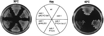
Our inability to detect significant expression from pHAL252 in the presence of a P1Cm lxc-2 prophage was unexpected (Table 1). This phage is able to suppress a temperature-sensitive E. coli mutant (Fig. 3) (42), and thus SSB-P1 is expected to be expressed at least in small amounts. A small difference in the expression of ssb-P1 between P1Cm and P1Cm lxc-2 was detected by using an X-Gal-based qualitative LacZ assay (Table 1), which is more sensitive to low levels of protein than a Miller-type LacZ assay (38) due to a much lower background level. Colonies of strain UT580(pHAL252, P1Cm lxc-2) appear blue when grown on X-Gal plates, indicating expression of the lacZ fusion construct, while colonies of the isogenic strain carrying the wild-type lysogen P1Cm remain white. This result shows that only very small amounts of SSB-P1 are expressed from a P1Cm lxc-2 lysogen. Similarly low levels of SSB-P1 are expressed during lytic growth of P1, as shown in Table 1. White colonies of the strains UT580(pHAL252, P1-15::Tn2680) and UT580(pHAL252, P1Cmclr.100) turn blue when the temperature-sensitive prophages are induced to grow lytically at 42°C.
FIG. 3.
Rescue of an E. coli ssb-1 mutant by ssb-P1. The bacterial strains KLC438 (wt), KLC436 (ssb-1) and derivatives of KLC436 (ssb-1) carrying either P1Cm lxc-2, P1Cm, pHAL251, or pACYC184 were grown in Luria broth at 30°C. Aliquots of cultures in logarithmic growth phase were spread onto prewarmed plates. Duplicate plates were incubated overnight at 30 and 42°C.
Rescue of an E. coli ssb(Ts) mutant.
In order to determine if ssb-P1 was essential and sufficient to rescue a temperature-sensitive E. coli ssb mutant at 42°C, we cloned the P1 gene under the control of its own promoter into the cloning vector pACYC184. The resulting plasmid, pHAL251, was then transformed into strain KLC436 (51) carrying the ssb-1 allele, conferring temperature-sensitive lethality due to rapid cessation of DNA replication (14). Overnight cultures of this strain and appropriate controls were grown at 30°C and then spread in duplicate in a sector of an agar plate. The duplicates were incubated overnight at 30 and 42°C. Figure 3 shows that all strains grew at the permissive temperature of 30°C. The KLC438 wild-type parent of KLC436 was not temperature sensitive, while KLC436, as expected, did not grow at 42°C. The lysogenic strain KLC436 (P1Cm), carrying a wild-type P1, was also temperature sensitive, while rescue at high temperature was observed in the presence of P1Cm lxc-2, confirming the results of Johnson (28) and Rosner (42). Growth at 42°C was observed in the presence of pHAL251, but not in the presence of the parent plasmid, pACYC184, indicating that the cloned P1 ssb gene was both essential and sufficient to rescue an E. coli mutant carrying the ssb-1 allele.
Alignment of SSB proteins.
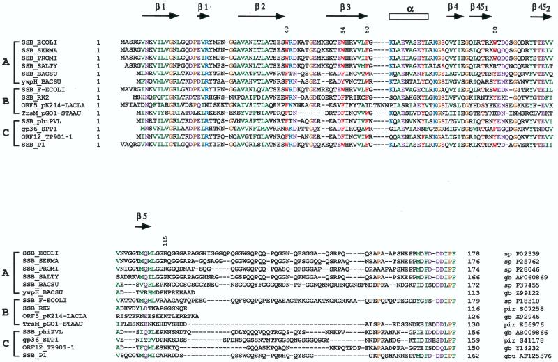
Searching the GenBank and Swiss-Prot databases we found many homologues of the P1 SSB protein. These homologues were encoded by bacteria, mitochondria, a number of broad- and narrow-host-range plasmids, and other bacteriophages with host spectra different from that of P1 (data not shown). A multiple sequence alignment including only a relevant subset of P1 SSB homologues is shown in Fig. 4.
FIG. 4.
Multiple sequence alignment of single-stranded DNA-binding proteins. The corresponding database accession number is given at the right side of each sequence (sp, Swiss-Prot; gb, GenBank; gbu, GenBankupdate; pir, Protein Identification Resource). The secondary-structure elements shown at the top of the figure are in accordance with the three-dimensional structure of the E. coli SSB protein (Protein Database Brookhaven PDB 1KAW) (41). Residues which are conserved in more than 70% of the sequences are highlighted by colors. Hydrophobic residues are shown in green, glycine and proline are in yellow, serine and threonine are in brown, aromatic residues important for DNA-binding are in red (with numbering according to the E. coli sequence), negatively charged residues and their amines are in violet, and positively charged residues are in blue. The brackets labeled A to C group sequences of bacterial, plasmid, and bacteriophage origin, respectively.
There are two highly conserved portions of the SSB proteins. The first includes the amino-terminal amino acids 1 to 115 (the numbers are in reference to the E. coli sequence [Fig. 4]) and corresponds to the DNA-binding domain (30, 56). This domain is also sufficient for the essential tetramerization of the SSB protein, as was demonstrated for an E. coli SSB proteolytic fragment, SSBc, and several deletion mutants (30, 56). The second conserved portion, located at the carboxy-terminal ends of the proteins, is a relatively short stretch of 15 to 23 amino acid residues, containing a remarkably conserved acidic patch located in the last five amino acid residues. According to recent data, this domain is involved in direct interactions between SSB and the χ subunit of the E. coli DNA polymerase III (29).
Intensive studies of a number of SSB homologues demonstrated that they were analogous in many ways (8, 36). The crystal structures of two members of the SSB family have been determined (41, 57), and their comparative analysis supported the idea that the members of the SSB family use common structural principles in order to bind to single-stranded DNA (41).
DISCUSSION
Several studies reporting mutational analyses of bacteriophage P1 failed to identify ssb-P1 (48, 49, 54, 55). Our result showing that the SSB proteins of P1 and E. coli are functionally interchangeable might account for this failure, as any mutations in ssb-P1 might well go unnoticed when assayed in E. coli. Also, several attempts to clone ssb-P1 failed (47), perhaps due to the close proximity of ssb-P1 and lysA, the gene coding for the P1 lysozyme (46). It was reported that even weak expression of lysA is very deleterious to host cells (46). While lysA in its natural context is expressed from a P1-specific late promoter sequence (33) and thus is not expressed in the absence of the phage-specific activator protein gp10 (32), indirect low-level expression from a promoter in the cloning vector might be sufficient to kill cells containing a plasmid carrying the lysA gene. In the presence of P1 repressor proteins, the inadvertent expression of the lysozyme might be prevented by the C1-Lxc repressor complex binding to Op21 (Fig. 2). However, under such conditions the ssb-P1 gene will not be expressed and thus might again go unnoticed. Only after we managed to separate ssb-P1 from lysA was it possible to analyze the function of the former gene.
The P1 ssb gene is located in close proximity to the resident IS1 element, and thus it can be speculated that P1 obtained the gene during a transposition event. However, the very strict and phage-specific regulation of ssb-P1 argues against a recent acquisition of the gene by the phage. The expression of SSB-P1 exclusively during lytic growth indicates a function of the protein related to vegetative DNA replication. Some bacteriophages, like T4, T7, and φ29, specify a complete set of replication proteins and are therefore independent of the host replication machinery (31). Unlike these phages, bacteriophage P1 does not specify a complete set of replication proteins, as its vegetative replication depends on DNA polymerase III (DnaE) and primase (DnaG) activities of the host (18). Nevertheless, P1 does specify several replication-associated proteins, like the lytic replication initiator protein RepL (7), a DnaB-like helicase (9), a Dam methyltransferase (13), and a homologue of the theta subunit of DNA polymerase III (34), in addition to SSB-P1. These proteins, with the exception of RepL, are homologous to the respective E. coli proteins and thus appear redundant. Indeed, it has been shown that the ssb genes of several conjugative plasmids are dispensable (11, 16, 23). However, the strong conservation of key residues important for SSB function indicates that the maintenance of ssb has to have some selective advantage for the phage or the plasmids. It is conceivable that subtle differences between the proteins of the episome and the host might allow the former to exert specific control over key regulatory steps during vegetative replication or conjugation. Alternatively, it cannot be excluded that P1 or the analyzed conjugative plasmids might encounter host bacteria which differ considerably from E. coli, and in such a host the SSB proteins expressed by the episomal genetic elements might well turn out to be essential.
That highly homologous ssb genes are encoded by both gram-negative and gram-positive bacteria, as well as by some of their plasmids and bacteriophages, raises some evolutionary questions about the possible origin of the gene and the mechanisms by which it is disseminated. A careful phylogenetic analysis might provide some answers to such questions.
ACKNOWLEDGMENTS
We thank J. Lee Rosner for personal communications and for the phage P1Cm lxc-2, Solvej Oestergaard and Finn K. Vogensen for making the complete nucleotide sequence of the ssb gene of TP901-1 available to us prior to publication, and Mathias Velleman and the late Heinz Schuster for personal communications.
This work was supported by a grant from the Statens Naturvidenskabelige Forskningsråd to H.L.
REFERENCES
- 1.Altschul S F, Madden T L, Schaffer A H, Zhang J, Zhang Z, Miller W, Lipman D J. Gapped BLAST and PSI-BLAST: a new generation of protein database search programs. Nucleic Acids Res. 1997;25:3389–3402. doi: 10.1093/nar/25.17.3389. [DOI] [PMC free article] [PubMed] [Google Scholar]
- 2.Berlyn M K B, Low K B, Rudd K E. Linkage map of Escherichia coli K-12. In: Neidhardt F C, Curtiss III R, Ingraham J L, Lin E C C, Low K B, Magasanik B, Reznikoff W S, Riley M, Schaechter M, Umbarger H E, editors. Escherichia coli and Salmonella: cellular and molecular biology. 2nd ed. Vol. 2. Washington, D.C.: ASM Press; 1996. pp. 1715–1902. [Google Scholar]
- 3.Bolivar F, Rodriquez R L, Greene P J, Betlach M C, Heyneker H L, Boyer H W. Construction and characterization of new cloning vehicles. II. A multipurpose cloning system. Gene. 1977;2:95–113. [PubMed] [Google Scholar]
- 4.Chang A C Y, Cohen S N. Construction and characterization of amplifiable multicopy DNA cloning vehicles derived from the p15A cryptic miniplasmid. J Bacteriol. 1978;134:1141–1156. doi: 10.1128/jb.134.3.1141-1156.1978. [DOI] [PMC free article] [PubMed] [Google Scholar]
- 5.Chase J W. The role of E. coli single-stranded DNA binding protein in DNA metabolism. Bioessays. 1984;1:218–222. [Google Scholar]
- 6.Citron M, Velleman M, Schuster H. Three additional operators, Op21, Op68, and Op88 of bacteriophage P1. J Biol Chem. 1989;264:3611–3617. [PubMed] [Google Scholar]
- 7.Cohen G, Sternberg N L. Genetic analysis of the lytic replication of bacteriophage P1. I. Isolation and partial characterization. J Mol Biol. 1989;207:99–109. doi: 10.1016/0022-2836(89)90443-9. [DOI] [PubMed] [Google Scholar]
- 8.Curth U, Urbanke C, Greipel J, Gerberding H, Tiranti V, Zeviani M. Single-stranded-DNA-binding proteins from human mitochondria and Escherichia coli have analogous physicochemical properties. Eur J Biochem. 1994;221:435–443. doi: 10.1111/j.1432-1033.1994.tb18756.x. [DOI] [PubMed] [Google Scholar]
- 9.D’Ari R, Jaffé-Brachet A, Touati-Schwartz D, Yarmolinsky M B. A dnaB analog specified by bacteriophage P1. J Mol Biol. 1975;94:341–366. doi: 10.1016/0022-2836(75)90207-7. [DOI] [PubMed] [Google Scholar]
- 10.Devereux J, Haeberli P, Smithies O. A comprehensive set of sequence analysis programs for the VAX. Nucleic Acids Res. 1984;12:387–395. doi: 10.1093/nar/12.1part1.387. [DOI] [PMC free article] [PubMed] [Google Scholar]
- 11.de Vries J, Wackernagel W. Cloning and sequencing of the Proteus mirabilis gene for a single-stranded DNA-binding protein (SSB) and complementation of Escherichia coli ssb point and deletion mutations. Microbiology. 1994;140:889–895. doi: 10.1099/00221287-140-4-889. [DOI] [PubMed] [Google Scholar]
- 12.Dreiseikelmann B, Velleman M, Schuster H. The c1 repressor of bacteriophage P1. J Biol Chem. 1988;263:1391–1397. [PubMed] [Google Scholar]
- 13.Eliason J L, Sternberg N L. Characterization of the binding sites of c1 repressor of bacteriophage P1. Evidence for multiple asymmetric sites. J Mol Biol. 1987;198:281–293. doi: 10.1016/0022-2836(87)90313-5. [DOI] [PubMed] [Google Scholar]
- 14.Glassberg J, Meyer R R, Kornberg A. Mutant single-strand DNA binding protein of Escherichia coli: genetic and physiological characterization. J Bacteriol. 1979;140:14–19. doi: 10.1128/jb.140.1.14-19.1979. [DOI] [PMC free article] [PubMed] [Google Scholar]
- 15.Golub E I, Low K B. Conjugative plasmids of enteric bacteria from many different incompatibility groups have similar genes for single-stranded DNA-binding proteins. J Bacteriol. 1985;162:235–241. doi: 10.1128/jb.162.1.235-241.1985. [DOI] [PMC free article] [PubMed] [Google Scholar]
- 16.Golub E I, Low K B. Derepression of single-stranded DNA-binding protein genes on plasmid derepressed for conjugation, and complementation of an E. coli ssb-mutation by these genes. Mol Gen Genet. 1986;204:410–416. doi: 10.1007/BF00331017. [DOI] [PubMed] [Google Scholar]
- 17.Hawley D K, McClure W R. Compilation and analysis of Escherichia coli promoter and DNA sequences. Nucleic Acids Res. 1983;11:2237–2255. doi: 10.1093/nar/11.8.2237. [DOI] [PMC free article] [PubMed] [Google Scholar]
- 18.Hay N, Cohen G. Requirement of E. coli DNA synthesis functions for the lytic replication of bacteriophage P1. Virology. 1983;131:193–206. doi: 10.1016/0042-6822(83)90545-7. [DOI] [PubMed] [Google Scholar]
- 19.Heinrich J, Riedel H-D, Baumstark B R, Kimura M, Schuster H. The c1 repressor of bacteriophage P1 operator-repressor interaction of wild-type and mutant repressor protein. Nucleic Acids Res. 1989;17:7681–7692. doi: 10.1093/nar/17.19.7681. [DOI] [PMC free article] [PubMed] [Google Scholar]
- 20.Heinrich J, Riedel H-D, Rückert B, Lurz R, Schuster H. The lytic replicon of bacteriophage P1 is controlled by an antisense RNA. Nucleic Acids Res. 1995;23:1468–1474. doi: 10.1093/nar/23.9.1468. [DOI] [PMC free article] [PubMed] [Google Scholar]
- 21.Heinzel T, Velleman M, Schuster H. ban operon of bacteriophage P1. Mutational analysis of the c1 repressor-controlled operator. J Mol Biol. 1989;205:127–135. doi: 10.1016/0022-2836(89)90370-7. [DOI] [PubMed] [Google Scholar]
- 22.Heinzel T, Velleman M, Schuster H. The c1 repressor inactivator protein Coi of bacteriophage P1. Cloning and expression of coi and its interference with C1 repressor function. J Biol Chem. 1990;265:17928–17934. [PubMed] [Google Scholar]
- 23.Howland C J, Rees C E D, Barth P T, Wilkins B M. The ssb gene of plasmid ColIb-P9. J Bacteriol. 1989;171:2466–2473. doi: 10.1128/jb.171.5.2466-2473.1989. [DOI] [PMC free article] [PubMed] [Google Scholar]
- 24.Hübner P, Haffter P, Iida S, Arber W. Bent DNA is needed for recombinational enhancer activity in the site-specific recombination system Cin of bacteriophage P1. The role of FIS protein. J Mol Biol. 1989;205:493–500. doi: 10.1016/0022-2836(89)90220-9. [DOI] [PubMed] [Google Scholar]
- 25.Iida S, Arber W. On the role of IS1 in the formation of hybrids between bacteriophage P1 and the R plasmid NR1. Mol Gen Genet. 1980;177:261–270. doi: 10.1007/BF00267437. [DOI] [PubMed] [Google Scholar]
- 26.Iida S, Arber W. Plaque forming, specialized transducing phage P1: isolation of P1CmSmSu, a precursor of P1Cm. Mol Gen Genet. 1977;153:259–269. doi: 10.1007/BF00431591. [DOI] [PubMed] [Google Scholar]
- 27.Iida S, Hiestand-Nauer R, Sandmeier H, Lehnherr H, Arber W. Accessory genes in the darA operon of bacteriophage P1 affect antirestriction function, generalized transduction, head morphogenesis, and host cell lysis. Virology. 1998;251:49–58. doi: 10.1006/viro.1998.9405. [DOI] [PubMed] [Google Scholar]
- 28.Johnson B F. Suppression of the lexC (ssbA) mutation of Escherichia coli by a mutant of bacteriophage P1. Mol Gen Genet. 1982;186:122–126. doi: 10.1007/BF00422923. [DOI] [PubMed] [Google Scholar]
- 29.Kelman Z, Yuzhakow A, Andjelkovic J, O’Donnell M. Devoted to the lagging strand—the χ subunit of DNA polymerase III holoenzyme contacts SSB to promote processive elongation and sliding clamp assembly. EMBO J. 1998;17:2436–2449. doi: 10.1093/emboj/17.8.2436. [DOI] [PMC free article] [PubMed] [Google Scholar]
- 30.Kinebuchi T, Shindo H, Nagai H, Shimamoto N, Shimizu M. Functional domains of Escherichia coli single-stranded DNA binding protein as assessed by analyses of the deletion mutants. Biochemistry. 1997;36:6732–6738. doi: 10.1021/bi961647s. [DOI] [PubMed] [Google Scholar]
- 31.Kornberg A, Baker T. DNA replication. 2nd ed. New York, N.Y: W. H. Freeman and Company; 1992. [Google Scholar]
- 32.Lehnherr H, Guidolin A, Arber W. Bacteriophage P1 gene 10 encodes a trans-activating factor required for late gene expression. J Bacteriol. 1991;173:6438–6445. doi: 10.1128/jb.173.20.6438-6445.1991. [DOI] [PMC free article] [PubMed] [Google Scholar]
- 33.Lehnherr H, Guidolin A, Arber W. Mutational analysis of the bacteriophage P1 late promoter sequence Ps. J Mol Biol. 1992;228:101–107. doi: 10.1016/0022-2836(92)90494-5. [DOI] [PubMed] [Google Scholar]
- 34.Lehnherr H, Maguin E, Jafri S, Yarmolinsky M B. Plasmid addiction genes of bacteriophage P1: doc, which causes cell death on curing of prophage, and phd, which prevents host death when prophage is retained. J Mol Biol. 1993;233:414–428. doi: 10.1006/jmbi.1993.1521. [DOI] [PubMed] [Google Scholar]
- 35.Lennox E S. Transduction of linked genetic characters of the host by bacteriophage P1. Virology. 1955;1:190–206. doi: 10.1016/0042-6822(55)90016-7. [DOI] [PubMed] [Google Scholar]
- 36.Lohmann T M, Ferrari M E. Escherichia coli single-stranded DNA-binding protein: multiple DNA-binding modes and cooperatives. Annu Rev Biochem. 1994;63:527–570. doi: 10.1146/annurev.bi.63.070194.002523. [DOI] [PubMed] [Google Scholar]
- 37.Meyer R R, Laine P S. The single-stranded DNA-binding protein of Escherichia coli. Microbiol Rev. 1990;54:342–380. doi: 10.1128/mr.54.4.342-380.1990. [DOI] [PMC free article] [PubMed] [Google Scholar]
- 38.Miller J H. Experiments in molecular genetics. Cold Spring Harbor, N.Y: Cold Spring Harbor Laboratory Press; 1972. [Google Scholar]
- 39.Minton N P. Improved plasmid vectors for the isolation of translational lac gene fusions. Gene. 1984;31:269–273. doi: 10.1016/0378-1119(84)90220-8. [DOI] [PubMed] [Google Scholar]
- 40.Mollet B, Clerget M, Meyer J, Iida S. Organization of the Tn6-related kanamycin resistance transposon Tn2680 carrying two copies of IS26 and an IS903 variant, IS903.B. J Bacteriol. 1985;163:55–60. doi: 10.1128/jb.163.1.55-60.1985. [DOI] [PMC free article] [PubMed] [Google Scholar]
- 40a.National Center for Biotechnology Information Website. copyright date. [Online.] National Center for Biotechnology Information. http://www.ucbi.ulm.uih.gov/BLAST. [16 April 1999, last date accessed.] 11 June 1996. [Google Scholar]
- 41.Raghunathan S, Ricard C S, Lohman T M, Waksman G. Crystal structure of the homo-tetrameric DNA binding domain of Escherichia coli single-stranded DNA-binding protein determined by multiwavelength x-ray diffraction on the selenomethionyl protein at 2.9 Å resolution. Proc Natl Acad Sci USA. 1997;94:6652–6657. doi: 10.1073/pnas.94.13.6652. [DOI] [PMC free article] [PubMed] [Google Scholar]
- 42.Rosner, J. L. Personal communication.
- 43.Rosner J L. Formation, induction, and curing of bacteriophage P1 lysogens. Virology. 1972;48:679–689. doi: 10.1016/0042-6822(72)90152-3. [DOI] [PubMed] [Google Scholar]
- 44.Sambrook J, Fritsch E F, Maniatis T. Molecular cloning: a laboratory manual. 2nd ed. Plainview, N.Y: Cold Spring Harbor Laboratory Press; 1989. [Google Scholar]
- 45.Sanger F, Nicklen S, Coulson A R. DNA sequencing with chain-terminating inhibitors. Proc Natl Acad Sci USA. 1977;74:5463–5467. doi: 10.1073/pnas.74.12.5463. [DOI] [PMC free article] [PubMed] [Google Scholar]
- 46.Schmidt C, Velleman M, Arber W. Three functions of bacteriophage P1 involved in cell lysis. J Bacteriol. 1996;178:1099–1104. doi: 10.1128/jb.178.4.1099-1104.1996. [DOI] [PMC free article] [PubMed] [Google Scholar]
- 47.Schuster, H., and M. Velleman. Personal communication.
- 48.Scott J R. Clear plaque mutants of phage P1. Virology. 1970;41:66–71. doi: 10.1016/0042-6822(70)90054-1. [DOI] [PubMed] [Google Scholar]
- 49.Scott J R. Phage P1 cryptic. II. Location and regulation of prophage genes. Virology. 1973;53:327–336. doi: 10.1016/0042-6822(73)90210-9. [DOI] [PubMed] [Google Scholar]
- 50.Touati-Schwartz D. A new pleiotropic bacteriophage P1 mutation, bof, affecting c1 repression activity, the expression of plasmid incompatibility and the expression of certain constitutive prophage genes. Mol Gen Genet. 1979;174:189–202. doi: 10.1007/BF00268355. [DOI] [PubMed] [Google Scholar]
- 51.Vales L D, Chase J W, Murphy J B. Effect of ssbA1 and lexC113 mutations on lambda prophage induction, bacteriophage growth, and cell survival. J Bacteriol. 1980;143:887–896. doi: 10.1128/jb.143.2.887-896.1980. [DOI] [PMC free article] [PubMed] [Google Scholar]
- 52.Velleman M, Dreiseikelmann B, Schuster H. Multiple repressor binding sites in the genome of bacteriophage P1. Proc Natl Acad Sci USA. 1987;84:5570–5574. doi: 10.1073/pnas.84.16.5570. [DOI] [PMC free article] [PubMed] [Google Scholar]
- 53.Velleman M, Heinzel T, Schuster H. The Bof protein of bacteriophage P1 exerts its modulating function by formation of a ternary complex with operator DNA and C1 repressor. J Biol Chem. 1992;267:12174–12181. [PubMed] [Google Scholar]
- 54.Walker D H, Walker J T. Coliphage P1 morphogenesis: analysis of mutants by electron microscopy. J Virol. 1983;45:1118–1139. doi: 10.1128/jvi.45.3.1118-1139.1983. [DOI] [PMC free article] [PubMed] [Google Scholar]
- 55.Walker D H J, Walker J T. Genetic studies of coliphage P1. III. Extended genetic map. J Virol. 1976;20:177–187. doi: 10.1128/jvi.20.1.177-187.1976. [DOI] [PMC free article] [PubMed] [Google Scholar]
- 56.Williams K R, Spicer E K, LoPresti M B, Guggenheimer R A, Chase J W. Limited proteolysis studies on the Escherichia coli single-stranded DNA binding protein. J Biol Chem. 1983;258:3346–3355. [PubMed] [Google Scholar]
- 57.Yang C, Curth U, Urbanke C, Kang C H. Crystal structure of human mitochondrial single-stranded DNA binding protein at 2.4 Å resolution. Nat Struct Biol. 1997;4:153–157. doi: 10.1038/nsb0297-153. [DOI] [PubMed] [Google Scholar]
- 58.Yanisch-Perron C, Vieira J, Messing J. Improved M13 phage cloning vectors and host strains: nucleotide sequences of the M13mp18 and pUC19 vectors. Gene. 1985;33:103–119. doi: 10.1016/0378-1119(85)90120-9. [DOI] [PubMed] [Google Scholar]
- 59.Yarmolinsky M B, Lobocka M B. Bacteriophage P1. In: O’Brian S J, editor. Locus maps of complex genomes. 6th ed. Cold Spring Harbor, N.Y: Cold Spring Harbor Laboratory Press; 1993. pp. 1.50–1.61. [Google Scholar]
- 60.Yarmolinsky M B, Sternberg N L. Bacteriophage P1. In: Calendar R, editor. The bacteriophages. Vol. 1. New York, N.Y: Plenum Press; 1988. pp. 291–438. [Google Scholar]


