Abstract
Foodborne pathogens cause many diseases and significantly impact human health and the economy. Foodborne pathogens mainly include Salmonella spp., Escherichia coli, Staphylococcus aureus, Shigella spp., Campylobacter spp. and Listeria monocytogenes, which are present in agricultural products, dairy products, animal-derived foods and the environment. Various pathogens in many different types of food and water can cause potentially life-threatening diseases and develop resistance to various types of antibiotics. The harm of foodborne pathogens is increasing, necessitating effective and efficient methods for early monitoring and detection. Traditional methods, such as real-time polymerase chain reaction (RT-PCR), enzyme-linked immunosorbent assay (ELISA) and culture plate, are time-consuming, labour-intensive and expensive and cannot satisfy the demands of rapid food testing. Therefore, new fast detection methods are urgently needed. Electrochemical biosensors provide consumer-friendly methods to quickly detect foodborne pathogens in food and the environment and achieve extensive accuracy and reproducible results. In this paper, by focusing on various mechanisms of electrochemical transducers, we present a comprehensive overview of electrochemical biosensors for the detection of foodborne pathogens. Furthermore, the review introduces the hazards of foodborne pathogens, risk analysis methods and measures of control. Finally, the review also emphasizes the recent research progress and solutions regarding the use of electrochemical biosensors to detect foodborne pathogens in food and the environment, evaluates limitations and challenges experienced during the development of biosensors to detect foodborne pathogens and discusses future possibilities.
Keywords: electrochemical biosensors, foodborne pathogens, food, environment, detection
1. Introduction
Foodborne pathogens are pathogenic bacteria that can cause food poisoning or use food as a transmission medium [1]. Pathogenic bacteria directly or indirectly contaminate food and water sources, and oral infection in humans can lead to the occurrence of intestinal infectious diseases, food poisoning and the prevalence of infectious diseases in livestock and poultry [1]. Salmonella spp., Escherichia coli, Staphylococcus aureus, Shigella spp., Campylobacter spp. and Listeria monocytogenes are the major bacterial agents that cause foodborne infections [2]. As foodborne pathogens harm people’s health, people pay special attention to food safety and food biosecurity. On the one hand, foodborne pathogens in livestock, poultry and aquatic animals are treated or prevented by antibiotics, but some antibiotics are resistant to pathogenic bacteria; as a result, these antibiotics are not effective against pathogenic bacteria [3]. To protect customers from crippling and sometimes lethal instances of pathogen outbreaks, the safety of foods from farm to fork across the supply chain continuum must be guaranteed. The method of hazard analysis critical control points (HACCPs) is one preventive strategy that can be used to ensure safety; however, its full potential will not be reached unless the necessary supporting tools are created [4]. Therefore, a rapid, sensitive and accurate detection method combined with HACCPs must be established to improve the safety of foods.
The presence of preliminaries in ready-to-eat (RTE) foods is a serious problem because these products usually have not received any further treatment before consumption. In fact, outbreaks of foodborne pathogens originate from undercooked or processed RTE meats, dairy products, fruits and vegetables [5,6,7]. Agricultural products (vegetables and fruits), animal-derived foods (meat, milk and eggs) and the environment (water and soil) are the most important reservoirs for many foodborne pathogens [8,9,10,11,12]. Therefore, fruits, vegetables, seafood, meat, eggs and milk products may carry Salmonella, Staphylococcus aureus, Campylobacter, Listeria, Shigella or Escherichia coli O157:H7 organisms. The traditional detection methods commonly used for foodborne pathogens include polymerase chain reaction (PCR) [13,14,15], enzyme-linked immunosorbent assay (ELISA) [16,17,18] and culture plate [19]. However, traditional detection techniques are limited by disadvantages, such as large time costs, low efficiency and complex equipment. The test paper method exhibits several advantages, as it is efficient, portable and convenient to operate; thus, multiple foodborne pathogens in food and the environment can be quantitatively detected by this method [20]. Biosensor detection technology exhibits several advantages, including strong selectivity, high accuracy and short detection time, and has attracted widespread attention and been applied to detect foodborne pathogens [21,22,23,24,25]. Compared with traditional detection technology, advanced methods (test strips and biosensors) offer technological innovation and can efficiently, quickly and conveniently detect foodborne pathogens in food and the environment [26].
Electrochemical biosensors detect foodborne pathogens based on potentiometry, conductometry and impedimetry [27]. Due to their advantages, including rapid processes, high sensitivity, high specificity, low cost, portability, miniaturization and point-of-care detection, electrochemical biosensors have been widely used in the fields of food, biology and life sciences [28,29]. Electrochemical biosensors provide a rapid, efficient and alternative method for detecting foodborne pathogens to ensure the safety of RTE foods and can be used as stand-alone devices for on-site monitoring. Nanomaterials (NMs) employed in the fabrication and nanobiosensors include metallic nanoparticles, carbon nanotubes (CNTs), organic nanoparticles, metal oxide nanoparticles and silica nanoparticles [30]. Furthermore, these nanomaterials can act as transduction elements, thereby improving the sensitivity and detection limit of the electrochemical biosensor method [31,32]. Therefore, the selection of a highly specific bioreceptor in combination with a nanomaterial is essential for electrochemical biosensor development, which can quickly and efficiently detect foodborne pathogens [33].
This review attempts to provide a comprehensive overview of the detection of foodborne pathogens through rapid, sensitive and accurate electrochemical biosensor methods for food and environmental research. In addition, this review introduces the principle of electrochemical biosensors, focuses on the hazards, risk analysis and control of foodborne pathogens, discusses the recent progress and limitations of electrochemical biosensors in foodborne pathogen detection and proposes some solutions and future challenges.
2. Principle of Electrochemical Biosensors
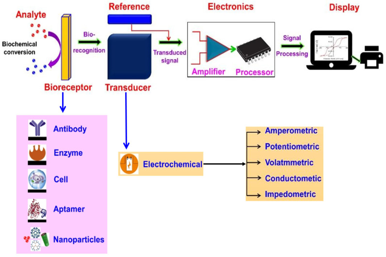
A typical electrochemical biosensor consists of an analyte (e.g., Salmonella, Staphylococcus aureus, Campylobacter and Listeria), bioreceptor (e.g., antibodies, enzymes, cells, aptamers and nanoparticles), electrochemical transducer, electronics and display [32,34]. The most extensively studied and applied class of biosensors, electrochemical biosensors, depend on the electrochemical nature of the analyte and the transducer for their operation [35]. The electrochemical biosensor is based on the principle that a bioreceptor and analyte interact electrochemically on the transducer surface, resulting in detectable electrochemical signals; this signal is measured in terms of voltage, current, impedance and capacitance, allowing for the quantitative or qualitative analysis of the analyte [36]. Figure 1 depicts the electrochemical biosensor’s working principle.
Figure 1.
Schematic diagram of a typical electrochemical biosensor consisting of a bioreceptor, transducer, electronic system (amplifier and processor) and display (PC or printer) for the detection of foodborne pathogens. Adapted with permission from Naresh and Lee [32]. Copyright 2021, MDPI.
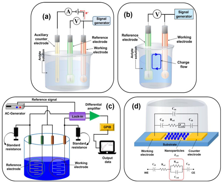
The combination of various bioreceptors (antibodies, DNA, enzymes, microbes or cells) and electrochemical converters (current, potential, voltage, conductance and impedance) can constitute a variety of electrochemical biosensors. Electrochemical biosensors are divided into amperometric, potentiometric, voltammetric, conductometric and impedimetric biosensors based on the transduction principle [37]. Figure 2 shows schematic designs for the following different types of biosensors: (a) amperometric/voltammetric, (b) potentiometric, (c) conductometric and (d) impedimetric biosensors. Compared with other biosensors, electrochemical biosensors exhibit several advantages, including high sensitivity, good selectivity, fast response, small sample dosage and easy-to-achieve multicomponent measurement [38]. At present, electrochemical biosensor technology has been widely used in the detection of foodborne pathogens.
Figure 2.
Schematic diagram of (a) amperometric/voltametric; (b) potentiometric; (c) conductometric biosensors; and (d) equivalent circuit of the impedimetric biosensor (Cdl = double-layer capacitance of the electrodes; Rsol = resistance of the solution; Cde = capacitance of the electrode; Zcell = impedance introduced by the bound nanoparticles; Rcell and Ccell = the resistance and capacitance in parallel, respectively). Adapted with permission from Naresh and Lee [32]. Copyright 2021, MDPI.
3. Foodborne Pathogens: Hazards, Risk Analysis and Control
Foodborne illness is a major cause of morbidity and continues to pose a serious danger to public health worldwide. Foodborne illnesses are most frequently caused by bacteria, which exhibit a range of sizes, varieties and characteristics. Foodborne illness starts with the production of breeding animals, vegetables and fruits during processing; it is then transported to the supermarket or farmer’s market and is finally passed to consumers. Therefore, based on the needs of consumers in production and processing, it is necessary to design an effective and safe food safety management system to control and reduce the harm and risks caused by foodborne bacteria. This review mainly introduces the hazards, risk analysis methods and measures used to control Salmonella spp., Escherichia coli, Staphylococcus aureus, Shigella spp., Campylobacter spp. and Listeria monocytogenes.
3.1. Salmonella spp.
Theobald Smith isolated Salmonella bacteria from pig intestines infected with classical swine fever in 1885 [39]. Salmonella is a flagellated Gram-negative, non-spore-forming bacillus and facultative anaerobe that thrives at temperatures from 35 to 37 °C [40]. Salmonella has a complex antigen structure, which can generally be divided into somatic antigen (O), flagella antigen (H) and surface antigen (Vi) [41]. This bacterium is well known as a foodborne pathogen because most infections are acquired through food. The bacteria cause salmonellosis, and the main symptoms include nausea, vomiting, abdominal pain, headache, chills and diarrhoea [42]. The people who are the most likely to be infected with Salmonella are infants or children under 5 years of age, elderly individuals and immune-damaged people [39]. Salmonellosis can be acquired from the ingestion of food and water contaminated with Salmonella or exposure to an environment contaminated with faeces containing Salmonella [43]. The consumption of undercooked food from infected animals in poultry products, other meats, raw milk, dairy products made from raw milk, RTE foods (such as fruits and vegetables contaminated with faeces of infected animals) or water contaminated with the faeces of infected people or animals could all be sources of contamination [43,44]. Salmonella infection is a common outbreak of diseases worldwide, including in European and American countries [45,46]. Therefore, the study of Salmonella has always been a hot topic. To prevent an outbreak of salmonellosis, we can take some preventive measures, including introducing sanitary environments at farms, treating faeces in a no-risk manner and treating feed and water [47]. During the breeding process, some antibiotics and vaccines can be used to inhibit the growth of Salmonella, but attention must be focused on the amount of antibiotics, the dosage period and elimination law [48,49]. Based on the growth temperature of Salmonella, pathogenic bacteria can be killed at high temperature [40]. To control Salmonella, people can use high-temperature cooking methods when preparing animal products, such as meat, milk and eggs.
3.2. Escherichia coli
Escherichia coli (E. coli) is a Gram-negative, facultative anaerobic rod that inhabits the intestinal tract of animals and humans from birth [50]. E. coli is a member of the natural microbial community of the animal and human gut. It produces useful vitamins and competes with and inhibits the growth of pathogenic bacteria that may be present or consumed with food and water, among other beneficial functions in the body [40]. Many of these E. coli strains are not pathogenic, and only a small part causes various diseases of animals and humans under certain conditions [51]. According to serological classification, E. coli strains can be divided into somatic antigen (O), flagellar antigen (H) and capsule antigen (K) [52]. Based on the mechanism by which the gastrointestinal pathogenic E. coli causes illnesses, it is divided into the following major foodborne diarrhoeagenic E. coli pathotypes: Shiga toxin-producing E. coli/enterohemorrhagic E. coli (STEC/EHEC), enteropathogenic E. coli (EPEC), enteroinvasive E. coli (EIEC), enterotoxigenic E. coli (ETEC) and enteroaggregative E. coli (EAEC) [53]. Pathogenic E. coli strains can cause intestinal gastroenteritis, urinary tract infections, meningitis infections and blood infections [52]. The sickness caused by the bacterium E. coli, which typically lives in the lower intestines of most warm-blooded mammals, is known as “colibacillosis.” It is mainly caused by infections, such as specific bacterial wool antigen and pathogenic toxins. E. coli has been utilized as a sign of faecal contamination for almost a century since it is one of the predominant enteric species in human faeces, in addition to anaerobic bacteria [40]. The concept of indicators is based on the premise that the presence of E. coli in food or water is proof that it has been faeces-contaminated and may also be evidence of the presence of pathogens. Although the use of E. coli as a faecal indicator has been criticized for being unreliable because it can be found in environmental sources, it is nevertheless used as an indicator of cleanliness throughout the world because no adequate replacement has been suggested. In recent years, pathogenic E. coli has caused many foodborne outbreaks in industrialized countries via the faecal–oral route because it is consumed in contaminated meat, vegetables, fruits and water [54]. O157:H7 and some of the other pathogenic E. coli families have been well documented for transmitting secondary infections through animal or person-to-person contact [55]. E. coli has been exposed to antibiotics for a long time in humans and animal intestines; as a result, E. coli is resistant to many antibiotics (β-lactams, quinolones, aminoglycosides, tetracyclines, sulphonamides and phenicols) [56]. From a One Health perspective, antimicrobial resistance in E. coli is a problem of the utmost concern because it affects both the human and animal sectors. Considering the causes of pathogenic E. coli and drug resistance, some measures can be taken to control the bacteria, such as sterilizing milk and juice through the Pakistani method, cooking meat and effectively washing RTE foods.
3.3. Staphylococcus aureus
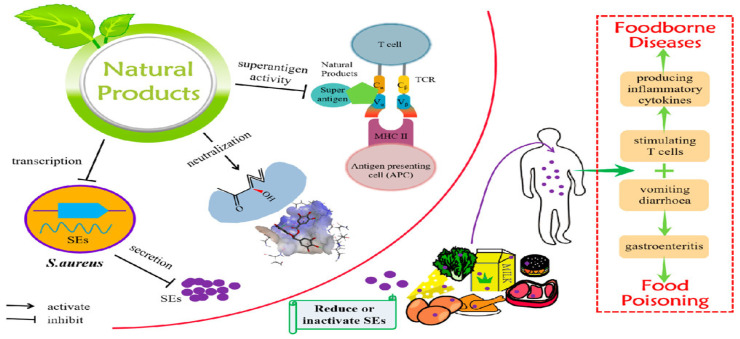
The genus Staphylococcus contains more than 30 species, of which Staphylococcus aureus (S. aureus) has the greatest effect on human health [57]. S. aureus is a common Gram-positive bacterium with a diameter of approximately 1 μm [58]. The temperature and pH range for the growth of S. aureus are 7–49 °C and 4–9, respectively, and the best growth temperature and pH are 30–37 °C and 7, respectively [44]. S. aureus is a serious bacterial pathogen that can lead to a wide range of illnesses, including food poisoning, toxic shock syndrome, wound infections and skin infections [59]. S. aureus is a common dweller (commensal) of the skin, nares, respiratory tracts and genitalia of both humans and animals [59]. However, as an opportunistic pathogen, it can cause invasive and deadly infections in a variety of organs. A significant amount of extracellular proteins and toxins are produced by S. aureus. Given that many S. aureus strains produce enterotoxins, the growth and spread of S. aureus in foods pose a potential risk to consumer health [60]. The most significant toxins are known as staphylococcal enterotoxins (SEs) and SE-like toxins (SEls), and these toxins have the following factors in common: they are structurally identical proteolytic enzymes that are resistant to heat, are superantigenic and exert emetic effects [61,62]. In addition, drug-resistant S. aureus strains have become one of the most common pathogens recovered from hospital-associated (nosocomial) infections, which is of particular public health concern [63]. Due to the medicinal resistance and heat resistance of these enterotoxins, the treatment and control of S. aureus remains a challenge. Therefore, the main goal should be to stop S. aureus from growing and contaminating food. According to the growth conditions of S. aureus, deep cooking can effectively prevent the harm caused by S. aureus. Additionally, a number of natural products can be employed to effectively lower the toxicity of SEs and the prevalence of foodborne diseases; these products can also serve as food antibacterial agents in place of antibiotics and chemical preservatives [64]. In Figure 3, Liu et al. [64] presented information on the toxicity of SEs, the types of food that are contaminated by SEs and the sources and methods by which SEs can contaminate food. This information will help to manage and lower the rate by which SEs contaminate food.
Figure 3.
Mechanism by which natural products prevent foodborne diseases induced by SEs. Adapted with permission from Liu et al. [64]. Copyright 2022, American Chemical Society.
3.4. Shigella spp.
Shigella are pathogens that originate in the Escherichia genus but are commonly categorized as a different genus [65]. Shigella spp. are Gram-negative bacteria that cause the intestinal infection known as shigellosis [66]. Shigella may grow at pH levels of 6 to 8 and in a wide range of temperatures (from 10 to 48 °C) [67]. It is possible to isolate Shigella spp. from a variety of food sources, and it causes several outbreaks and sporadic cases of foodborne diseases worldwide. Typically, moist items touched with bare hands, such as salads, uncooked veggies, fruits, shellfish and water, are linked to shigellosis [68]. The most common symptoms of shigellosis are diarrhoea, fever, nausea, vomiting, gastrointestinal bloating and constipation [69]. Shigella species and EIEC both produce diarrhoeal illnesses using the same invasive mechanism [70]. Shigella spp. can cause many people to develop and even show high mortality, which seriously endangers public health [71,72,73]. Shigella infection therapy with antibiotics is crucial for lowering the disease’s prevalence and fatality rates [74]. Ciprofloxacin is recommended by the World Health Organization (WHO) as a first-line treatment for shigellosis, and second-line treatments include azithromycin, ceftriaxone or pivmecillinam [75]. However, many antibiotics have caused the strains of Shigella to produce multidrug resistance, including β-lactams, fluoroquinolones, macrolides, tetracyclines and phenicols, thereby limiting the effects of their antibiotic resistance to severe infection [76,77,78,79,80,81,82]. Some preventive measures for foodborne shigellosis include the removal of faeces, ensuring safe drinking water, developing good personal hygiene habits, avoiding cross-infection of RTE foods and using appropriate water–chloride-washed vegetables for salad and refrigerated food. WHO, a global institution that has extensively focused on this subject, has emphasized the significance of creating an effective vaccination against Shigella. Due to the multidrug resistance of Shigella spp., scientific researchers are developing vaccines to produce corresponding antibodies by activating the body’s immune system, thereby effectively controlling these pathogenic strains of Shigella [63,83]. It is believed that these vaccines developed for Shigella can pass clinical trials in the future and reduce mortality.
3.5. Campylobacter spp.
Campylobacter (C.) spp., Gram-negative bacteria, are responsible for human acute gastroenteritis (campylobacteriosis) worldwide, with most cases being caused by C. jejuni and C. coli [84,85]. The optimal pH and temperature range of Campylobacter growth are 6.5–7.5 and 37–42 °C, respectively. Compared to Salmonella or pathogenic E. coli, the number of cases caused by Campylobacter is much greater [86]. Human infection mainly manifests as symptoms of acute enteritis, such as diarrhoea, discomfort, fever, abdominal pain and blood in stools. Current Campylobacteriosis outbreaks have been linked to meat, raw milk, fruits and vegetables [87,88]. The main source of Campylobacter transmission in humans is poultry, specifically broiler chickens, which contain the highest concentrations of this bacteria [89,90]. One of the primary public health policies in the EU aimed at preventing campylobacteriosis is to manage this disease in poultry and poultry meat [91]. This suggests that controlling Campylobacter in chickens at the farm level can reduce the danger of human exposure to this virus and significantly improve food safety. It would be very interesting to see how biosecurity measures could reduce environmental exposure [92]. In slaughterhouses, waste can be disinfected when chickens are slaughtered, and the packaging carton can be disinfected to prevent transmission to humans by transportation. To control the spread of Campylobacter on farms, either the prevalence of infected broiler flocks must be reduced or the amount of the pathogen in the broilers’ intestines must be reduced before slaughter [93]. Although Campylobacter exhibits resistance to some antibiotics, antibiotic use remains an effective measure to control Campylobacter spp. [94]. In addition, reuterin is a broad-spectrum antimicrobial system produced by specific strains of Lactobacillus reuteri during the anaerobic metabolism of glycerol, which can effectively inhibit the potential of Campylobacter spp. [95]. Furthermore, antibiotics are anticipated to be replaced by plant-, animal-, bacterial- and marine-derived antimicrobials to suppress Campylobacter spp. [96]. The comprehensive approach (longitudinally integrated safety assurance model, LISA) across the farm–slaughterhouse–processing–retail–consumer continuum is the suggested method for preventing and controlling Campylobacter along the poultry meat chain [93].
3.6. Listeria monocytogenes
Listeria monocytogenes (Lm) is a Gram-positive, rod-shaped and psychotropic bacterium that causes listeriosis, a very uncommon but potentially fatal gastrointestinal illness [97]. The temperature and pH range for the growth of Lm are 0–45 °C and 4.1–9.6, respectively, and the optimum growth temperature is 30–35 °C [53]. The bacterium Lm has been found in a variety of environments and foods, including water, soil, sewage, silage, pasteurized milk, various fruits and vegetables and several meat products. With a high foodborne proportion of up to 99%, the consumption of infected food products is the primary method by which listeriosis is transmitted to humans [98]. Lm bacteria usually lead to intestinal infection, causing patients to show symptoms such as fever, muscle soreness, nausea and vomiting. It can also invade the nervous system and circulatory system, causing severe meningitis and sepsis [99]. The outbreak of listeriosis seriously endangers human health and causes economic losses. Therefore, the European Union has developed food safety criteria (Commission Regulation (EC) 2073/2005) for Lm in RTE foods. Although some antibiotics show resistance to Lm, the use of antibiotics remains one of the most common methods for treating Lm. Amoxicillin or ampicillin, frequently in conjunction with gentamicin, is the mainstay therapy for severe infections caused by Lm, and cotrimoxazole, fluoroquinolones, rifampicin and linezolid are alternatives to aminopenicillins [100]. To reduce Lm infection, regular disinfection must be performed in breeding environments, including the pollution-free treatment of faeces [101]. In addition, some natural or synthetic compounds can inhibit the formation of Lm biofilms, which is also a novel strategy [102]. Utilizing natural antimicrobial agents, which can serve as a viable replacement for synthetic preservatives for the production of organic food products, is among the alluring and efficient ways to limit the growth of Lm in food items [103].
In summary, the use of antibiotics or natural antibacterial agents can inhibit foodborne pathogens. To prevent the infection of foodborne pathogens, the consumption of RTE foods should be minimized, and the products should be disinfected and sealed during the entire food production chain. In addition, it is important to develop effective, fast and sensitive analysis methods to quickly identify foodborne pathogens in food and the environment.
4. Electrochemical Biosensors for the Detection of Foodborne Pathogens in Food and the Environment
This review focuses on different bioreceptors combined with electrochemical transducers to measure six types of foodborne pathogens in food and the environment. Enzymes, DNAs/RNAs, aptamers and antibodies are frequently used in bioreceptor applications [104]. In addition, numerous studies have employed nanomaterial-based biosensors for the detection of foodborne pathogens [105]. Based on different bioreceptors, we summarized the development of electrochemical biosensors for the detection of foodborne pathogens in the past ten years (2013–2023), aiming to provide the latest trends in this research field. Table 1 summarizes some published electrochemical biosensor methods for the detection of foodborne pathogens in food and the environment.
Table 1.
An overview of some reported electrochemical biosensors used for the detection of foodborne pathogens in food and the environment.
| Target Pathogen | Bioreceptor | Detection Method | Assay Strategy | Material Type | LOD | Linear Range | Matrix | Ref. |
|---|---|---|---|---|---|---|---|---|
| Salmonella spp. | DNA probe | SWV–CV–EIS | SPIA-based biosensors | AuNPs/GCE | 68 CFU/mL | 6.8 × 101–6.8 × 10⁸ CFU/mL | Animal meat | [106] |
| Salmonella spp. | DNA probe | SWV | SRCA-CRISPR/Cas12a signal amplification strategy | AuNPs/GCE | 2.08 fg/μL | 5.8 fg/μL–5.8 ng/μL | Chicken and pork | [107] |
| Salmonella spp. | Aptamer | DPV | Aptasensor | Gold nanoparticles | 200 CFU/mL | 2 × 10²–2 × 10⁶ CFU/mL | Milk | [108] |
| Salmonella spp. | Aptamer | CV–EIS–DPV | Aptasensor | rGO-AuNPs | 200 CFU/mL | 6 × 10²–6 × 10⁷ CFU/mL | Pork and beef | [109] |
| Salmonella spp. | Antibody | EIS | Immunosensors | Multilayer graphene | 13 CFU/mL | 101–105 CFU/mL | Chicken broth | [110] |
| Salmonella spp. | Antibody | DPV | Immunosensor | CoFe-MOFs-graphene | 1.2 × 10² CFU/mL | 2.4 × 10²–2.4 × 10⁸ CFU/mL | Milk | [111] |
| S. enteritidis | Bacteriophages as new molecular probes | EIS | Phage-based biosensor | GDE-AuNPs-Cys-Phage SEP37 | 1 CFU/mL | 2 × 10²–2 × 10⁵ CFU/mL | Chicken breast meat | [112] |
| S. pullorum and S. gallinarum | Antibody | CV | Immunosensor | SPCE | 16.1 CFU/mL | 101–109 CFU/mL | Chicken and eggs | [113] |
| S. typhi | DNA probe | DPV | DNA biosensor | SPE/P-Cys@AuNPs | 1 CFU/mL | 1.8–1.8 × 10⁵ CFU/mL | Blood, poultry faeces, eggs and milk | [114] |
| S. typhimurium | Magnetosome-anti-Salmonella antibody complex | EIS | Magnetosome-based biosensors | SPCE | 101 CFU/mL | 101–107 CFU/mL | Water and milk | [115] |
| S. typhimurium | Antibody | CV–EIS | Immunosensor | AuNPs/PAMAM-MWCNT-Chi/GCE | 5.0 × 10² CFU/mL | 1.0 × 10³–1.0 × 10⁷ CFU/mL |
Milk | [116] |
| S. typhimurium | Aptamer | DPV | Aptasensor | rGO-TiO2 nanocomposite | 101 CFU/mL | 101–108 CFU/mL | Chicken meat | [117] |
| S. typhimurium | DNA probe | SWV–CV–EIS | SRCA-based ratiometric electrochemical biosensor | SH-β-CD/AuNPs/GCE | 15.8 fg/μL | 30 fg/μL–30 ng/μL | Animal meat, eggs and dairy products | [118] |
| S. typhimurium | Antibody | SWV | Immunosensor | SPCE | 4 CFU/mL | 4–36 CFU/mL | Milk | [119] |
| S. typhimurium | Aptamer | CV–EIS | Aptasensor | AuNPs/GCE | 1 CFU/mL | 6.5 × 10²–6.5 × 10⁸ CFU/mL | Eggs | [120] |
| E. coli | Engineered phage | DPV | Bacteriophage-based biosensors | SWCNT-SPE | 1 CFU/mL | 1–104 CFU/mL | Spinach leaves | [121] |
| E. coli | L-cysteine | CV | Amino functionalized iron nanoparticles-based biosensors | L-Cyst-Fe3O4 NPs | 10 CFU/mL | 101–105 CFU/mL | Tap water | [122] |
| E. coli | PNA probe | Conductometry | DNA biosensor | AuNPs | 102 CFU/mL | 103–108 CFU/mL | Water | [123] |
| E. coli | Aptamer-primer probe | CV–DPV | RCA coupled DNAzyme amplification-based biosensor | Au | 8 CFU/mL | 9.4–9.4 × 10⁵ CFU/mL | Milk | [124] |
| E. coli | Antibody | CV–EIS | Immunosensor | AuSPEs | 30 CFU/mL | 101–108 CFU/mL | Drinking water | [125] |
| E. coli | Aptamer | DPV | Aptasensor | Au | 80 CFU/mL | 5.0 × 10²–5.0 × 10⁷ CFU/mL | Licorice extract | [126] |
| E. coli | Antibody | EIS | MOF based biosensor | Ab/Cu3(BTC)2-PANI/ITO | 2 CFU/mL | 2.0–2 × 10⁸ CFU/mL | Lake water | [127] |
| E. coli | Aptamer-NanoZyme | CV | Aptamer-NanoZyme based biosensor | AuNPs | 10 CFU/mL | 101–109 CFU/mL | Apple juice | [128] |
| E. coli O157:H7 | DNA probe | DPV | CRISPR/Cas12a- and immuno-RCA-based biosensors | Au | 10 CFU/mL | 101–107 CFU/mL | Milk | [129] |
| E. coli O157:H7 | Aptamer | CV–EIS–DPV | Aptasensor | Au | 10 CFU/mL | 101–106 CFU/mL | Milk | [130] |
| E. coli O157:H7 | Dual-DNA probe | CV–EIS–DPV | Dual-DW biosensor | Au | 30 aM | 10−7–10−1 nM | Peach juice and milk | [131] |
| E. coli O157:H7 | Dual-DNA probe | SWV | Dual-DW biosensor | Polyaniline nanopillar array | 10 CFU/mL | 101–105 CFU/mL | Milk | [132] |
| E. coli O157:H7 | Aptamer | Impedimetry | Aptasensor | MNPs-AuNPs | 10 CFU/mL | 101–105 CFU/mL | Milk | [133] |
| E. coli O157:H7 | DNA probe | DPV | DNA hybridization biosensors | CD/ZnO/PANI | 1.3 × 10⁻¹⁸ M | 1.3 × 10⁻¹⁸–5.2 × 10⁻¹² M | Water | [134] |
| E. coli O157:H7 | Aptamer | EIS | Aptasensor | 3D-IDEA | 2.9 × 10² CFU/mL | 101–105 CFU/mL | Drinking water | [135] |
| E. coli O157:H7 | Antibody | CV | Immunosensor | SPCE-PANI-AuNPs | 2.84 × 10³ CFU/mL | 8.9 × 10³–8.9 × 10⁹ CFU/mL | Milk and pork | [136] |
| S. aureus | DNA probe | SWV | SRCA-CRISPR/Cas12a -based E-DNA biosensor |
AuNPs/GCE | 3 CFU/mL | 3.9 × 101–3.9 × 10⁷ CFU/mL | Milk | [137] |
| S. aureus | DNA probe | EIS | Aptasensor | rGO-AuNPs | 10 CFU/mL | 10–106 CFU/mL | Fish and water | [138] |
| S. aureus | DNA probe | DPV | SDA reaction and triple-helix molecular switch based biosensor | Au | 8 CFU/mL | 30–3 × 10⁸ CFU/mL | Lake water, tap water and honey | [139] |
| S. aureus | IgG | EIS | Label-free ECL biosensor | Carboxyl graphene/porcin IgG/GCE | 3.1 × 10² CFU/mL | 103–109 CFU/mL | Milk, lake water, human saliva and human urine | [140] |
| S. aureus | Aptamer | CV–EIS | Aptasensor | AuNPs/CNPs/CNFs | 1 CFU/mL | 1.2 × 101–1.2 × 10⁸ CFU/mL |
Human serum | [141] |
| S. aureus | DNA probe | DPV | DNA biosensor | MWCNT-Chi-Bi | 3.17×10⁻¹⁴ M | 3.87 × 10⁻¹⁴–1.22 × 10⁻¹⁵ M | Beef | [142] |
| S. aureus | Antibody | CV–DPV | Paper-based immunosensor | SWCNT | 13 CFU/mL | 10–107 CFU/mL | Milk | [143] |
| S. aureus | Aptamer | DPV | Aptasensor | AgNPs | 1 CFU/mL | 10–106 CFU/mL | Tap and river water | [144] |
| S. aureus | Dual-DNA probe | DPV | DNA walker and DNA nanoflowers based biosensor | Au | 9 CFU/mL | 60–6 × 10⁷ CFU/mL | Lake water, tap water and honey | [145] |
| Shigella flexneri | DNA probe | CV–EIS–DPV | DNA biosensor | ITO/P-Mel/PGA/DSS | 10 cells/mL | 80–8 × 10¹⁰ Cells/mL | Meat, milk, bread, tape water and salad | [146] |
| Shigella dysenteriae | Aptamer | EIS | Aptasensor | GCE/AuNPs | 1 CFU/mL | 101–106 CFU/mL | Water and milk | [147] |
| Campylobacter spp. | DNA probe | CV–SWV | Genosensor | COP/Au | 90 pM | 1–25 nM | Raw poultry meat | [148] |
| L. monocytogenes | Antibody | CV–EIS | Immunosensor | SAM/Au | 102 CFU/mL | 103–106 CFU/mL | Milk | [149] |
| L. monocytogenes | DNA probe | CV | DNA biosensor | CNF/AuNPs | 82 fg/6 µL | 0–0.234 ng/6 μL | Milk | [150] |
| L. monocytogenes | Antibody | CV | Immunosensor | MWCNT fibres | 1.07 × 10² CFU/mL | 102–105 CFU/mL | Milk | [151] |
| L. monocytogenes | Antibody | EIS | Immunosensor | IDE/MBs-AuNPs | 30 CFU/mL | 3.0 × 101–3.0 × 10⁴ CFU/mL | Lettuce | [152] |
| L. monocytogenes | DNA probe | CV | DNA biosensor | MNPs | 102 CFU/mL | 2 × 10²–2 × 10⁷ CFU/mL | Ham | [153] |
| L. monocytogenes | DNA probe | SWV | CRISPR/Cas12a-based biosensor | Au | 9.4 × 10² CFU/g | 9.4 × 100–9.4 × 10⁷ CFU/mL |
Flammulina velutipes | [154] |
| L. monocytogenes | Ferric ammonium citrate and esculin | Amperometry | SCC based biosensor | Pt | - | 102–108 CFU/mL |
Milk | [155] |
| L. monocytogenes | Antibody | EIS | Immunosensor | IDE Au | 5.5 CFU/mL | 1 × 10²–2.2 × 10³ CFU/mL | Milk | [156] |
| L. monocytogenes | DNA probe | DPV | DNA biosensor | ssDNA/RGO/AuNPs/CILE | 3.17 × 10⁻¹⁴ M | 10–13–10–6 M | Fish meat | [157] |
| L. monocytogenes | Polyclonal antibody | EIS | Impedance biosensor | MNP(MAb)-Lm-AuNPs (urease-PAb)/SPIE | 1.6 × 10³ CFU/mL | 1.9 × 10³–1.9 × 10⁶ CFU/mL | Lettuce | [158] |
Abbreviations: limit of detection, LOD; differential pulse voltammetry, DPV; poly cysteine, P-Cys; colony-forming units, CFU; electrochemical impedance spectroscopy, EIS; screen-printed carbon electrode, SPCE; single primer isothermal amplification, SPIA; square wave voltammetry, SWV; cyclic voltammetry, CV; glassy carbon electrode, GCE; saltatory rolling circle amplification, SRCA; clustered regularly interspaced short palindromic repeats-associated, CRISPR–Cas; reduced graphene oxide, rGO; thiol-modified β-cyclodextrin, SH-β-CD; immuno-rolling circle amplification, immuno-RCA; single-wall carbon nanotube-modified screen-printed electrode, SWCNT-SPE; L-cysteine, L-Cyst; Nanoparticles, NPs; peptide nucleic acid, PNA; dual-DNA walker, dual-DW; gold screen printed electrodes, AuSPEs; magnetic nanoparticles, MNPs; carbon dot, CD; polymerizing aniline, PANI; metal–organic frameworks, MOFs; indium–tin oxide, ITO; three-dimensional interdigitated electrode array, 3D-IDEA; polyaniline, PANI; strand displacement amplification, SDA; electrochemiluminescent, ECL; immunoglobulin G, IgG; carbon nanoparticles, CNPs; cellulose nanofibers nanocomposite, CNFs; multiwalled carbon nanotubes–chitosan–bismuth, MWCNT-Chi-Bi; poly melamine, P-Mel; poly-glutamic acid, PGA; disuccinimidyl suberate, DSS; cyclo Olefin Polymer, COP; self-assembled monolayers, SAM; interdigitated electrode, IDE; magnetic beads, MBs; magnetic nanoparticles, MNPs; somatic cell count, SCC; interdigitated electrode, IDE; carbon ionic liquid electrode, CILE; monoclonal antibody, MAb; polyclonal antibody, PAbs; screen-printed interdigitated electrode, SPIE.
4.1. DNA-Based Electrochemical Biosensors
Genosensors are DNA biosensors that utilize hybridization processes to identify certain nucleic acids in bacterial cells and detect the analyte [159]. Over the past ten years, DNA probe diagnostic testing has emerged as a technology with great potential for pathogen identification and analysis in food samples. Since there is no chance of detecting antigens or antibodies, as is typically performed in physiological samples, direct detection in the genetic fragment is achievable by nanosensors with probes containing nucleic acids, which are strongly advised against ingestion [160]. To increase sensitivity and specificity, DNA-coated nanomaterials are frequently used in probes, which can frequently detect bacterial RNA without amplifying it [161].
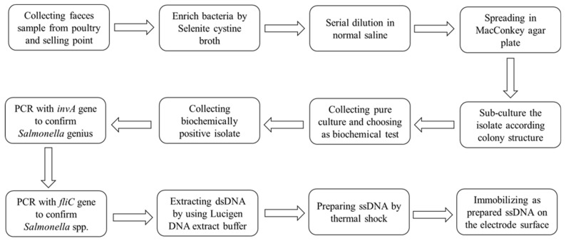
Bacchu et al. [114] developed a DNA-based biosensor for detecting Salmonella Typhi (S. typhi) in blood, poultry faeces, eggs and milk by using DNA-immobilized modified SPE. The biosensor was created by immobilizing an amine-labelled single-stranded DNA (ssDNA) probe specific to S. Typhi on the surface of the P-Cys@AuNP-modified SPE. They introduced a process to quickly and efficiently extract ssDNA, and the whole process lasted approximately 2 h (Figure 4). This biosensor uses the DPV technique to determine S. Typhi complementary-target DNA sequences. The linear response in the actual sample was 1.8–1.8 × 10⁵ CFU/mL, and the LOD value was 1 CFU/mL. The excellent recoveries in the spiked sample were 96.54–103.47%, indicating that the biosensor could detect S. Typhi in food and clinical samples. The combination of ssDNA probes and nanomaterials provides the selectivity, stability, reproducibility and regeneration of electrochemical biosensors, which should be applied to detect other foodborne pathogens.
Figure 4.
Detailed work flow for S. Typhi sample collection and identification. Adapted with permission from Bacchu et al. [114]. Copyright 2022, Elsevier.
As DNA-based biosensors have advanced in the detection of food pathogens, Pangajam et al. [134] developed a novel electrochemical sensor based on a CDs/ZnO nanoroad/PANI nanoassembly for the detection of E. coli O157:H7 in water samples. It was discovered that the exceptional electrical conductivity of CD/ZnO/PANI increased the sensitivity for the detection of E. coli. The successful detection of E. coli O157:H7 in water samples was achieved using the developed electrochemical biosensor, which also showed good selectivity and had a detection limit of 1.3 × 10⁻¹⁸ M. A rapid and sensitive analysis for LM in ham samples was achieved by Li et al. [153] through a phosphatase (ALP)-mediated magnetic relaxation DNA biosensor. This magnetic biosensor demonstrated great sensitivity for LM detection with a linear range from 2 × 10² to 2 × 10⁷ CFU/mL and an LOD of 102 CFU/mL without requiring any DNA amplification steps.
4.2. Electrochemical Immunosensors
Antibody–antigen biosensors, commonly referred to as immunosensors, are frequently used analytical instruments for the detection of foodborne pathogens in food [162]. The immobilization of a particular anti-pathogen antibody on the surface of a transducer serves as the basis for this biosensor’s operation. When an antigen is coupled to the antibody, an immunochemical reaction occurs that serves as the signal for biosensor detection [163]. Recently, electrochemical immunosensors have been widely used in the detection of different foodborne pathogens in foods.
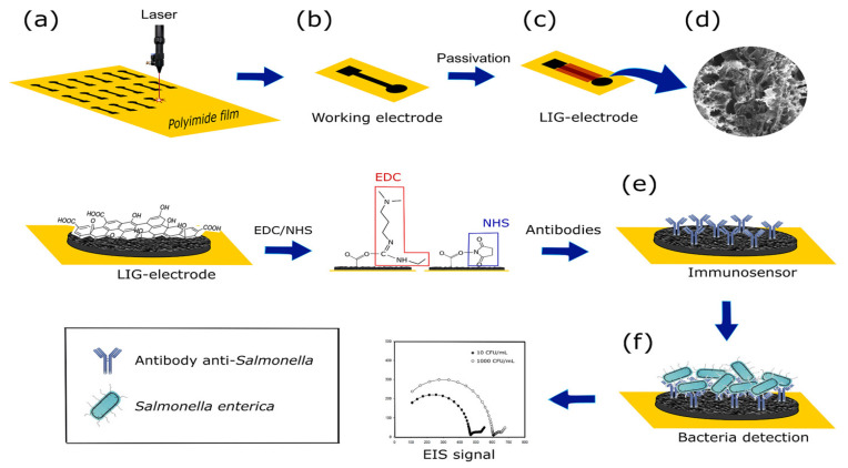
A label-free biosensor was developed by Soares et al. [110] for the detection of Salmonella enterica in chicken broth. To identify S. enterica Typhimurium by carbodiimide cross-linking, a bare laser-induced graphene (LIG) electrode was functionalized with polyclonal antibodies on its surface (Figure 5). This study used the EIS method to determine S. enterica Typhimurium in chicken broth. The analysis time was 22 min, the linear range of the method was 101–105 CFU/mL and the detection limit was 13 CFU/mL. In this research, a low-cost, sensitive and selective electrochemical immunosensor method was developed to determine foodborne pathogens in food, which provides an important contribution to food safety.
Figure 5.
Fabrication, biofunctionalization and sensing scheme of the LIG immunosensor. The fabrication and biofunctionalization steps included: (a) LIG processing onto a polyimide (Kapton) sheet to create the working electrode; (b) working electrode; (c) passivation of the working electrode with lacquer; (d) SEM image showing the LIG surface; (e) biofunctionalization with Salmonella antibodies immobilized on the working electrode via carbodiimide cross-linking chemistry; and (f) Salmonella binding to the electrode and the resultant Nyquist plot generated during electrochemical sensing. Adapted with permission from Soares et al. [110]. Copyright 2020, American Chemical Society.
Mo et al. [136] described a novel sensitive and quantitative sandwich electrochemical immunosensor technique for the detection of E. coli O157:H7 using immune gold@platinum nanoparticles (Au@Pt), neutral red (NR), rGO nanocomposite and regenerative leucoemeraldine-based PANI/AuNP-modified SPCE. Although the SPCE’s disposable nature was replaced by the potential for reuse, its batch-manufacturing benefits were still present. Based on electrochemical detection of E. coli O157:H7, the linear range of the method was 8.9 × 10³–8.9 × 10⁹ CFU/mL, with an LOD of 2.84 × 10³ CFU/mL. To further evaluate the quantitative detection capacity of biosensors, this study conducted a spiked recovery experiment on milk and pork samples. The recovery of spiked milk and pork samples exceeded 78.6%, showing the good precision and reliability of the immunosensor. Similarly, Lu et al. [151] developed an enzyme-labelled amperometric immunosensor for the detection of Lm by immobilizing an HRP-labelled antibody against Lm onto the surface of novel MWCNT fibres. Milk samples were spiked with Lm bacteria, and qualitative results detecting contamination were presented. The linear range of the method was from 102 to 105 CFU/mL (𝑅2 = 0.993), and the LOD was 1.07 × 10² CFU/mL. The potential use of the immunosensor for the quick detection of LM was further demonstrated by its good storage stability and reproducibility (RSD < 6.5%).
4.3. Electrochemical Aptasensors
Short DNA or RNA molecules known as aptamers exhibit a high affinity and selectivity when binding to their target molecules, which can include drugs, proteins, toxins, sugar, antibiotics and bacteria [164]. Compared to RNA aptamers, DNA aptamers are more stable and are widely used in electrochemical aptasensors to detect foodborne pathogens in food and the environment. Compared to the manufacture of antibodies, the synthesis of aptamers exhibits numerous advantages because it is quick, inexpensive, does not involve animal products and does not generate batch-to-batch fluctuations. DNA aptamers typically have a high affinity for their target, are resistant to high temperatures, are stable over time and are simple to modify by chemical groups for immobilization or labelling purposes.
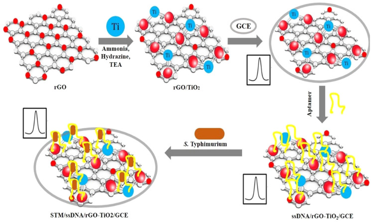
S. typhimurium was detected by Muniandy et al. [117] in chicken meat samples using rGO-TiO2 nanocomposite-based electrochemical aptasensors (Figure 6). The bacterial cells are linked to the DNA aptamer that has been adsorbed on the rGO-TiO2 surface, creating a physical barrier that prevents electron transmission. This study used the DPV method to identify S. typhimurium. The optimized aptasensor demonstrated good selectivity for Salmonella bacteria, great sensitivity, a wide detection range (101–108 CFU/mL) and an LOD of 10 CFU/mL. Wang et al. [133] developed an electrochemical aptasensor using a coaxial capillary with magnetic nanoparticles, urease catalysis and a PCB electrode for the rapid and sensitive detection of E. coli O157:H7 in milk samples. This aptasensor obtained a good recovery (>99.7%) and precision (RSD, 1.4%–4.3%), and the LOD was 10 CFU/mL. In another study, Abbaspour et al. [144] introduced a sensitive and highly selective dual-aptamer-based sandwich immunosensor for the detection of S. aureus. Due to its short detection time, high sensitivity and low cost, this proposed aptasensor offers the potential for practical applications in the detection of foodborne pathogens.
Figure 6.
A schematic diagram of the stepwise fabrication of rGO-TiO2 electrodes and electrochemical detection of bacteria. Adapted with permission from Muniandy et al. [117]. Copyright 2019, Elsevier.
4.4. CRISPR/Cas-Based Electrochemical Biosensor
As a method for genome editing, CRISPR is employed to treat numerous diseases [27]. However, with advancements in research, CRISPR combined with electrochemical biosensors has been utilized for the detection of Salmonella, E. coli O157:H7, S. aureus and Lm in food [107,129,137,154]. Recently, CRISPR/Cas-based methods for the detection of Salmonella, E. coli O157:H7, S. aureus and Lm were created, although these methods remain in their very early stages and need to be further developed. To our knowledge, the Cas9, Cas12a, Cas12b, Cas13a and Cas13b proteins are mainly used in the detection of foodborne pathogens [165,166].
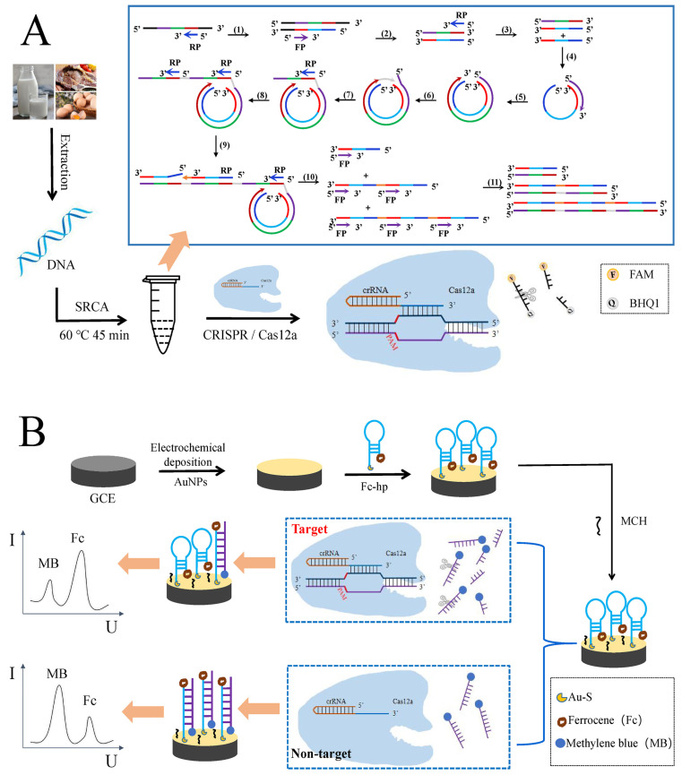
Zheng et al. [107] first reported a ratiometric electrochemical biosensor based on the SRCA-CRISPR/Cas12a system for the detection of Salmonella (Figure 7). This strategy can effectively use the target’s particular Cas12a-crRNA binding and eliminate nonspecific amplification. The specificity and sensitivity of traditional SRCA responses are greatly improved by the combination of SRCA and CRISPR/Cas12a. The linear range of the method was 5.8 fg/μL–5.8 ng/μL, and the LOD was 2.08 fg/μL. For the detection of actual samples (chicken and pork), this biosensor exhibited good sensitivity, precision and specificity, and the detection results of this biosensor were consistent with real-time fluorescent quantitative PCR (RT-qPCR). Overall, the biosensor offers a useful platform for the extremely accurate and sensitive detection of Salmonella in food, with the potential to also monitor other foodborne pathogens. In another study, Chen et al. [129] developed an electrochemical biosensor based on CRISPR/Cas12a combined with immuno-RCA for the detection of E. coli O157:H7. The developed biosensor presented a broad linear range from 10 to 107 CFU/mL, with an LOD of 10 CFU/mL. Compared to traditional electrochemical DNA sensors, the CRISPR Cas system based on electrochemical DNA sensors is higher in terms of sensitivity and precision [167]. It also exhibits complementarity between CRISPR and electrochemical-sensing technology.
Figure 7.
(A) Schematic diagram of the SRCA-CRISPR/Cas12a assay; (B) schematic diagram of the biosensor for the detection of Salmonella. Adapted with permission from Zheng et al. [107]. Copyright 2023, Elsevier.
Recently, Huang et al. [137] introduced a novel electrochemical biosensor based on SRCA combined with the CRISPR/Cas12a system for the accurate detection of S. aureus. In the presence of S. aureus, the target DNA double strands obtained by SRCA can be specifically identified with the Cas12a/crRNA complex. The accidental cleavage characteristic of Cas12a is activated by this combination, which amplifies the reporting signal. The sensitivity and specificity of the method is significantly enhanced by this step. The linear range of the method was 3.9 × 101–3.9 × 10⁷ CFU/mL, and the LOD was 3 CFU/mL. For the detection of actual milk samples, the recoveries were 98.8–117.1%, which justified the good accuracy of this biosensor. It provides a highly specific and ultrasensitive detection platform for foodborne pathogens. Similarly, Li et al. [154] developed an ultrasensitive CRISPR/Cas12a-based electrochemical biosensor (E-CRISPR) combined with recombinase-assisted amplification (RAA) for the detection of Lm. The results indicated that this method has a good linearity (9.4 × 100–9.4 × 10⁷ CFU/mL) and sensitivity (LOD, 9.4 × 10² CFU/g). Compared to previous Cas12a-based signal amplification strategies, the RAA-based E-CRISPR platform not only took full advantage of the specific RNA recognition ability of Cas12a to achieve high specificity, but also converted the target recognition activity into a detectable electrochemical signal to improve the sensitivity.
5. Conclusions and Outlooks
Foodborne pathogenic microorganisms in food and the environment are an issue that warrants attention and are related to human health and safety. To ensure the food safety of consumers, it is very important to develop rapid and efficient biosensor technology to effectively determine food pathogens in food and the environment. Therefore, we provided a comprehensive review of electrochemical biosensors for the detection of food pathogens in food and the environment (2013–2023). Compared with traditional detection methods (RT-PCR, ELISA and culture plates), electrochemical biosensors exhibit several advantages, as the technique exhibits high sensitivity, achieves real-time detection and is selective, rapid and inexpensive. This review focused on the detection principle behind electrochemical biosensors, the hazards of food pathogens, risk analysis and control measures and recent progress. Various combinations of materials and methods have been used to develop different bioreceptor-based sensors for the detection of six kinds of food pathogens. As shown in Table 1, this review of bioreceptors, detection methods, assay strategies and material types analysed the current advanced electrochemical biosensors for the detection of food pathogens in different sample matrices. With the development of nanomaterials, good nanomaterials are fixed to the electrode to improve the sensitivity, selectivity and stability of electrochemical biosensors. Due to some of the shortcomings of bioreceptors, there are certain limitations of electrochemical biosensors, such as low stability for antibodies, restriction to DNA targets for nucleic acids and sensitivity to nuclease for aptamers. However, the combination of CRISPR technology and electrochemical DNA sensors successfully improved the sensitivity and precision. Biosensor-based devices have become an important part of the equipment used in laboratories to detect biological responses. In spite of having created a variety of biosensors for detecting foodborne pathogens, it is still difficult to design biosensors for the reliable and effective determination of microorganisms in real food samples. In practical applications, some electrochemical biosensors can only detect single food samples, which may result from the complexity of the animal-derived food matrix. In addition, available research reports indicate that these electrochemical biosensors still have a problem with simultaneously detecting the number of food pathogens. Overall, a biosensor’s essential characteristics include sensitivity, specificity, stability, detection time, sample processing, size and the capacity to function under a variety of settings without the need for specialized training. Although electrochemical biosensors must be further developed to solve these problems, cooperation between scientific researchers and enterprises can pave the way for the development of a desirable, portable product. The development of food safety biosensors will significantly improve people’s quality of life and health.
Author Contributions
Conceptualization, Z.Y.; investigation, B.W., H.W., X.L. and X.Z.; data curation, B.W.; writing—original draft preparation, B.W.; writing—review and editing, B.W. and Z.Y.; funding acquisition, B.W., X.L., X.Z. and Z.Y. All authors have read and agreed to the published version of the manuscript.
Institutional Review Board Statement
Not applicable.
Informed Consent Statement
Not applicable.
Data Availability Statement
Not applicable.
Conflicts of Interest
The authors declare no conflict of interest.
Funding Statement
This research was financially supported by the Jiangsu Funding Program for Excellent Postdoctoral Talent, the China Postdoctoral Science Foundation (2023M732999), the National Natural Science Foundation of China (NSFC) grant (No. 31901801), the Foundation of China National Key Research & Development Program (2016YFC1300201), the Jiangsu Key Research & Development Program (BE2019436-5), and the Natural Science Foundation of Jiangsu Province (BK20210814).
Footnotes
Disclaimer/Publisher’s Note: The statements, opinions and data contained in all publications are solely those of the individual author(s) and contributor(s) and not of MDPI and/or the editor(s). MDPI and/or the editor(s) disclaim responsibility for any injury to people or property resulting from any ideas, methods, instructions or products referred to in the content.
References
- 1.Bintsis T. Foodborne pathogens. AIMS Microbiol. 2017;3:529–563. doi: 10.3934/microbiol.2017.3.529. [DOI] [PMC free article] [PubMed] [Google Scholar]
- 2.Gousia P., Economou V., Sakkas H., Leveidiotou S., Papadopoulou C. Antimicrobial resistance of major foodborne pathogens from major meat products. Foodborne Pathog. Dis. 2011;8:27–38. doi: 10.1089/fpd.2010.0577. [DOI] [PubMed] [Google Scholar]
- 3.Mayrhofer S., Paulsen P., Smulders F.J., Hilbert F. Antimicrobial resistance profile of five major food-borne pathogens isolated from beef, pork and poultry. Int. J. Food Microbiol. 2004;97:23–29. doi: 10.1016/j.ijfoodmicro.2004.04.006. [DOI] [PubMed] [Google Scholar]
- 4.Bhunia A.K. Biosensors and bio-based methods for the separation and detection of foodborne pathogens. Adv. Food Nutr. Res. 2008;54:1–44. doi: 10.1016/S1043-4526(07)00001-0. [DOI] [PubMed] [Google Scholar]
- 5.European Food Safety Authority. European Centre for Disease Prevention and Control The European Union summary report on trends and sources of zoonoses, zoonotic agents and food-borne outbreaks in 2017. EFSA J. 2018;16:e05500. doi: 10.2903/j.efsa.2018.5500. [DOI] [PMC free article] [PubMed] [Google Scholar]
- 6.Pietzka A., Allerberger F., Murer A., Lennkh A., Stöger A., Rosel A.C., Huhulescu S., Maritschnik S., Springer B., Lepuschitz S. Whole genome sequencing based surveillance of L. monocytogenes for early detection and investigations of listeriosis outbreaks. Front. Public Health. 2019;7:139. doi: 10.3389/fpubh.2019.00139. [DOI] [PMC free article] [PubMed] [Google Scholar]
- 7.European Food Safety Authority. European Centre for Disease Prevention and Control Multi-country outbreak of Salmonella Agona infections possibly linked to ready-to-eat food. EFSA Support. Publ. 2018;15:1465E. doi: 10.2903/sp.efsa.2018.EN-1465. [DOI] [Google Scholar]
- 8.Strawn L.K., Fortes E.D., Bihn E.A., Nightingale K.K., Gröhn Y.T., Worobo R.W., Wiedmann M., Bergholz P.W. Landscape and meteorological factors affecting prevalence of three food-borne pathogens in fruit and vegetable farms. Appl. Environ. Microbiol. 2013;79:588–600. doi: 10.1128/AEM.02491-12. [DOI] [PMC free article] [PubMed] [Google Scholar]
- 9.Miceli A., Settanni L. Influence of agronomic practices and pre-harvest conditions on the attachment and development of Listeria monocytogenes in vegetables. Ann. Microbiol. 2019;69:185–199. doi: 10.1007/s13213-019-1435-6. [DOI] [Google Scholar]
- 10.Paudyal N., Pan H., Liao X., Zhang X., Li X., Fang W., Yue M. A meta-analysis of major foodborne pathogens in Chinese food commodities between 2006 and 2016. Foodborne Pathog. Dis. 2018;15:187–197. doi: 10.1089/fpd.2017.2417. [DOI] [PubMed] [Google Scholar]
- 11.Heredia N., García S. Animals as sources of food-borne pathogens: A review. Anim. Nutr. 2018;4:250–255. doi: 10.1016/j.aninu.2018.04.006. [DOI] [PMC free article] [PubMed] [Google Scholar]
- 12.Aijuka M., Buys E.M. Persistence of foodborne diarrheagenic Escherichia coli in the agricultural and food production environment: Implications for food safety and public health. Food Microbiol. 2019;82:363–370. doi: 10.1016/j.fm.2019.03.018. [DOI] [PubMed] [Google Scholar]
- 13.Bai Y., Song M., Cui Y., Shi C., Wang D., Paoli G.C., Shi X. A rapid method for the detection of foodborne pathogens by extraction of a trace amount of DNA from raw milk based on amino-modified silica-coated magnetic nanoparticles and polymerase chain reaction. Anal. Chim. Acta. 2013;787:93–101. doi: 10.1016/j.aca.2013.05.043. [DOI] [PubMed] [Google Scholar]
- 14.Cremonesi P., Cortimiglia C., Picozzi C., Minozzi G., Malvisi M., Luini M., Castiglioni B. Development of a droplet digital polymerase chain reaction for rapid and simultaneous identification of common foodborne pathogens in soft cheese. Front. Microbiol. 2016;7:1725. doi: 10.3389/fmicb.2016.01725. [DOI] [PMC free article] [PubMed] [Google Scholar]
- 15.Bonilauri P., Bardasi L., Leonelli R., Ramini M., Luppi A., Giacometti F., Merialdi G. Detection of food hazards in foods: Comparison of real time polymerase chain reaction and cultural methods. Ital. J. Food Saf. 2016;5:5641. doi: 10.4081/ijfs.2016.5641. [DOI] [PMC free article] [PubMed] [Google Scholar]
- 16.Ma Z., Yang X., Fang Y., Tong Z., Lin H., Fan H. Detection of Salmonella infection in chickens by an indirect enzyme-linked immunosorbent assay based on presence of PagC antibodies in sera. Foodborne Pathog. Dis. 2018;15:109–113. doi: 10.1089/fpd.2017.2322. [DOI] [PubMed] [Google Scholar]
- 17.Lv X., Huang Y., Liu D., Liu C., Shan S., Li G., Duan M., Lai W. Multicolor and ultrasensitive enzyme-linked immunosorbent assay based on the fluorescence hybrid chain reaction for simultaneous detection of pathogens. J. Agric. Food Chem. 2019;67:9390–9398. doi: 10.1021/acs.jafc.9b03414. [DOI] [PubMed] [Google Scholar]
- 18.Wangman P., Surasilp T., Pengsuk C., Sithigorngul P., Longyant S. Development of a species-specific monoclonal antibody for rapid detection and identification of foodborne pathogen Vibrio vulnificus. J. Food Saf. 2021;41:e12939. doi: 10.1111/jfs.12939. [DOI] [Google Scholar]
- 19.Ayala D.I., Cook P.W., Franco J.G., Bugarel M., Kottapalli K.R., Loneragan G.H., Brashears M.M., Nightingale K.K. A systematic approach to identify and characterize the effectiveness and safety of novel probiotic strains to control foodborne pathogens. Front. Microbiol. 2019;10:1108. doi: 10.3389/fmicb.2019.01108. [DOI] [PMC free article] [PubMed] [Google Scholar]
- 20.Qin X., Liu J., Zhang Z., Li J., Yuan L., Zhang Z., Chen L. Microfluidic paper-based chips in rapid detection: Current status, challenges, and perspectives. TrAC Trends Anal. Chem. 2021;143:116371. doi: 10.1016/j.trac.2021.116371. [DOI] [Google Scholar]
- 21.Ravindran N., Kumar S., Yashini M., Rajeshwari S., Mamathi C.A., Thirunavookarasu S.N., Sunil C.K. Recent advances in surface plasmon resonance (SPR) biosensors for food analysis: A review. Crit. Rev. Food Sci. Nutr. 2023;63:1055–1077. doi: 10.1080/10408398.2021.1958745. [DOI] [PubMed] [Google Scholar]
- 22.Dong X., Qi S., Khan I.M., Sun Y., Zhang Y., Wang Z. Advances in riboswitch-based biosensor as food samples detection tool. Compr. Rev. Food Sci. Food Saf. 2023;22:451–472. doi: 10.1111/1541-4337.13077. [DOI] [PubMed] [Google Scholar]
- 23.Lu L., Chee G., Yamada K., Jun S. Electrochemical impedance spectroscopic technique with a functionalized microwire sensor for rapid detection of foodborne pathogens. Biosens. Bioelectron. 2013;42:492–495. doi: 10.1016/j.bios.2012.10.060. [DOI] [PubMed] [Google Scholar]
- 24.Han E., Li X., Zhang Y., Zhang M.N., Cai J.R., Zhang X.N. Electrochemical immunosensor based on self-assembled gold nanorods for label-free and sensitive determination of Staphylococcus aureus. Anal. Biochem. 2020;611:113982. doi: 10.1016/j.ab.2020.113982. [DOI] [PubMed] [Google Scholar]
- 25.Feng K.W., Li T., Ye C.Z., Gao X.Y., Yue X.L., Ding S.Y., Dong Q.L., Yang M.Q., Huang G.H., Zhang J.S. A novel electrochemical immunosensor based on Fe3O4@graphene nanocomposite modified glassy carbon electrode for rapid detection of Salmonella in milk. J. Dairy Sci. 2022;105:2108–2118. doi: 10.3168/jds.2021-21121. [DOI] [PubMed] [Google Scholar]
- 26.Umesha S., Manukumar H.M. Advanced molecular diagnostic techniques for detection of food-borne pathogens: Current applications and future challenges. Crit. Rev. Food Sci. Nutr. 2018;58:84–104. doi: 10.1080/10408398.2015.1126701. [DOI] [PubMed] [Google Scholar]
- 27.Mahari S., Gandhi S. Recent advances in electrochemical biosensors for the detection of Salmonellosis: Current prospective and challenges. Biosensors. 2022;12:365. doi: 10.3390/bios12060365. [DOI] [PMC free article] [PubMed] [Google Scholar]
- 28.Melo A.M.A., Alexandre D.L., Furtado R.F., Borges M.F., Figueiredo E.A.T., Biswas A., Cheng H.N., Alves C.R. Electrochemical immunosensors for Salmonella detection in food. Appl. Microbiol. Biotechnol. 2016;100:5301–5312. doi: 10.1007/s00253-016-7548-y. [DOI] [PubMed] [Google Scholar]
- 29.Long W., Patra I., Alhachami F.R., Sherbekov U.A., Majdi A., Abed S.A. Aptamer based nanoprobes for detection of foodborne virus in food and environment samples: Recent progress and challenges. Crit. Rev. Anal. Chem. 2022 doi: 10.1080/10408347.2022.2114785. [DOI] [PubMed] [Google Scholar]
- 30.Kurmendra Nanomaterial gas sensors for biosensing applications: A review. Recent Pat. Nanotechnol. 2023;17:104–118. doi: 10.2174/1872210515666211129115229. [DOI] [PubMed] [Google Scholar]
- 31.Hussain B., Yüce M., Ullah N., Budak H. Bioconjugated nanomaterials for monitoring food contamination. In: Grumezescu A.M., editor. Nanobiosensors. Elsevier; Amsterdam, The Netherlands: 2017. pp. 93–127. [Google Scholar]
- 32.Naresh V., Lee N. A review on biosensors and recent development of nanostructured materials-enabled biosensors. Sensors. 2021;21:1109. doi: 10.3390/s21041109. [DOI] [PMC free article] [PubMed] [Google Scholar]
- 33.Awang M.S., Bustami Y., Hamzah H.H., Zambry N.S., Najib M.A., Khalid M.F., Aziah I., Manaf A.A. Advancement in Salmonella detection methods: From conventional to electrochemical-based sensing detection. Biosensors. 2021;11:346. doi: 10.3390/bios11090346. [DOI] [PMC free article] [PubMed] [Google Scholar]
- 34.Gu N., Liu S. Introduction to biosensors. J. Mater. Chem. B. 2020;8:3168–3170. doi: 10.1039/D0TB90051F. [DOI] [PubMed] [Google Scholar]
- 35.Kimmel D.W., LeBlanc G., Meschievitz M.E., Cliffel D.E. Electrochemical sensors and biosensors. Anal. Chem. 2012;84:685–707. doi: 10.1021/ac202878q. [DOI] [PMC free article] [PubMed] [Google Scholar]
- 36.Zhang Z., Zhou J., Du X. Electrochemical biosensors for detection of foodborne pathogens. Micromachines. 2019;10:222. doi: 10.3390/mi10040222. [DOI] [PMC free article] [PubMed] [Google Scholar]
- 37.Grieshaber D., MacKenzie R., Vörös J., Reimhult E. Electrochemical biosensors-sensor principles and architectures. Sensors. 2008;8:1400–1458. doi: 10.3390/s80314000. [DOI] [PMC free article] [PubMed] [Google Scholar]
- 38.Kozitsina A.N., Svalova T.S., Malysheva N.N., Okhokhonin A.V., Vidrevich M.B., Brainina K.Z. Sensors based on bio and biomimetic receptors in medical diagnostic, environment, and food analysis. Biosensors. 2018;8:35. doi: 10.3390/bios8020035. [DOI] [PMC free article] [PubMed] [Google Scholar]
- 39.Eng S.K., Pusparajah P., Mutalib N.S.A., Ser H.L., Chan K.G., Lee L.H. Salmonella: A review on pathogenesis, epidemiology and antibiotic resistance. Front. Life Sci. 2015;8:284–293. doi: 10.1080/21553769.2015.1051243. [DOI] [Google Scholar]
- 40.Labbé R.G., García S. Guide to Foodborne Pathogens. John Wiley & Sons; Chichester, UK: 2013. [Google Scholar]
- 41.Baron S. Medical Microbiology. University of Texas Medical Branch at Galveston; Galveston, TX, USA: 1996. [PubMed] [Google Scholar]
- 42.Neto W.S., Leotti V.B., Pires S.M., Hald T., Corbellini L.G. Non-typhoidal human Salmonellosis in Rio Grande do Sul, Brazil: A combined source attribution study of microbial subtyping and outbreak data. Int. J. Food Microbiol. 2021;338:108992. doi: 10.1016/j.ijfoodmicro.2020.108992. [DOI] [PubMed] [Google Scholar]
- 43.Harris A.M. Travel-related infectious diseases. In: Centers for Disease Control and Prevention, editor. CDC Yellow Book. Oxford University Press; New York, NY, USA: 2020. pp. 169–394. [Google Scholar]
- 44.Jay J.M., Loessner M.J., Golden D.A. Modern Food Microbiology. Springer; New York, NY, USA: 2008. [Google Scholar]
- 45.Chlebicz A., Śliżewska K. Campylobacteriosis, salmonellosis, yersiniosis, and listeriosis as zoonotic foodborne diseases: A review. Int. J. Environ. Res. Public Health. 2018;15:863. doi: 10.3390/ijerph15050863. [DOI] [PMC free article] [PubMed] [Google Scholar]
- 46.Li R., Peng K., Huang W., Sun X., Huang Y., Lei G., Lv H., Wang Z., Yang X. The genomic epidemiology of mcr-positive Salmonella enterica in clinical patients from 2014 to 2017 in Sichuan, China and global epidemiological features. J. Infect. 2022;85:702–769. doi: 10.1016/j.jinf.2022.08.042. [DOI] [PubMed] [Google Scholar]
- 47.Carrique-Mas J.J., Bedford S., Davies R.H. Organic acid and formaldehyde treatment of animal feeds to control Salmonella: Efficacy and masking during culture. J. Appl. Microbiol. 2007;103:88–96. doi: 10.1111/j.1365-2672.2006.03233.x. [DOI] [PubMed] [Google Scholar]
- 48.Jing W., Liu J., Wu S., Li X., Liu Y. Role of cpxA mutations in the resistance to aminoglycosides and β-lactams in Salmonella enterica serovar Typhimurium. Front. Microbiol. 2021;12:604079. doi: 10.3389/fmicb.2021.604079. [DOI] [PMC free article] [PubMed] [Google Scholar]
- 49.Hu J., Che C., Zuo J., Niu X., Wang Z., Lian L., Jia Y., Zhang H., Zhang T., Yu F. Effect of antibiotics on the colonization of live attenuated Salmonella enteritidis vaccine in chickens. Front. Vet. Sci. 2021;8:784160. doi: 10.3389/fvets.2021.784160. [DOI] [PMC free article] [PubMed] [Google Scholar]
- 50.Nataro J.P., Kaper J.B. Diarrheagenic Escherichia coli. Clin. Microbiol. Rev. 1998;11:142–201. doi: 10.1128/CMR.11.1.142. [DOI] [PMC free article] [PubMed] [Google Scholar]
- 51.Kim J.S., Lee M.S., Kim J.H. Recent updates on outbreaks of Shiga toxin-producing Escherichia coli and its potential reservoirs. Front. Cell Infect. Microbiol. 2020;10:273. doi: 10.3389/fcimb.2020.00273. [DOI] [PMC free article] [PubMed] [Google Scholar]
- 52.Vila J., Sáez-López E., Johnson J.R., Römling U., Dobrindt U., Cantón R., Giske C.G., Naas T., Carattoli A., Martínez-Medina M. Escherichia coli: An old friend with new tidings. FEMS Microbiol. Rev. 2016;40:437–463. doi: 10.1093/femsre/fuw005. [DOI] [PubMed] [Google Scholar]
- 53.Gourama H. Foodborne pathogens. In: Demirci A., Feng H., Krishnamurthy K., editors. Food Safety Engineering. Springer; Cham, Switzerland: 2020. pp. 25–49. [Google Scholar]
- 54.Yang S.C., Lin C.H., Aljuffali I.A., Fang J.Y. Current pathogenic Escherichia coli foodborne outbreak cases and therapy development. Arch. Microbiol. 2017;199:811–825. doi: 10.1007/s00203-017-1393-y. [DOI] [PubMed] [Google Scholar]
- 55.Saeedi P., Yazdanparast M., Behzadi E., Salmanian A.H., Mousavi S.L., Nazarian S., Amani J. A review on strategies for decreasing E. coli O157: H7 risk in animals. Microb. Pathog. 2017;103:186–195. doi: 10.1016/j.micpath.2017.01.001. [DOI] [PubMed] [Google Scholar]
- 56.Poirel L., Madec J.Y., Lupo A., Schink A.K., Kieffer N., Nordmann P., Schwarz S. Antimicrobial resistance in Escherichia coli. Microbiol. Spectr. 2018;6:ARBA-0026-2017. doi: 10.1128/microbiolspec.ARBA-0026-2017. [DOI] [PMC free article] [PubMed] [Google Scholar]
- 57.Kadariya J., Smith T.C., Thapaliya D. Staphylococcus aureus and staphylococcal food-borne disease: An ongoing challenge in public health. BioMed Res. Int. 2014;2014:827965. doi: 10.1155/2014/827965. [DOI] [PMC free article] [PubMed] [Google Scholar]
- 58.Pinchuk I.V., Beswick E.J., Reyes V.E. Staphylococcal enterotoxins. Toxins. 2010;2:2177–2197. doi: 10.3390/toxins2082177. [DOI] [PMC free article] [PubMed] [Google Scholar]
- 59.Lowy F.D. Staphylococcus aureus infections. N. Engl. J. Med. 1998;339:520–532. doi: 10.1056/NEJM199808203390806. [DOI] [PubMed] [Google Scholar]
- 60.Argudín M.Á., Mendoza M.C., Rodicio M.R. Food poisoning and Staphylococcus aureus enterotoxins. Toxins. 2010;2:1751–1773. doi: 10.3390/toxins2071751. [DOI] [PMC free article] [PubMed] [Google Scholar]
- 61.Bhunia A.K. Foodborne Microbial Pathogens: Mechanisms and Pathogenesis. Springer; New York, NY, USA: 2018. [Google Scholar]
- 62.Tarisse C.F., Goulard-Huet C., Nia Y., Devilliers K., Marcé D., Dambrune C., Lefebvre D., Hennekinne J.A., Simon S. Highly sensitive and specific detection of staphylococcal enterotoxins SEA, SEG, SEH, and SEI by immunoassay. Toxins. 2021;13:130. doi: 10.3390/toxins13020130. [DOI] [PMC free article] [PubMed] [Google Scholar]
- 63.Assis L.M., Nedeljković M., Dessen A. New strategies for targeting and treatment of multi-drug resistant Staphylococcus aureus. Drug Resist. Updates. 2017;31:1–14. doi: 10.1016/j.drup.2017.03.001. [DOI] [PubMed] [Google Scholar]
- 64.Liu C., Shen Y., Yang M., Chi K., Guo N. Hazard of staphylococcal enterotoxins in food and promising strategies for natural products against virulence. J. Agric. Food Chem. 2022;70:2450–2465. doi: 10.1021/acs.jafc.1c06773. [DOI] [PubMed] [Google Scholar]
- 65.Seferbekova Z., Zabelkin A., Yakovleva Y., Afasizhev R., Dranenko N.O., Alexeev N., Gelfand M.S., Bochkareva O.O. High rates of genome rearrangements and pathogenicity of Shigella spp. Front. Microbiol. 2021;12:628622. doi: 10.3389/fmicb.2021.628622. [DOI] [PMC free article] [PubMed] [Google Scholar]
- 66.Nosrati M., Hajizade A., Nazarian S., Amani J., Vansofla A.N., Tarverdizadeh Y. Designing a multi-epitope vaccine for cross-protection against Shigella spp: An immunoinformatics and structural vaccinology study. Mol. Immunol. 2019;116:106–116. doi: 10.1016/j.molimm.2019.09.018. [DOI] [PubMed] [Google Scholar]
- 67.Zaidi M.B., Estrada-García T. Shigella: A highly virulent and elusive pathogen. Curr. Trop. Med. Rep. 2014;1:81–87. doi: 10.1007/s40475-014-0019-6. [DOI] [PMC free article] [PubMed] [Google Scholar]
- 68.Dudley E.G. Food microbiology: Fundamentals and frontiers. Emerg. Infect. Dis. 2022;28:267. doi: 10.3201/eid2801.211862. [DOI] [Google Scholar]
- 69.Kotloff K.L., Riddle M.S., Platts-Mills J.A., Pavlinac P., Zaidi A.K. Shigellosis. Lancet. 2018;391:801–812. doi: 10.1016/S0140-6736(17)33296-8. [DOI] [PubMed] [Google Scholar]
- 70.Van Den Beld M.J.C., Warmelink E., Friedrich A.W., Reubsaet F.A.G., Schipper M., De Boer R.F., Notermans D.W., Petrignani M.W.F., Van Zanten E., Rossen J.W.A. Incidence, clinical implications and impact on public health of infections with Shigella spp. and entero-invasive Escherichia coli (EIEC): Results of a multicenter cross-sectional study in the Netherlands during 2016–2017. BMC Infect. Dis. 2019;19:1037. doi: 10.1186/s12879-019-4659-y. [DOI] [PMC free article] [PubMed] [Google Scholar]
- 71.Karimi-Yazdi M., Ghalavand Z., Shabani M., Houri H., Sadredinamin M., Taheri M., Eslami G. High rates of antimicrobial resistance and virulence gene distribution among Shigella spp. isolated from pediatric patients in Tehran, Iran. Infect. Drug Resist. 2020;13:485–492. doi: 10.2147/IDR.S238559. [DOI] [PMC free article] [PubMed] [Google Scholar]
- 72.Guglielmino C.J.D., Kakkanat A., Forde B.M., Rubenach S., Merone L., Stafford R., Graham R., Beatson S.A., Jennison A.V. Outbreak of multi-drug-resistant (MDR) Shigella flexneri in Northern Australia due to an endemic regional clone acquiring an IncFII plasmid. Eur. J. Clin. Microbiol. Infect. Dis. 2021;40:279–286. doi: 10.1007/s10096-020-04029-w. [DOI] [PMC free article] [PubMed] [Google Scholar]
- 73.Charles H., Prochazka M., Thorley K., Crewdson A., Greig D.R., Jenkins C., Painset A., Fifer H., Browning L., Cabrey P. Outbreak of sexually transmitted, extensively drug-resistant Shigella sonnei in the UK, 2021–22: A descriptive epidemiological study. Lancet Infect. Dis. 2022;22:1503–1510. doi: 10.1016/S1473-3099(22)00370-X. [DOI] [PubMed] [Google Scholar]
- 74.Ranjbar R., Farahani A. Shigella: Antibiotic-resistance mechanisms and new horizons for treatment. Infect. Drug Resist. 2019;12:3137–3167. doi: 10.2147/IDR.S219755. [DOI] [PMC free article] [PubMed] [Google Scholar]
- 75.World Health Organization . Guidelines for the Control of Shigellosis, Including Epidemics Due to Shigella dysenteriae Type 1. World Health Organization; Geneva, Switzerland: 2005. [Google Scholar]
- 76.Puzari M., Sharma M., Chetia P. Emergence of antibiotic resistant Shigella species: A matter of concern. J. Infect. Public Health. 2018;11:451–454. doi: 10.1016/j.jiph.2017.09.025. [DOI] [PubMed] [Google Scholar]
- 77.Shahin K., Bouzari M., Wang R., Yazdi M. Prevalence and molecular characterization of multidrug-resistant Shigella species of food origins and their inactivation by specific lytic bacteriophages. Int. J. Food Microbiol. 2019;305:108252. doi: 10.1016/j.ijfoodmicro.2019.108252. [DOI] [PubMed] [Google Scholar]
- 78.Tang S.S., Biswas S.K., Tan W.S., Saha A.K., Leo B.F. Efficacy and potential of phage therapy against multidrug resistant Shigella spp. PeerJ. 2019;7:e6225. doi: 10.7717/peerj.6225. [DOI] [PMC free article] [PubMed] [Google Scholar]
- 79.Bardsley M., Jenkins C., Mitchell H.D., Mikhail A.F.W., Baker K.S., Foster K., Hughes G., Dallman T.J. Persistent transmission of shigellosis in England is associated with a recently emerged multidrug-resistant strain of Shigella sonnei. J. Clin. Microbiol. 2020;58:e01692-19. doi: 10.1128/JCM.01692-19. [DOI] [PMC free article] [PubMed] [Google Scholar]
- 80.Pakbin B., Amani Z., Allahyari S., Mousavi S., Mahmoudi R., Brück W.M., Peymani A. Genetic diversity and antibiotic resistance of Shigella spp. isolates from food products. Food Sci. Nutr. 2021;9:6362–6371. doi: 10.1002/fsn3.2603. [DOI] [PMC free article] [PubMed] [Google Scholar]
- 81.Phiri A.F.N.D., Abia A.L.K., Amoako D.G., Mkakosya R., Sundsfjord A., Essack S.Y., Simonsen G.S. Burden, antibiotic resistance, and clonality of Shigella spp. implicated in community-acquired acute diarrhoea in Lilongwe, Malawi. Trop. Med. Infect. Dis. 2021;6:63. doi: 10.3390/tropicalmed6020063. [DOI] [PMC free article] [PubMed] [Google Scholar]
- 82.Elkenany R., Eltaysh R., Elsayed M., Abdel-Daim M., Shata R. Characterization of multi-resistant Shigella species isolated from raw cow milk and milk products. J. Vet. Med. Sci. 2022;84:890–897. doi: 10.1292/jvms.22-0018. [DOI] [PMC free article] [PubMed] [Google Scholar]
- 83.Farhani I., Nezafat N., Mahmoodi S. Designing a novel multi-epitope peptide vaccine against pathogenic Shigella spp. based immunoinformatics approaches. Int. J. Pept. Res. Ther. 2019;25:541–553. doi: 10.1007/s10989-018-9698-5. [DOI] [Google Scholar]
- 84.Śmiałek M., Kowalczyk J., Koncicki A. The use of probiotics in the reduction of Campylobacter spp. prevalence in poultry. Animals. 2021;11:1355. doi: 10.3390/ani11051355. [DOI] [PMC free article] [PubMed] [Google Scholar]
- 85.Morales-Partera A.M., Cardoso-Toset F., Luque I., Astorga R.J., Maldonado A., Herrera-León S., Hernández M., Gómez-Laguna J., Tarradas C. Prevalence and diversity of Salmonella spp., Campylobacter spp., and Listeria monocytogenes in two free-range pig slaughterhouses. Food Control. 2018;92:208–215. doi: 10.1016/j.foodcont.2018.04.053. [DOI] [Google Scholar]
- 86.Facciolà A., Riso R., Avventuroso E., Visalli G., Delia S.A., Laganà P. Campylobacter: From microbiology to prevention. J. Prev. Med. Hyg. 2017;58:E79–E92. [PMC free article] [PubMed] [Google Scholar]
- 87.Perez-Arnedo I., Gonzalez-Fandos E. Prevalence of Campylobacter spp. in poultry in three Spanish farms, a slaughterhouse and a further processing plant. Foods. 2019;8:111. doi: 10.3390/foods8030111. [DOI] [PMC free article] [PubMed] [Google Scholar]
- 88.Mohammadpour H., Berizi E., Hosseinzadeh S., Majlesi M., Zare M. The prevalence of Campylobacter spp. in vegetables, fruits, and fresh produce: A systematic review and meta-analysis. Gut Pathog. 2018;10:41. doi: 10.1186/s13099-018-0269-2. [DOI] [PMC free article] [PubMed] [Google Scholar]
- 89.El-Hack A., Mohamed E., El-Saadony M.T., Shehata A.M., Arif M., Paswan V.K., Batiha G.E.S., Khafaga A.F., Elbestawy A.R. Approaches to prevent and control Campylobacter spp. colonization in broiler chickens: A review. Environ. Sci. Pollut. Res. 2021;28:4989–5004. doi: 10.1007/s11356-020-11747-3. [DOI] [PubMed] [Google Scholar]
- 90.Syarifah I.K., Latif H., Basri C., Rahayu P. Identification and differentiation of Campylobacter isolated from chicken meat using real-time polymerase chain reaction and high resolution melting analysis of hipO and glyA genes. Vet. World. 2020;13:1875–1883. doi: 10.14202/vetworld.2020.1875-1883. [DOI] [PMC free article] [PubMed] [Google Scholar]
- 91.European Food Safety Authority. European Centre for Disease Prevention and Control The European Union one health 2019 zoonoses report. EFSA J. 2021;19:e06406. doi: 10.2903/j.efsa.2021.6406. [DOI] [PMC free article] [PubMed] [Google Scholar]
- 92.Lin J. Novel approaches for Campylobacter control in poultry. Foodborne Pathog. Dis. 2009;6:755–765. doi: 10.1089/fpd.2008.0247. [DOI] [PMC free article] [PubMed] [Google Scholar]
- 93.Nastasijevic I., Proscia F., Boskovic M., Glisic M., Blagojevic B., Sorgentone S., Kirbis A., Ferri M. The European Union control strategy for Campylobacter spp. in the broiler meat chain. J. Food Saf. 2020;40:e12819. doi: 10.1111/jfs.12819. [DOI] [Google Scholar]
- 94.Tang M., Zhou Q., Zhang X., Zhou S., Zhang J., Tang X., Lu J., Gao Y. Antibiotic resistance profiles and molecular mechanisms of Campylobacter from chicken and pig in China. Front. Microbiol. 2020;11:592496. doi: 10.3389/fmicb.2020.592496. [DOI] [PMC free article] [PubMed] [Google Scholar]
- 95.Asare P.T., Zurfluh K., Greppi A., Lynch D., Schwab C., Stephan R., Lacroix C. Reuterin demonstrates potent antimicrobial activity against a broad panel of human and poultry meat Campylobacter spp. isolates. Microorganisms. 2020;8:78. doi: 10.3390/microorganisms8010078. [DOI] [PMC free article] [PubMed] [Google Scholar]
- 96.Balta I., Linton M., Pinkerton L., Kelly C., Stef L., Pet I., Stef D., Criste A., Gundogdu O., Corcionivoschi N. The effect of natural antimicrobials against Campylobacter spp. and its similarities to Salmonella spp, Listeria spp., Escherichia coli, Vibrio spp., Clostridium spp. and Staphylococcus spp. Food Control. 2021;121:107745. doi: 10.1016/j.foodcont.2020.107745. [DOI] [Google Scholar]
- 97.Cheng C., Wang H., Ma T., Han X., Yang Y., Sun J., Chen Z., Yu H., Hang Y., Liu F. Flagellar basal body structural proteins FlhB, FliM, and FliY are required for flagellar-associated protein expression in Listeria monocytogenes. Front. Microbiol. 2018;9:208. doi: 10.3389/fmicb.2018.00208. [DOI] [PMC free article] [PubMed] [Google Scholar]
- 98.Scallan E., Hoekstra R.M., Angulo F.J., Tauxe R.V., Widdowson M.A., Roy S.L., Jones J.L., Griffin P.M. Foodborne illness acquired in the United States—Major pathogens. Emerg. Infect. Dis. 2011;17:7–15. doi: 10.3201/eid1701.P11101. [DOI] [PMC free article] [PubMed] [Google Scholar]
- 99.Schlech W.F. Epidemiology and clinical manifestations of Listeria monocytogenes infection. Microbiol. Spectr. 2019;7:601–608. doi: 10.1128/microbiolspec.GPP3-0014-2018. [DOI] [PMC free article] [PubMed] [Google Scholar]
- 100.Baquero F., Lanza V.F., Duval M., Coque T.M. Ecogenetics of antibiotic resistance in Listeria monocytogenes. Mol. Microbiol. 2020;113:570–579. doi: 10.1111/mmi.14454. [DOI] [PubMed] [Google Scholar]
- 101.Hafner L., Pichon M., Burucoa C., Nusser S.H., Moura A., Garcia-Garcera M., Lecuit M. Listeria monocytogenes faecal carriage is common and depends on the gut microbiota. Nat. Commun. 2021;12:6826. doi: 10.1038/s41467-021-27069-y. [DOI] [PMC free article] [PubMed] [Google Scholar]
- 102.Oloketuyi S.F., Khan F. Inhibition strategies of Listeria monocytogenes biofilms—Current knowledge and future outlooks. J. Basic Microbiol. 2017;57:728–743. doi: 10.1002/jobm.201700071. [DOI] [PubMed] [Google Scholar]
- 103.Darvish H.B., Bahrami A., Jafari S.M., Williams L. Micro/nanoencapsulation strategy to improve the efficiency of natural antimicrobials against Listeria monocytogenes in food products. Crit. Rev. Food Sci. Nutr. 2021;61:1241–1259. doi: 10.1080/10408398.2020.1755950. [DOI] [PubMed] [Google Scholar]
- 104.Riu J., Giussani B. Electrochemical biosensors for the detection of pathogenic bacteria in food. TrAC Trends Anal. Chem. 2020;126:115863. doi: 10.1016/j.trac.2020.115863. [DOI] [Google Scholar]
- 105.Zhang R., Belwal T., Li L., Lin X., Xu Y., Luo Z. Nanomaterial-based biosensors for sensing key foodborne pathogens: Advances from recent decades. Compr. Rev. Food Sci. Food Saf. 2020;19:1465–1487. doi: 10.1111/1541-4337.12576. [DOI] [PubMed] [Google Scholar]
- 106.Yin C., Yuan N., Zhang Y., Guo W., Liu J., Yang Q., Zhang W. Electrochemical biosensor based on single primer isothermal amplification (SPIA) for sensitive detection of Salmonella in food. Food Anal. Methods. 2022;15:3270–3282. doi: 10.1007/s12161-022-02387-z. [DOI] [Google Scholar]
- 107.Zheng S., Yang Q., Yang H., Zhang Y., Guo W., Zhang W. An ultrasensitive and specific ratiometric electrochemical biosensor based on SRCA-CRISPR/Cas12a system for detection of Salmonella in food. Food Control. 2023;146:109528. doi: 10.1016/j.foodcont.2022.109528. [DOI] [Google Scholar]
- 108.Li X., Fu H., He Y., Zhai Q., Guo J., Qing K., Yi G. Electrochemical aptasensor for rapid and sensitive determination of Salmonella based on target-induced strand displacement and gold nanoparticle amplification. Anal. Lett. 2016;49:2405–2417. doi: 10.1080/00032719.2016.1151888. [DOI] [Google Scholar]
- 109.Chai M. Aptamer based rGO-AuNPs electrochemical sensors and its application for detection of Salmonella anatum in food. Int. J. Electrochem. Sci. 2022;17:220117. doi: 10.20964/2022.02.20. [DOI] [Google Scholar]
- 110.Soares R.R.A., Hjort R.G., Pola C.C., Parate K., Reis E.L., Soares N.F.F., McLamore E.S., Claussen J.C., Gomes C.L. Laser-induced graphene electrochemical immunosensors for rapid and label-free monitoring of Salmonella enterica in chicken broth. ACS Sens. 2020;5:1900–1911. doi: 10.1021/acssensors.9b02345. [DOI] [PubMed] [Google Scholar]
- 111.Feng K., Li T., Ye C., Gao X., Yang T., Liang X., Yue X., Ding S., Dong Q., Yang M. A label-free electrochemical immunosensor for rapid detection of Salmonella in milk by using CoFe-MOFs-graphene modified electrode. Food Control. 2021;130:108357. doi: 10.1016/j.foodcont.2021.108357. [DOI] [Google Scholar]
- 112.Wang J., Li H., Li C., Ding Y., Wang Y., Zhu W., Wang J., Shao Y., Pan H., Wang X. EIS biosensor based on a novel Myoviridae bacteriophage SEP37 for rapid and specific detection of Salmonella in food matrixes. Food Res. Int. 2022;158:111479. doi: 10.1016/j.foodres.2022.111479. [DOI] [PubMed] [Google Scholar]
- 113.Wang D., Dou W., Chen Y., Zhao G. Enzyme-functionalized electrochemical immunosensor based on electrochemically reduced graphene oxide and polyvinyl alcohol-polydimethylsiloxane for the detection of Salmonella pullorum & Salmonella gallinarum. RSC Adv. 2014;4:57733–57742. doi: 10.1039/C4RA09901J. [DOI] [Google Scholar]
- 114.Bacchu M.S., Ali M.R., Das S., Akter S., Sakamoto H., Suye S.I., Rahman M.M., Campbell K., Khan M.Z.H. A DNA functionalized advanced electrochemical biosensor for identification of the foodborne pathogen Salmonella enterica serovar Typhi in real samples. Anal. Chim. Acta. 2022;1192:339332. doi: 10.1016/j.aca.2021.339332. [DOI] [PubMed] [Google Scholar]
- 115.Sannigrahi S., Arumugasamy S.K., Mathiyarasu J., Suthindhiran K. Magnetosome-anti-Salmonella antibody complex based biosensor for the detection of Salmonella typhimurium. Mater. Sci. Eng. C. 2020;114:111071. doi: 10.1016/j.msec.2020.111071. [DOI] [PubMed] [Google Scholar]
- 116.Dong J., Zhao H., Xu M., Ma Q., Ai S. A label-free electrochemical impedance immunosensor based on AuNPs/PAMAM-MWCNT-Chi nanocomposite modified glassy carbon electrode for detection of Salmonella typhimurium in milk. Food Chem. 2013;141:1980–1986. doi: 10.1016/j.foodchem.2013.04.098. [DOI] [PubMed] [Google Scholar]
- 117.Muniandy S., Teh S.J., Appaturi J.N., Thong K.L., Lai C.W., Ibrahim F., Leo B.F. A reduced graphene oxide-titanium dioxide nanocomposite based electrochemical aptasensor for rapid and sensitive detection of Salmonella enterica. Bioelectrochemistry. 2019;127:136–144. doi: 10.1016/j.bioelechem.2019.02.005. [DOI] [PubMed] [Google Scholar]
- 118.Yu H., Yuan N., Zhang Y., Guo W., Lu X., Yang Q., Zhang W. Saltatory rolling circle amplification-based ratiometric electrochemical biosensor for rapid detection of Salmonella enterica serovar Typhimurium in food. Food Anal. Methods. 2022;15:820–832. doi: 10.1007/s12161-021-02150-w. [DOI] [Google Scholar]
- 119.Murasova P., Kovarova A., Kasparova J., Brozkova I., Hamiot A., Pekarkova J., Dupuy B., Drbohlavova J., Bilkova Z., Korecka L. Direct culture-free electrochemical detection of Salmonella cells in milk based on quantum dots-modified nanostructured dendrons. J. Electroanal. Chem. 2020;863:114051. doi: 10.1016/j.jelechem.2020.114051. [DOI] [Google Scholar]
- 120.Ranjbar S., Shahrokhian S., Nurmohammadi F. Nanoporous gold as a suitable substrate for preparation of a new sensitive electrochemical aptasensor for detection of Salmonella typhimurium. Sens. Actuators B Chem. 2018;255:1536–1544. doi: 10.1016/j.snb.2017.08.160. [DOI] [Google Scholar]
- 121.El-Moghazy A.Y., Wisuthiphaet N., Yang X., Sun G., Nitin N. Electrochemical biosensor based on genetically engineered bacteriophage T7 for rapid detection of Escherichia coli on fresh produce. Food Control. 2022;135:108811. doi: 10.1016/j.foodcont.2022.108811. [DOI] [Google Scholar]
- 122.Panhwar S., Hassan S.S., Mahar R.B., Carlson K., Talpur M.Y. Highly sensitive and selective electrochemical sensor for detection of Escherichia coli by using L-cysteine functionalized iron nanoparticles. J. Electrochem. Soc. 2019;166:B227. doi: 10.1149/2.0691904jes. [DOI] [Google Scholar]
- 123.Zhang J., Wang J., Zhang X., He F. Rapid detection of Escherichia coli based on 16S rDNA nanogap network electrochemical biosensor. Biosens. Bioelectron. 2018;118:9–15. doi: 10.1016/j.bios.2018.07.041. [DOI] [PubMed] [Google Scholar]
- 124.Guo Y., Wang Y., Liu S., Yu J., Wang H., Wang Y., Huang J. Label-free and highly sensitive electrochemical detection of E. coli based on rolling circle amplifications coupled peroxidase-mimicking DNAzyme amplification. Biosens. Bioelectron. 2016;75:315–319. doi: 10.1016/j.bios.2015.08.031. [DOI] [PubMed] [Google Scholar]
- 125.Cimafonte M., Fulgione A., Gaglione R., Papaianni M., Capparelli R., Arciello A., Censi S.B., Borriello G., Velotta R., Ventura B.D. Screen printed based impedimetric immunosensor for rapid detection of Escherichia coli in drinking water. Sensors. 2020;20:274. doi: 10.3390/s20010274. [DOI] [PMC free article] [PubMed] [Google Scholar]
- 126.Wang H., Zhao Y., Bie S., Suo T., Jia G., Liu B., Ye R., Li Z. Development of an electrochemical biosensor for rapid and effective detection of pathogenic Escherichia coli in licorice extract. Appl. Sci. 2019;9:295. doi: 10.3390/app9020295. [DOI] [Google Scholar]
- 127.Gupta A., Bhardwaj S.K., Sharma A.L., Kim K.H., Deep A. Development of an advanced electrochemical biosensing platform for E. coli using hybrid metal-organic framework/polyaniline composite. Environ. Res. 2019;171:395–402. doi: 10.1016/j.envres.2019.01.049. [DOI] [PubMed] [Google Scholar]
- 128.Das R., Chaterjee B., Kapil A., Sharma T.K. Aptamer-NanoZyme mediated sensing platform for the rapid detection of Escherichia coli in fruit juice. Sens. Bio Sens. Res. 2020;27:100313. doi: 10.1016/j.sbsr.2019.100313. [DOI] [Google Scholar]
- 129.Chen Z., Ma L., Bu S., Zhang W., Chen J., Li Z., Hao Z., Wan J. CRISPR/Cas12a and immuno-RCA based electrochemical biosensor for detecting pathogenic bacteria. J. Electroanal. Chem. 2021;901:115755. doi: 10.1016/j.jelechem.2021.115755. [DOI] [Google Scholar]
- 130.Bai H., Bu S., Liu W., Wang C., Li Z., Hao Z., Wan J., Han Y. An electrochemical aptasensor based on cocoon-like DNA nanostructure signal amplification for the detection of Escherichia coli O157: H7. Analyst. 2020;145:7340–7348. doi: 10.1039/D0AN01258K. [DOI] [PubMed] [Google Scholar]
- 131.Zhang X., Wu T., Yang Y., Wen Y., Wang S., Xu L.P. Superwettable electrochemical biosensor based on a dual-DNA walker strategy for sensitive E. coli O157: H7 DNA detection. Sens. Actuators B Chem. 2020;321:128472. doi: 10.1016/j.snb.2020.128472. [DOI] [Google Scholar]
- 132.Park Y.M., Lim S.Y., Jeong S.W., Song Y., Bae N.H., Hong S.B., Choi B.G., Lee S.J., Lee K.G. Flexible nanopillar-based electrochemical sensors for genetic detection of foodborne pathogens. Nano Converg. 2018;5:15. doi: 10.1186/s40580-018-0147-0. [DOI] [PMC free article] [PubMed] [Google Scholar]
- 133.Wang L., Huang F., Cai G., Yao L., Zhang H., Lin J. An electrochemical aptasensor using coaxial capillary with magnetic nanoparticle, urease catalysis and PCB electrode for rapid and sensitive detection of Escherichia coli O157: H7. Nanotheranostics. 2017;1:403. doi: 10.7150/ntno.22079. [DOI] [PMC free article] [PubMed] [Google Scholar]
- 134.Pangajam A., Theyagarajan K., Dinakaran K. Highly sensitive electrochemical detection of E. coli O157: H7 using conductive carbon dot/ZnO nanorod/PANI composite electrode. Sens. Bio Sens. Res. 2020;29:100317. doi: 10.1016/j.sbsr.2019.100317. [DOI] [Google Scholar]
- 135.Brosel-Oliu S., Ferreira R., Uria N., Abramova N., Gargallo R., Munoz-Pascual F.X., Bratov A. Novel impedimetric aptasensor for label-free detection of Escherichia coli O157: H7. Sens. Actuators B Chem. 2018;255:2988–2995. doi: 10.1016/j.snb.2017.09.121. [DOI] [Google Scholar]
- 136.Mo X., Wu Z., Huang J., Zhao G., Dou W. A sensitive and regenerative electrochemical immunosensor for quantitative detection of Escherichia coli O157: H7 based on stable polyaniline coated screen-printed carbon electrode and rGO-NR-Au@ Pt. Anal. Methods. 2019;11:1475–1482. doi: 10.1039/C8AY02594K. [DOI] [Google Scholar]
- 137.Huang L., Yuan N., Guo W., Zhang Y., Zhang W. An electrochemical biosensor for the highly sensitive detection of Staphylococcus aureus based on SRCA-CRISPR/Cas12a. Talanta. 2023;252:123821. doi: 10.1016/j.talanta.2022.123821. [DOI] [PubMed] [Google Scholar]
- 138.Jia F., Duan N., Wu S., Ma X., Xia Y., Wang Z., Wei X. Impedimetric aptasensor for Staphylococcus aureus based on nanocomposite prepared from reduced graphene oxide and gold nanoparticles. Microchim. Acta. 2014;181:967–974. doi: 10.1007/s00604-014-1195-8. [DOI] [Google Scholar]
- 139.Cai R., Zhang Z., Chen H., Tian Y., Zhou N. A versatile signal-on electrochemical biosensor for Staphylococcus aureus based on triple-helix molecular switch. Sens. Actuators B Chem. 2021;326:128842. doi: 10.1016/j.snb.2020.128842. [DOI] [Google Scholar]
- 140.Yue H., Zhou Y., Wang P., Wang X., Wang Z., Wang L., Fu Z. A facile label-free electrochemiluminescent biosensor for specific detection of Staphylococcus aureus utilizing the binding between immunoglobulin G and protein A. Talanta. 2016;153:401–406. doi: 10.1016/j.talanta.2016.03.043. [DOI] [PubMed] [Google Scholar]
- 141.Ranjbar S., Shahrokhian S. Design and fabrication of an electrochemical aptasensor using Au nanoparticles/carbon nanoparticles/cellulose nanofibers nanocomposite for rapid and sensitive detection of Staphylococcus aureus. Bioelectrochemistry. 2018;123:70–76. doi: 10.1016/j.bioelechem.2018.04.018. [DOI] [PubMed] [Google Scholar]
- 142.Abdalhai M.H., Fernandes A.M., Bashari M., Ji J., He Q., Sun X. Rapid and sensitive detection of foodborne pathogenic bacteria (Staphylococcus aureus) using an electrochemical DNA genomic biosensor and its application in fresh beef. J. Agric. Food Chem. 2014;62:12659–12667. doi: 10.1021/jf503914f. [DOI] [PubMed] [Google Scholar]
- 143.Bhardwaj J., Devarakonda S., Kumar S., Jang J. Development of a paper-based electrochemical immunosensor using an antibody-single walled carbon nanotubes bio-conjugate modified electrode for label-free detection of foodborne pathogens. Sens. Actuators B Chem. 2017;253:115–123. doi: 10.1016/j.snb.2017.06.108. [DOI] [Google Scholar]
- 144.Abbaspour A., Norouz-Sarvestani F., Noori A., Soltani N. Aptamer-conjugated silver nanoparticles for electrochemical dual-aptamer-based sandwich detection of Staphylococcus aureus. Biosens. Bioelectron. 2015;68:149–155. doi: 10.1016/j.bios.2014.12.040. [DOI] [PubMed] [Google Scholar]
- 145.Cai R., Zhang S., Chen L., Li M., Zhang Y., Zhou N. Self-assembled DNA nanoflowers triggered by a DNA walker for highly sensitive electrochemical detection of Staphylococcus aureus. ACS Appl. Mater. Interfaces. 2021;13:4905–4914. doi: 10.1021/acsami.0c22062. [DOI] [PubMed] [Google Scholar]
- 146.Ali M.R., Bacchu M.S., Das S., Akter S., Rahman M.M., Aly M.A.S., Khan M.Z.H. Label free flexible electrochemical DNA biosensor for selective detection of Shigella flexneri in real food samples. Talanta. 2023;253:123909. doi: 10.1016/j.talanta.2022.123909. [DOI] [PubMed] [Google Scholar]
- 147.Zarei S.S., Soleimanian-Zad S., Ensafi A.A. An impedimetric aptasensor for Shigella dysenteriae using a gold nanoparticle-modified glassy carbon electrode. Microchim. Acta. 2018;185:538. doi: 10.1007/s00604-018-3075-0. [DOI] [PubMed] [Google Scholar]
- 148.Morant-Miñana M.C., Elizalde J. Microscale electrodes integrated on COP for real sample Campylobacter spp. detection. Biosens. Bioelectron. 2015;70:491–497. doi: 10.1016/j.bios.2015.03.063. [DOI] [PubMed] [Google Scholar]
- 149.Cheng C., Peng Y., Bai J., Zhang X., Liu Y., Fan X., Ning B., Gao Z. Rapid detection of Listeria monocytogenes in milk by self-assembled electrochemical immunosensor. Sens. Actuators B Chem. 2014;190:900–906. doi: 10.1016/j.snb.2013.09.041. [DOI] [Google Scholar]
- 150.Saini K., Kaushal A., Gupta S., Kumar D. PlcA-based nanofabricated electrochemical DNA biosensor for the detection of Listeria monocytogenes in raw milk samples. 3 Biotech. 2020;10:327. doi: 10.1007/s13205-020-02315-0. [DOI] [PMC free article] [PubMed] [Google Scholar]
- 151.Lu Y., Liu Y., Zhao Y., Li W., Qiu L., Li L. A novel and disposable enzyme-labeled amperometric immunosensor based on MWCNT fibers for Listeria monocytogenes detection. J. Nanomater. 2016;2016:3895920. doi: 10.1155/2016/3895920. [DOI] [Google Scholar]
- 152.Chen Q., Lin J., Gan C., Wang Y., Wang D., Xiong Y., Lai W., Li Y., Wang M. A sensitive impedance biosensor based on immunomagnetic separation and urease catalysis for rapid detection of Listeria monocytogenes using an immobilization-free interdigitated array microelectrode. Biosens. Bioelectron. 2015;74:504–511. doi: 10.1016/j.bios.2015.06.007. [DOI] [PubMed] [Google Scholar]
- 153.Li Y., Wu L., Wang Z., Tu K., Pan L., Chen Y. A magnetic relaxation DNA biosensor for rapid detection of Listeria monocytogenes using phosphatase-mediated Mn (VII)/Mn (II) conversion. Food Control. 2021;125:107959. doi: 10.1016/j.foodcont.2021.107959. [DOI] [Google Scholar]
- 154.Li F., Ye Q., Chen M., Zhou B., Zhang J., Pang R., Xue L., Wang J., Zeng H., Wu S. An ultrasensitive CRISPR/Cas12a based electrochemical biosensor for Listeria monocytogenes detection. Biosens. Bioelectron. 2021;179:113073. doi: 10.1016/j.bios.2021.113073. [DOI] [PubMed] [Google Scholar]
- 155.Huang Y.M., Hsu H.Y., Hsu C.L. Development of electrochemical method to detect bacterial count, Listeria monocytogenes, and somatic cell count in raw milk. J. Taiwan Inst. Chem. Eng. 2016;62:39–44. doi: 10.1016/j.jtice.2016.01.030. [DOI] [Google Scholar]
- 156.Chiriacò M.S., Parlangeli I., Sirsi F., Poltronieri P., Primiceri E. Impedance sensing platform for detection of the food pathogen Listeria monocytogenes. Electronics. 2018;7:347. doi: 10.3390/electronics7120347. [DOI] [Google Scholar]
- 157.Niu X., Zheng W., Yin C., Weng W., Li G., Sun W., Men Y. Electrochemical DNA biosensor based on gold nanoparticles and partially reduced graphene oxide modified electrode for the detection of Listeria monocytogenes hly gene sequence. J. Electroanal. Chem. 2017;806:116–122. doi: 10.1016/j.jelechem.2017.10.049. [DOI] [Google Scholar]
- 158.Wang D., Chen Q., Huo H., Bai S., Cai G., Lai W., Lin J. Efficient separation and quantitative detection of Listeria monocytogenes based on screen-printed interdigitated electrode, urease and magnetic nanoparticles. Food Control. 2017;73:555–561. doi: 10.1016/j.foodcont.2016.09.003. [DOI] [Google Scholar]
- 159.Paniel N., Baudart J., Hayat A., Barthelmebs L. Aptasensor and genosensor methods for detection of microbes in real world samples. Methods. 2013;64:229–240. doi: 10.1016/j.ymeth.2013.07.001. [DOI] [PubMed] [Google Scholar]
- 160.Tessaro L., Aquino A., De Almeida Rodrigues P., Joshi N., Ferrari R.G., Conte-Junior C.A. Nucleic acid-based nanobiosensor (NAB) used for Salmonella detection in Foods: A systematic review. Nanomaterials. 2022;12:821. doi: 10.3390/nano12050821. [DOI] [PMC free article] [PubMed] [Google Scholar]
- 161.Zhu D., Yan Y., Lei P., Shen B., Cheng W., Ju H., Ding S. A novel electrochemical sensing strategy for rapid and ultrasensitive detection of Salmonella by rolling circle amplification and DNA–AuNPs probe. Anal. Chim. Acta. 2014;846:44–50. doi: 10.1016/j.aca.2014.07.024. [DOI] [PubMed] [Google Scholar]
- 162.Zhu F., Zhao G., Dou W. A non-enzymatic electrochemical immunoassay for quantitative detection of Escherichia coli O157: H7 using Au@ Pt and graphene. Anal. Biochem. 2018;559:34–43. doi: 10.1016/j.ab.2018.08.016. [DOI] [PubMed] [Google Scholar]
- 163.Gizeli E., Lowe C.R. Immunosensors. Curr. Opin. Biotechnol. 1996;7:66–71. doi: 10.1016/S0958-1669(96)80097-8. [DOI] [PubMed] [Google Scholar]
- 164.Paniel N., Noguer T. Detection of Salmonella in food matrices, from conventional methods to recent aptamer-sensing technologies. Foods. 2019;8:371. doi: 10.3390/foods8090371. [DOI] [PMC free article] [PubMed] [Google Scholar]
- 165.Bonini A., Poma N., Vivaldi F., Kirchhain A., Salvo P., Bottai D., Tavanti A., Di Francesco F. Advances in biosensing: The CRISPR/Cas system as a new powerful tool for the detection of nucleic acids. J. Pharm. Biomed. Anal. 2021;192:113645. doi: 10.1016/j.jpba.2020.113645. [DOI] [PMC free article] [PubMed] [Google Scholar]
- 166.Aman R., Mahas A., Mahfouz M. Nucleic acid detection using CRISPR/Cas biosensing technologies. ACS Synth. Biol. 2020;9:1226–1233. doi: 10.1021/acssynbio.9b00507. [DOI] [PubMed] [Google Scholar]
- 167.Xu W., Jin T., Dai Y., Liu C.C. Surpassing the detection limit and accuracy of the electrochemical DNA sensor through the application of CRISPR Cas systems. Biosens. Bioelectron. 2020;155:112100. doi: 10.1016/j.bios.2020.112100. [DOI] [PubMed] [Google Scholar]
Associated Data
This section collects any data citations, data availability statements, or supplementary materials included in this article.
Data Availability Statement
Not applicable.


