Abstract
The prevalence of the tick-borne spotted fever group rickettsioses pathogens in ticks collected in Barnaul, the administrative center of Altai Krai, Western Siberia, was studied. The causative agent of tick-borne lymphadenopathy (TIBOLA) Rickettsia raoultii was revealed to be present in 61.9% of the samples from Dermacentor ticks. Moreover, Rickettsia helvetica has been identified in 5.1% of Ixodes ticks.
Keywords: ticks, spotted fever group Rickettsia, phylogenetic analysis, Russia
1. Introduction
Altai Krai is endemic with tick-borne rickettsioses (TBRs) in the Russian Federation. This Asian region, as well as the adjacent areas of Russia (Novosibirsk and Kemerovo Regions, Altai Republic), and neighboring Kazakhstan are hotspots for tick-borne infections, particularly for TBR [1]. Several human pathogenic genospecies of the tick-borne spotted fever group rickettsioses (SFGRs) can be causative agents of TBR. The endemicity of this region is due to the abundance and diversity of the local ixodid tick fauna. Epidemiological surveillance and control of TBR is a difficult task. In general, the disease is diagnosed in humans by considering a combination of clinical symptoms, the most important of which are a fever and a rash, as well as taking into account epidemiological data (seasonality and/or tick infestation/attachment). It should be noted that a rash is not always observed in TBR, which is a feature of rickettsiosis commonly found in Siberia, known as North Asian tick typhus (NATT, synonym: Siberian tick typhus), caused by Rickettsia sibirica subsp. sibirica (R. sibirica sensu stricto). Until recently, NATT was considered the disease with the highest incidence among rickettsioses in Russia [2].
The role of R. raoultii (synonym: R. conorii subsp. raoultii, https://lpsn.dsmz.de/subspecies/rickettsia-conorii-raoultii) in the etiology of the infection known as tick-borne lymphadenopathy (TIBOLA/DEBONEL/SENLAT) in patients affected by tick bites has been demonstrated recently in the Novosibirsk region. Clinically, the disease presents with an intermittent fever of up to 37.7 °C and the absence of a rash [3]. It should be emphasized that NATT and tick-borne lymphadenopathy have different clinical manifestations. In the latter case, rickettsiosis often occurs without a rash. Patients without a rash who have been bitten by a tick are often not noticed by physicians. In addition, the same patients may have been infected with other SFGR genospecies/genovariants [4]. The difficulty in determining the etiologic agent from clinical material is also due to the fact that serologic tests are group-specific and molecular biology tests are time-limited in patients who respond to antibiotic use. There is a clear need to study the diversity of Rickettsia species in vectors in areas endemic with tick-borne infections.
The aim of this study was to determine the prevalence of SFGR in ticks from natural biotopes of Barnaul, the administrative center of Altai Krai, with a population of approximately 630,000 people (30% of the region’s population).
2. Materials and Methods
2.1. Study Area and Ticks
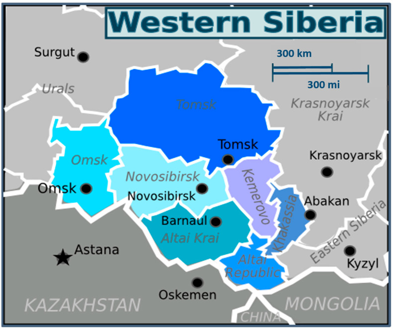
Barnaul (geographic coordinates of center: 53°20′ N 83°45′ E) is the largest city and administrative center of Altai Krai, the Asian part of Russia, located in the forest steppe zone of the West Siberian Plain on the left bank of the Ob River. The border with Kazakhstan is 345 km (210 mi) to the south, which makes Barnaul the closest major city to the Altai Mountains. The city is relatively close to the Russian borders with Mongolia and China (Figure 1).
Figure 1.
Geographical position of Barnaul and Altai Krai, Western Siberia (modified from https://commons.wikimedia.org/wiki/File:Western_Siberia_WV_map_PNG.png; accessed on 3 July 2023).
The climate of Barnaul is continental with a short, warm, and humid summer. The relative humidity during the warm period is around 62%. The native vegetation of Barnaul and its suburbs belongs to the southern forest steppe zone and is represented by steppe, forest, and floodplain-meadow types. Grasses and motley grasses are common here: narrow-leaved bluegrass, Russian brome grass, silver cinquefoil, sickle alfalfa, etc.
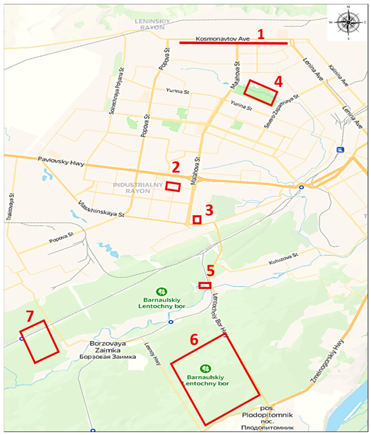
All 300 ticks of the three genera of the Ixodidae family were collected in the urban parks, squares, and wastelands within the city of Barnaul, Altai Krai. In total, there were seven zones for tick collection: urban ecosystem with grass plantations (1, 2), urban ecosystem near buildings next to coniferous forest (3), urban forest park area (4), boundary between aquatic and forest ecosystems (5), and boundary between urban and forest ecosystems (6, 7). Figure 2 depicts the collection zones and geographic coordinates. Ticks were collected by flagging the vegetation between April and June 2022. Briefly, ticks were collected in the daylight hours by dragging a flag 1.5 × 2.0 m in size over vegetation in above-mentioned zones including both coniferous and broad-leaved trees with a moderate herb layer. All ticks were unengorged (unfed). Ticks attached to the flag were removed, placed into individual Eppendorf tubes, and stored at −70 °C until transportation to the laboratory. Transportation to the laboratory was carried out by air in a thermal container with enclosed ice packs for 3 days. Homogenization and DNA extraction were carried out within a week from the day that they arrived at the laboratory. The isolated DNA and the remains of the homogenates were stored at −20 °C during the month when PCR and sequencing were performed. Subsequently, all nucleic acid residues and homogenates were transferred for long-term storage at −70 °C.
Figure 2.
The map of tick collection zones in Barnaul. Zones for tick collection with geographic coordinates: urban ecosystem with grass plantations (1 [53°23′12.0′′ N 83°42′57.7′′ E], 2 [53°20′27.3′′ N 83°41′07.4′′ E]), urban ecosystem near buildings next to coniferous forest (3 [53°19′40.1′′ N 83°41′39.0′′ E]), urban forest park area (4 [53°22′10.0′′ N 83°43′15.8′′ E]), boundary between aquatic and forest ecosystems (5 [53°18′26.7′′ N 83°41′59.1′′ E]), boundary between urban and forest ecosystems (6 [53°16′32.1′′ N 83°42′15.4′′ E], 7 [53°17′16.4′′ N 83°37′34.9′′ E]).
2.2. DNA Extraction and Quantitative PCR
After morphological identification, each tick was individually washed with 96% ethanol and then 0.15 M NaCl solution. Ticks were homogenized in a 2.0 mL Eppendorf tube in 300 μL 0.15 M NaCl solution with tungsten carbide beads in a TissueLyser LT homogenizer (Qiagen, Hilden, Germany) at 50 Hz/s for 10 min. Total DNA was extracted using «AmpliSens® RIBO-prep» kit (CRIE, Moscow, Russia). qPCR screening for Rickettsia spp. was performed using Rotor-Gene Q (Qiagen, Hilden, Germany) and «AmpliSens® Rickettsia spp. The SFG-FL» kit targeted the ompB gene (CRIE, Moscow, Russia) according to the manufacturer’s instructions.
2.3. Sequence Analysis
The SFGR species were determined by Sanger sequencing amplified by conventional PCR to identify citrate synthase gltA, outer membrane protein A ompA, outer membrane protein B ompB, 17 kDa protein htrA, and 16S rRNA gene fragments on two DNA strands using primers listed in Table 1. BLASTN 2.13.0 was used to search against the GenBank non-redundant database using default parameters. Dendrograms were constructed in MEGA 6.06 using the maximum likelihood method on aligned gene fragment sequences with a bootstrap value of 1000. Homologous DNA sequences from the complete genomes of the corresponding representative SFGR obtained from GenBank were used for comparison and the R. bellii An04 genome (NZ_CP015010) was used as an outgroup where possible.
Table 1.
List of primers used in the study.
| Primer Name | Sequence (5′-3′) | Annealing Temperature (°C) |
Amplicon Size (bp) | Reference |
|---|---|---|---|---|
| Rp877p | GGGGACCTGCTCACGGCGG | 58 | 382 | [5] |
| Rp1258n | ATTGCAAAAAGTACCGTGAACA | |||
| Rr.190.70p | ATGGCGAATATTTCTCCAAAA | 55 | 532 | [6] |
| Rr.190.602n | AGTGCAGCATTCGCTCCCCCT | |||
| rompB SFG IF | GTTTAATACGTGCTGCTAACCAA | 57 | 426 | [7] |
| rompB SFG/TG IR | GGTTTTGCCCATATACCGTAAG | |||
| Rr17k.90p | GCTCTTGCAGCTTCTATGTT | 55 | 450 | [8] |
| Rr17k.539n | TCAATTCACAACTTGCCATT | |||
| 16S3 | GATGGATGAGCCCGCGTCAG | 65 | 772 | [3] |
| 16S4 | GCATCTCTGCGATCCGCGAC |
Confidence intervals were calculated by using the modified Wald method in QuickCalcs (GraphPad, San Diego, CA, USA).
The sequences from this study are available in GenBank (OQ723808-OQ723947, OQ727556-OQ727559).
3. Results
Ticks from three genera (n = 300) were collected in this study. After morphological identification at the genus level, the majority of ticks were found to belong to the genus Dermacentor (n = 202), mostly D. nuttalli and D. silvarum. There were significantly fewer ticks belonging to the genus Ixodes (n = 97) and one tick belonging to the genus Haemaphysalis.
For primary screening, we used SFGR-specific qPCR. Positive qPCR results were obtained in 43.3% of samples (130/300). A total of 96% (125/130) of all positive qPCR results were obtained from Dermacentor ticks. A total of 115 of 125 rickettsial DNA samples with Ct (cycle threshold) values ≤ 31 isolated from ticks were subsequently determined by Sanger sequencing.
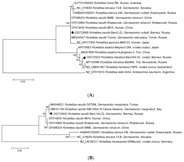
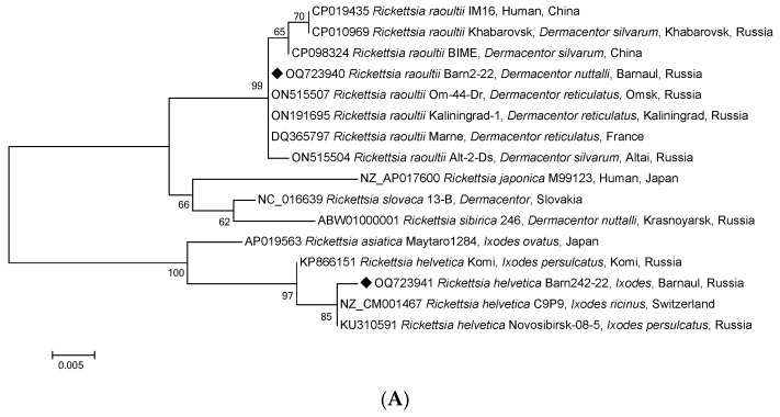
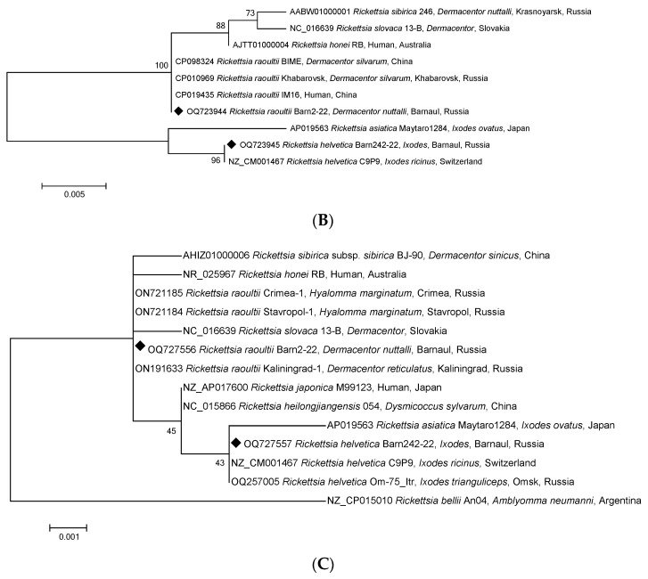
To identify Rickettsia species, each sample was additionally tested by conventional PCR with electrophoretic detection, and fragments of the gltA, ompA, ompB, htrA, and 16S rRNA genes of Rickettsia spp. SFG were sequenced. GenBank analysis of rickettsial DNA sequences found in 115 Dermacentor ticks confirmed the species of the ticks themselves with 100% identity and the gltA gene fragment of 69 strains of R. raoultii, including three complete genomes of Khabarovsk, IM16, and BIME strains (Figure 3A). There was also confirmation with 100% identity of the ompA gene fragments of the R. raoultii isolates from D. marginatus in Turkey and Italy (Figure 3B). Table 2 shows the distribution of SFGR-positive samples among the three tick genera. For all five genes, the isolates obtained were part of the R. raoultii cluster but differed slightly from the three complete genome strains of the ompB gene fragment (Figure 3 and Figure 4A–C). Therefore, we concluded that 100% of Rickettsia spp. SFG identified in Dermacentor ticks belonged to the same species, R. raoultii. Furthermore, the sequences of the fragments of all the five genes mentioned above were 100% identical.
Figure 3.
Phylogenetic trees constructed using the maximum likelihood method based on nucleotide sequences of Rickettsia spp. from ticks, including ones from this study (Barnaul, black diamonds), and reference sequences of the (A) gltA (384 bp) and (B) ompA (532 bp) gene fragments. The R. bellii An04 (NZ_CP015010) and R. monacensis IrR/Munich (NZ_LN794217) sequences were used as an outgroup. The GenBank accession numbers for reference sequences are shown with the sequence name, tick species, and country. The branch numbers indicate bootstrap support (1000 replicates). The scale bar indicates the phylogenetic distance.
Table 2.
Prevalence of tick-borne rickettsioses pathogens in ticks, Barnaul, 2022.
| Genus | Collecting Zones | Number of Ticks | Number of Ticks Infected by SFGR (%, 95% CI) |
|
|---|---|---|---|---|
| R. raoultii | R. helvetica | |||
| Dermacentor | 1 | 71 | 37 (52.1, 40.7–63.3) | 0 |
| 2 | 60 | 33 (55.0, 42.5–66.9) | 0 | |
| 3 | 11 | 11 (100.0, 70.0–100.0) | 0 | |
| 4 | 1 | 0 | 0 | |
| 5 | 57 | 42 (73.7, 60.9–83.4) | 0 | |
| 6 | 2 | 2 (100.0, 29.0–100.0) | 0 | |
| 7 | - | - | - | |
| Subtotal | 202 | 125 (61.9, 55.0–68.3) | 0 | |
| Ixodes | 1 | - | - | - |
| 2 | - | - | - | |
| 3 | - | - | - | |
| 4 | 11 | 0 | 0 | |
| 5 | 5 | 0 | 2 (40.0, 11.6–77.1) | |
| 6 | 39 | 0 | 2 (5.1, 0.5–17.8) | |
| 7 | 42 | 0 | 1 (2.4, <0.01–13.4) | |
| Subtotal | 97 | 0 | 5 (5.1, 1.9–11.8) | |
| Haemaphysalis | 7 | 1 | 0 | 0 |
| Total | 300 | 125 (41.7, 36.2–47.3) | 5 (1.7, 0.6–4.0) | |
Figure 4.
Phylogenetic tree constructed using the maximum likelihood method based on nucleotide sequences of Rickettsia spp. from ticks, Barnaul (black diamonds), and reference sequences of the (A) ompB (427 bp), (B) htrA (450 bp), and (C) 16S rRNA (768 bp) gene fragments. The GenBank accession numbers for reference sequences are shown with the sequence name. The branch numbers indicate bootstrap support (1000 replicates). The scale bar indicates the phylogenetic distance.
Notably, DNA samples of Rickettsia isolated from three Ixodes spp. ticks, amplified and sequenced by fragments of the gltA gene, showed the highest identity with the homologous gene of strain R. helvetica C9P9 (99.74%) (Figure 3A) and the fragments of ompB, htrA, and 16S rRNA (Figure 4A–C). As shown in Figure 4A–C, R. helvetica was 100% identical to R. helvetica C9P9 for htrA and 16S RNA gene fragments and 99.77% homologous to the ompB gene. Similar to some SFGR species, R. helvetica also lacks the ompA gene.
4. Discussion
In Russia, the incidence of the population with NATT and Astrakhan spotted fever, the etiological sources of which are R. sibirica subsp. sibirica and R. conorii subsp. caspia, respectively, is subject to official recording. The regions of Siberia are endemic with NATT, which occurs with a rash and a fever. However, the absence of a rash among tick-bite patients in the region does not rule out Rickettsia infection. According to official data from Rospotrebnadzor, in 2021, 9952 people suffered from tick bites in the Altai Krai (436.06 per 100,000 population) and NATT was diagnosed for 460 people (including 299 people from rural areas) [9]. There is no registration of rickettsioses without a rash. However, such rickettsiosis cannot be excluded.
Barnaul’s recreational zones are located on the territory of the Ob Plateau, which is mostly covered with grassy meadows and feather-grass steppes. As a rule, Dermacentor ticks live in such areas. Most often, the hosts for these ticks are small mammals.
In this study, the SFGR pathogens were found in ticks in Barnaul at a high prevalence. Ticks of the genus Dermacentor are the main host and natural reservoir of R. raoultii not only in Europe [10] but also in some Asian countries [11]. All rickettsial DNA isolated from D. reticularis in Europe was found to belong to R. raoultii, with an average infection rate of 47.9% [12]. In a previous study, the percentage of R. raoultii in questing Dermacentor ticks in Altai Krai was relatively low (15.0%) [13]. In our study, all R. raoultii isolates were detected in the vast majority (61.9%, 95% CI 55.0%–68.3%) of the Dermacentor spp. ticks. Considering that this Rickettsia species is thought to be the cause of DEBONEL/TIBOLA/SENLAT syndrome and does not cause the classic rash clinic typical for most TBRs registered in Russia (NATT, Far Eastern tick-borne rickettsiosis, Astrakhan spotted fever), further monitoring studies are necessary.
R. helvetica has also been identified in 5.1% (95% CI 1.9–11.8%) of Ixodes ticks. It was previously recognized as non-pathogenic in humans, but several cases described in Sweden suggest that infection may be accompanied by non-specific human fever, meningitis, and perimyocarditis [14,15,16]. Speck et al. [17] classified R. helvetica as a low-pathogenic microorganism. We did not find the NATT causative agent, R. sibirica, in tick vectors in this study. Although Altai Krai is endemic with TBR, it has been understudied in terms of understanding the genetic diversity of human pathogens in ixodid ticks. Information about the prevalence of Rickettsia species/subspecies in this particular area should be taken into account by clinicians since rickettsioses can occur without a rash.
5. Conclusions
Analysis of the amplified sequences of the gltA, ompA, ompB, htrA, and 16S rRNA regions of Rickettsia spp. suggests that the detected R. raoultii and R. helvetica represent homogeneous populations but are genetically distinct from previously described genotypes isolated from ticks in other regions of Russia. It is necessary to continue the study of the species diversity of pathogens contained in ticks from the Russian regions endemic with tick-borne infections.
Author Contributions
Conceptualization, A.V.R. and T.A.C.; methodology, A.V.R., K.P. and A.V.V.; software, A.V.R.; validation, A.V.R., T.A.C. and K.P.; formal analysis, A.V.R.; investigation, A.V.R.; resources, A.V.T., S.V.S. and N.V.L.; data curation, A.V.R.; writing—original draft preparation, A.V.R.; writing—review and editing, A.V.R. and T.A.C.; visualization, A.V.R., T.A.C. and A.V.T.; supervision, T.A.C.; project administration, T.A.C. and A.V.T.; funding acquisition, V.G.A. All authors have read and agreed to the published version of the manuscript.
Institutional Review Board Statement
Not applicable.
Informed Consent Statement
Not applicable.
Data Availability Statement
The sequences from this study are available in the NCBI GenBank under accession numbers OQ723808-OQ723947 and OQ727556-OQ727559.
Conflicts of Interest
The authors declare no conflict of interest.
Funding Statement
This work was supported by the state assignment topic «Improvement of the epidemiological monitoring system in the Russian Federation for natural focal vector-borne infections of bacterial nature» (Reg. No.: AAAA-A21-121011890133-8).
Footnotes
Disclaimer/Publisher’s Note: The statements, opinions and data contained in all publications are solely those of the individual author(s) and contributor(s) and not of MDPI and/or the editor(s). MDPI and/or the editor(s) disclaim responsibility for any injury to people or property resulting from any ideas, methods, instructions or products referred to in the content.
References
- 1.Rudakov N.V., Pen’evskaya N.A., Kumpan L.V., Blokh A.I., Shpynov S.N., Trankvilevsky D.V., Shtrek S.V. Epidemiological Situation on Tick-Borne Spotted Fever Group Rickettsioses in the Russian Federation in 2012–2021, Prognosis for 2022–2026. Probl. Part. Danger. Infect. 2022;1:54–63. doi: 10.21055/0370-1069-2022-1-54-63. [DOI] [Google Scholar]
- 2.Beskhlebova O.V., Granitov V.M., Shpynov S.N., Dedkov V.G., Arseneva I.V., Pantyukhina A.N. Rickettsioses of spotted fever group in the Altai region. Infect. Dis. News Opin. Train. 2017;19:73–78. doi: 10.24411/2305-3496-2017-00037. [DOI] [Google Scholar]
- 3.Igolkina Y., Krasnova E., Rar V., Savelieva M., Epikhina T., Tikunov A., Khokhlova N., Provorova V., Tikunova N. Detection of causative agents of tick-borne rickettsioses in Western Siberia, Russia: Identification of Rickettsia raoultii and Rickettsia sibirica DNA in clinical samples. Clin. Microbiol. Infect. 2018;24:199.e9–199.e12. doi: 10.1016/j.cmi.2017.06.003. [DOI] [PubMed] [Google Scholar]
- 4.Rudakov N., Samoylenko I., Shtrek S., Igolkina Y., Rar V., Zhirakovskaia E., Tkachev S., Kostrykina Y., Blokhina I., Lentz P., et al. A fatal case of tick-borne rickettsiosis caused by mixed Rickettsia sibirica subsp. sibirica and “Candidatus Rickettsia tarasevichiae” infection in Russia. Ticks Tick Borne Dis. 2019;10:101278. doi: 10.1016/j.ttbdis.2019.101278. [DOI] [PubMed] [Google Scholar]
- 5.Roux V., Rydkyna E., Eremeeva M., Raoult D. Citrate synthase gene comparison, a new tool for phylogenetic analysis, and its application for the rickettsiae. Int. J. Syst. Bacteriol. 1997;47:252–261. doi: 10.1099/00207713-47-2-252. [DOI] [PubMed] [Google Scholar]
- 6.Regnery R.L., Spruill C.L., Plikaytis B.D. Genotypic identification of rickettsiae and estimation of intraspecies sequence divergence for portions of two rickettsial genes. J. Bacteriol. 1991;173:1576–1589. doi: 10.1128/jb.173.5.1576-1589.1991. [DOI] [PMC free article] [PubMed] [Google Scholar]
- 7.Choi Y.J., Jang W.J., Kim J.H., Ryu J.S., Lee S.H., Park K.H., Paik H.S., Koh Y.S., Choi M.S., Kim I.S. Spotted fever group and typhus group rickettsioses in humans, South Korea. Emerg. Infect. Dis. 2005;11:237–244. doi: 10.3201/eid1102.040603. [DOI] [PMC free article] [PubMed] [Google Scholar]
- 8.Ishikura M., Ando S., Shinagawa Y., Matsuura K., Hasegawa S., Nakayama T., Fujita H., Watanabe M. Phylogenetic analysis of spotted fever group rickettsiae based on gltA, 17-kDa, and rOmpA genes amplified by nested PCR from ticks in Japan. Microbiol. Immunol. 2003;47:823–832. doi: 10.1111/j.1348-0421.2003.tb03448.x. [DOI] [PubMed] [Google Scholar]
- 9.Comparison of Infectious Disease Rates. [(accessed on 29 June 2023)]. Available online: https://www.iminfin.ru/areas-of-analysis/health/perechen-zabolevanij/sravnenie?territory=01000000.
- 10.Mediannikov O., Matsumoto K., Samoylenko I., Drancourt M., Roux V., Rydkina E., Davoust B., Tarasevich I., Brouqui P., Fournier P.E. Rickettsia raoultii sp. nov., a spotted fever group rickettsia associated with Dermacentor ticks in Europe and Russia. Int. J. Syst. Evol. Microbiol. 2008;58:1635–1639. doi: 10.1099/ijs.0.64952-0. [DOI] [PubMed] [Google Scholar]
- 11.Seo M.G., Kwon O.D., Kwak D. High prevalence of Rickettsia raoultii and associated pathogens in canine ticks, South Korea. Emerg. Infect. Dis. 2020;26:2532. doi: 10.3201/eid2610.191649. [DOI] [PMC free article] [PubMed] [Google Scholar]
- 12.Balážová A., Földvári G., Bilbija B., Nosková E., Široký P. High prevalence and low diversity of Rickettsia in Dermacentor reticulatus ticks, Central Europe. Emerg. Infect. Dis. 2022;28:893–895. doi: 10.3201/eid2804.211267. [DOI] [PMC free article] [PubMed] [Google Scholar]
- 13.Dedkov V.G., Simonova E.G., Beshlebova O.V., Safonova M.V., Stukolova O.A., Verigina E.V., Savinov G.V., Karaseva I.P., Blinova E.A., Granitov V.M., et al. The burden of tick-borne diseases in the Altai region of Russia. Ticks Tick Borne Dis. 2017;8:787–794. doi: 10.1016/j.ttbdis.2017.06.004. [DOI] [PubMed] [Google Scholar]
- 14.Nilsson K., Elfving K., Pahlson C. Rickettsia helvetica in patient with meningitis, Sweden, 2006. Emerg. Infect. Dis. 2010;16:490–492. doi: 10.3201/eid1603.090184. [DOI] [PMC free article] [PubMed] [Google Scholar]
- 15.Nilsson K. Septicaemia with Rickettsia helvetica in a patient with acute febrile illness, rash and myasthenia. J. Infect. 2009;58:79–82. doi: 10.1016/j.jinf.2008.06.005. [DOI] [PubMed] [Google Scholar]
- 16.Nilsson K., Lindquist O., Påhlson C. Association of Rickettsia helvetica with chronic perimyocarditis in sudden cardiac death. Lancet. 1999;9185:1169–1173. doi: 10.1016/S0140-6736(99)04093-3. [DOI] [PubMed] [Google Scholar]
- 17.Speck S., Kern T., Aistleitner K., Dilcher M., Dobler G., Essbauer S. In vitro studies of Rickettsia-host cell interactions: Confocal laser scanning microscopy of Rickettsia helvetica-infected eukaryotic cell lines. PLoS Negl. Trop. Dis. 2018;12:e0006151. doi: 10.1371/journal.pntd.0006151. [DOI] [PMC free article] [PubMed] [Google Scholar]
Associated Data
This section collects any data citations, data availability statements, or supplementary materials included in this article.
Data Availability Statement
The sequences from this study are available in the NCBI GenBank under accession numbers OQ723808-OQ723947 and OQ727556-OQ727559.


