



Abstract
Extracorporeal membrane oxygenation (ECMO) is a type of extracorporeal life support (ECLS) in which the function of the heart and/or lungs is partially or completely replaced by a portable system that provides prolonged support to critically ill patients with respiratory or cardiac failure. There are two major variants of ECMO: veno-venous (VV) ECMO and veno-arterial (VA) ECMO. VV ECMO replaces the function of the lung in which it uses a cannula to remove venous blood and oxygenates it using the extracorporeal system, and returns the blood to the right atrium to be pumped to the body. VA ECMO is slightly different in that it replaces the function of the heart and lungs by returning oxygenated blood to the aorta. As a therapy for respiratory failure, ECMO minimizes hypoxia, diminishes lung stress and strain, and allows lung protective mechanical ventilation. As a support for acute and terminal heart failure, ECMO reduces preload, increases aortic flow, and allows for end-organ perfusion. Due to its physiological support and advantages, it is used for a variety of chronic and acute support purposes such as bridge therapy for heart/lung transplant, durable ventricular assist devices, and intermediate-term mechanical support postoperatively. Our review gives a broad overview of the two main types of ECMO strategies and their clinical indications, cannulation strategies, unique clinical utility, and their limitations.
Keywords: Cardiothoracic surgery, ECMO, Cannulation
Introduction
Extracorporeal membrane oxygenation (ECMO) is a modified form of cardiopulmonary bypass utilized to provide adequate oxygen delivery to the tissues for patients with respiratory and/or cardiac failure while the heart or lung recovers [1]. Two main forms of ECMO are veno-arterial ECMO (VA ECMO) and veno-venous ECMO (VV ECMO). VA ECMO is primarily utilized for circulatory support where blood is drained from the right side of the heart, passes through an oxygenator, and then returned to arterial circulation, hence, partially bypassing the heart and lungs. In VV ECMO, venous blood is drained, passes through an oxygenator, and returned to the right side of the heart. There are a variety of factors that can be related to predicting survival for patients cannulated for VA ECMO described in the Survival After Veno-arterial ECMO (SAVE) score, such as age, weight, diagnoses prior to cannulation, and hemodynamics. Similarly, survival can be predicted for patients cannulated for VV ECMO using the Respiratory ECMO Survival Prediction (RESP) score using factors such as age, central nervous system (CNS) dysfunction, and partial pressure of carbon dioxide (PaCO2) [2].
As is the case with other major surgical procedures, ECMO outcomes correlate directly with the volume of a performing center, which speaks to the importance of performing VA ECMO in high-volume centers. ECMO is frequently instituted at other centers, although it is recommended that once the patient is stable, the patient should be transferred to a center with significant ECMO experience, leveraging a so-called hub and spoke model [3]. A large team of experts is required for excellent care of these patients and includes a surgeon, a perfusionist or ECMO specialist, a heart failure cardiologist, intensive care specialists, a physical therapist, a respiratory therapist, a pharmacist, a dietician, an infectious disease physician, a neurologist, and a palliative care specialist.
ECMO circuit
Basic components of the ECMO circuit include arterial and venous cannulae, tubing, a pump head, an oxygenator, a gas source, a heat exchanger, and a flow meter. Cannulae utilized are often the same as would be used for central access cardiopulmonary bypass or peripheral access minimally invasive cardiac surgery. There are some specialized cannulae utilized exclusively for ECMO, as will be discussed. The oxygenator typically consists of a microporous hollow fiber design using fibers made from polymethylpentene. These are more durable (weeks) as compared to the microporous hollow fiber oxygenators made from polypropylene used in cardiopulmonary bypass. The most commonly used pump is a centrifugal, non-pulsatile pump. These pumps are both preload dependent and afterload sensitive; therefore, there is no fixed relationship between pump speed and blood flow. For this reason, a flow meter is required within the circuit to accurately assess the blood flow. The gas sweep flow rate is utilized to increase carbon dioxide removal by increasing the rate of gas flow through the oxygenator; however, it is important to note that increasing gas sweep does not affect the oxygenation [1]. To increase oxygen delivery to the tissues, one must increase the ECMO flow. The native lungs can contribute to the ventilation and oxygenation of the blood to either a large or small extent depending on the underlying pulmonary pathology and ventilator settings.
Shock
Shock is a state which is defined by an inadequate oxygen delivery to tissues. The etiologies of shock vary, with ECMO being utilized to manage primarily cardiogenic shock. The stages of shock have been described in a multitude of forms, but the Society for Cardiovascular Angiography and Intervention (SCAI) outlines the stages of cardiogenic shock as an indication for severity and comprises a component of a mortality risk [4]. The stages are outlined from A to E as viewed in Fig. 1. We recommend the initiation of ECMO prior to a patient reaching extremis, although use of ECMO during cardiopulmonary resuscitation (CPR) is not uncommon.
Fig. 1.

Society for Cardiovascular Angiography and Intervention (SCAI) classification for shock [5]
Veno-arterial (VA) ECMO
Introduction
VA ECMO provides biventricular support to the heart as well as the lungs by partially replacing their function to a variable extent in providing gas exchange and cardiac output [6]. The VA ECMO circuit does not have a reservoir, in contrast to the cardiopulmonary bypass (CPB) machine. This provides certain advantages and limitations. The ECMO circuit is unable to handle a significant amount of air and thus must remain air free. However, the absence of the blood-air interface permits significant reduction in anticoagulation goals and subsequent decrease in bleeding risk. VA ECMO has a significant effect on hemodynamics. VA ECMO frequently can cause an increase in left ventricular end-diastolic pressure (LVEDP). Without venting, as discussed below, left ventricle (LV) distention in the setting of LV dysfunction and higher afterload from the VA ECMO flow can lead to pressurization of the pulmonary circuit.
VA ECMO is not the only choice for individuals who fall into these parameters of refractory cardiogenic shock (RCS) as other short-term circulatory support devices are likewise available. The advantages of VA ECMO include a short lead-time to initiation, bedside initiation, and ability for biventricular and pulmonary support.
VA ECMO is limited by the complexity of issues and pitfalls that may arise during the care of these patients. The most common of these include the management of worsening of aortic insufficiency, possible left ventricular distension requiring a vent, inability to initiate peripheral VA ECMO in the case of peripheral arterial disease, titration of anticoagulation, and subsequent complications. Additionally, hemolysis and distal arterial ischemia are also not uncommon. Our recommended management of each of these issues, should they arise, is described separately throughout the remainder of this paper.
Indications
VA ECMO is indicated in cases of RCS due to a number of etiologies. Primary cardiac dysfunction may be due to acute coronary syndrome, post-cardiotomy or failure to wean from CPB, CPR, primary graft failure following transplant, fulminant myocarditis, chronic decompensated heart failure, or ventricular tachycardia storm. Secondary cardiac dysfunction may be caused by pulmonary hypertension or pulmonary emboli, sepsis-associated cardiomyopathy, peripartum cardiomyopathy, drug overdose or poisoning, and pheochromocytoma-induced catecholamine crisis [7, 8].
Inotropes and vasoconstrictors used in the setting of cardiogenic shock to support cardiac output and tissue perfusion do so at the cost of increased myocardial oxygen demand and ischemia, arrhythmogenicity, and microcirculation compromise [1, 9]. In this setting, VA ECMO increases tissue perfusion while concomitantly decreasing myocardial oxygen demand and ischemia.
Objective parameters of RCS include a cardiac index of < 1.8 L/min/m2 without hemodynamic support, cardiac index of < 2.2 L/min/m2 with hemodynamic support, systolic blood pressure < 90 mmHg for > 30 min, drop in mean arterial pressure > 30 mmHg below baseline, or pulmonary capillary wedge pressure > 15 mmHg. Cardiogenic shock with a rising serum lactate despite optimal medical management and inadequate support with an intra-aortic balloon pump (IABP) or Impella device likewise suggest that ECMO is indicated [1, 10, 11].
VA ECMO functions as a short-term bridge to definitive therapy, bridge to recovery, or as a bridge to decision if the prognosis is uncertain. Specifically in cardiac arrest settings where the patient’s neurological status is not clear with acute pulmonary edema, VA ECMO can serve to buy time for a patient to declare their longer-term clinical prognosis [11].
Contraindications
VA ECMO functions as a short-term bridge; therefore, contraindications are primarily based upon whether the patient has a condition which precludes any hope of future recovery or the inability to tolerate VA ECMO. This includes inability to be anticoagulated, the patient cannot be reasonably cannulated either centrally or peripherally, the patient is not a candidate for a ventricular assist device (VAD), do not resuscitate status, non-recoverable cardiac dysfunction, and the patient is not a candidate for transplantation, advanced malignancy (except in occasions of chemotherapy-induced cardiomyopathy in an otherwise curable malignancy), and moderate to severe aortic insufficiency (unless an Impella device is placed first as a vent) [9, 11, 12].
Cannulation
The most commonly utilized adult arterial cannulae range from 15 to 21 Fr and the venous 19–25 Fr. Depending on the size of the cannulae, VA ECMO can provide over 6 L/min of blood flow. Flow of VA ECMO is measured by cardiac index and is recommended to not fall below 2.2 to 2.5 L/min/m2 as to not clot the cannulae.
VA ECMO can be initiated centrally, peripherally, or through a combination of both. If needed, any combination of central and peripheral cannula sites may be used (for example: femoral venous drainage with ascending aortic arterial outflow). If there are any issues with the femoral vessels, right atrium, or ascending aorta, other potential cannulation sites include the internal jugular (IJ) vein, superior vena cava (SVC), inferior vena cava (IVC), carotid artery, and the axillary or subclavian artery. In all cases of cannulation, the cannula positioning should be verified with chest and abdominal radiographs as soon as possible [13].
Central cannulation
Central cannulation is commonly used in post-cardiotomy settings by placing the arterial cannula in the ascending aorta and the venous cannula in the right atrium or percutaneously via the femoral vein [7]. In cases of central cannulation in a post-cardiotomy setting, leaving a LV vent is advisable as described below. As bleeding from the cannulation site is the most common complication, it is important to ensure adequate hemostasis of the cannulation sites prior to leaving the operating room (Fig. 2).
Fig. 2.

Schematic of cannulation of central VA ECMO in a post-cardiotomy setting. Legend: VA, veno-arterial; SVC, superior vena cava; IVC, inferior vena cava; RA, right atrium; RV, right ventricle; PA, pulmonary artery; LA, left atrium; LV, left ventricle; O2, oxygen
If this is difficult to achieve, heparin can be held temporarily until hemostasis can be achieved within a reasonable amount of time, balancing bleeding risks with circuit-related thrombo-emboli. Techniques to improve hemostasis include the use of red rubber tourniquet to allow adequate snaring of the purse string sutures. Tension can also be held in place via securing a polypropylene suture to the red rubber snare with multiple ligaclips, which allow for no instruments to be left in the chest. Multiple silk sutures can be applied to secure the tourniquet to the arterial and venous cannulae. In cases of anticipated longer ECMO runs, tunneling the cannulae to enable interval chest closure is recommended.
Another central cannulation strategy is sewing a chimney graft to the ascending aorta and tunneling this through the suprasternal notch. An arterial cannula is then placed through this graft with a percutaneous femoral venous cannula.
Peripheral cannulation
In emergent circumstances, peripheral cannulation is the most expeditious way to initiate VA ECMO [12]. Peripheral cannulation may be performed percutaneously at bedside or in the hybrid operating room (OR) suite with fluoroscopic guidance. Femoral vessels are most commonly used, although the axillary artery can similarly be utilized (Fig. 3). For peripheral VA ECMO cannulation, invasive arterial blood pressure monitoring is preferred to be in the right upper extremity for optimal oxygenation monitoring for the early detection of differential oxygenation.
Fig. 3.

Schematic of peripheral VA ECMO cannulation via femoral vessels
Peripheral cannulation can be performed percutaneously via the Seldinger technique, or in an open manner where a cutdown is employed to place the cannulae under direct visualization. In cases of a cut down, cannulae can be placed under direct visualization. Purse string sutures can be placed prior to cannula placement to allow for ease of closure after decannulation. Limited distal dissection is advised to manually palpate the distal perfusion cannula in the superficial femoral artery. Inadvertently placing the cannula in the profunda femoris artery results in limb ischemia.
Alternatively, a chimney graft may be sewn to the vessel. However, this is associated with higher rates of needle hole bleeding after reaching therapeutic anticoagulation. Additionally, the risk to using a chimney graft is that it can lead to limb hyper-perfusion syndrome which occasionally requires distal banding. However, the benefit to a chimney graft is that it eliminates the need for a distal perfusion cannula.
If the cannulae were placed percutaneously via the Seldinger technique without concomitant percutaneous closure device, the groin with the arterial cannula is usually explored at the time of decannulation, in order to close or repair the arteriotomy. Although this may also be performed for the venotomy site, it is also possible to simply remove the venous cannula and hold pressure to the site, followed by closure of the skin site with suture.
Special considerations
Distal limb ischemia
Femoral arterial cannulation carries the risk of distal limb ischemia due to obstruction of distal arterial flow. To mitigate this risk, an antegrade perfusion catheter is placed in the superficial femoral artery (SFA) distal to the femoral arterial cannula. In more elective settings, it is technically easier to visualize the SFA via ultrasound prior to the insertion of the femoral arterial cannula as it may mask ultrasound imaging.
A 4- or 5-Fr micro-puncture kit is used for access. Size permitting, we recommend using of a 9-Fr Femflex metal-braided introducer, as plastic introducers can kink leading to severe hemolysis or flow obstruction and a smaller cannula often clots off. Placing an interposition quarter-inch tubing can be utilized to “Y” the distal perfusion cannula (DPC) to the arterial femoral cannula to enable flow measurement since the smallest flow probe can only fit on quarter-inch tubing. A minimum of 100 mL/min is recommended to prevent limb ischemia.
Another tip to prevent distal limb ischemia is utilizing smaller femoral cannulae. Often, in an average adult, a 17-Fr arterial cannula would suffice and up to 330 mmHg arterial pressure line can be tolerated. It is essential to maintain frequent neurovascular checks of the cannulated leg to detect any distal limb ischemia. Ultrasound Doppler and spectroscopy can also be used to assess distal limb vessel patency and flow.
If limb ischemia still occurs, a second DPC may be placed via cut down on the posterior tibial artery and may also be “Y”-ed to the femoral arterial cannula. This can be achieved by cutting down on the posterior tibial artery with a vertical incision behind the medial malleolus towards the posterior edge of the tibia, locating the artery under the medial retinaculum, and following this proximally to find a location in which an 8-Fr pediatric aortic cannula may be inserted. Careful cannulation to avoid retrograde dissection is key [13].
Some patients present in cardiogenic shock with an Impella device in place, which cannot be removed as it is being utilized as an LV vent. However, its large caliber sheath can be flow limiting, leading to limb ischemia. In this case, a DPC can be placed in the SFA and “Y”-ed to the femoral arterial cannula to provide bilateral distal limb perfusion (one to the ECMO side and one to the Impella side). This allows for utilization of the Impella as a direct LV vent, typically termed “ECPELLA”.
Differential oxygenation (Harlequin syndrome)
In case of peripheral VA ECMO cannulation, a retrograde column of hyper-oxygenated blood travels up the descending aorta. Prolonged cardiac arrest or severe LV dysfunction causes pulmonary edema, and when the heart starts to recover, this edema may still be present and lungs could act as a large shunt. Blood ejected from the heart in this case could potentially be low in oxygen, leading to a mixing cloud between retrograde hyper-oxygenated blood from ECMO circuit and the antegrade low oxygen blood from the native heart ejection due to pulmonary edema.
The first arteries that reflect this low oxygen because of the differential oxygen mixing are the coronaries, followed by arch vessels. The right innominate artery and its branches are the most distant arterial points from the femoral arterial cannula; therefore, monitoring oxygenation from a right radial or brachial arterial line is essential.
Differential oxygenation is when the patient has normal oxygen saturation on plethysmograph on lower extremities, and low oxygen saturation on the upper body, also known as “Harlequin syndrome” or “North-South Mixing Syndrome” (Fig. 4). Usually, this represents a positive predictor for myocardial recovery, as the heart is able to pump against retrograde ECMO flow, but it also reflects continued pulmonary edema.
Fig. 4.

Clinical scenario of Harlequin syndrome (North-South mixing syndrome) in a patient with large anterior wall myocardial infarction after percutaneous intervention on peripheral VA ECMO. Chest X-ray (CXR) indicating severe pulmonary edema (A) with accompanying arterial blood gases (ABGs) and (B) identifying differential oxygen saturation in femoral vessels versus radial artery as depicted in our review. C CXR indicating resolution of differential oxygenation in radial artery and pulmonary edema post-IJ cannula (superior arrow) placement 13 cm from venous cannula (inferior arrow). Legend: pCO2, partial pressure of carbon dioxide; pO2, partial pressure of oxygen
To mitigate this situation, the lungs may be utilized more by optimizing the ventilator settings, decreasing the heart rate (which may be accomplished with amiodarone or other sedation agents), and increasing ECMO flow. Increasing ECMO flow deprives the heart from venous return and subsequently results in decreased ejection. It is also important to note that tachycardia is occasionally desirable when weaning from ECMO as cardiac output is created by heart rate times stroke volume (slowing the heart rate should only be selectively utilized).
If there is inadequately oxygenated blood flowing to the upper body, an additional cannula may be placed in an IJ to the SVC — right atrial junction and connected to the arterial limb of the VA ECMO circuit. In this manner, oxygenated blood is delivered to the right atrium and eventually to the LV and ascending aorta (despite some shunting through the lung). This configuration is referred to as veno-arterio-venous (VAV) ECMO (Fig. 4C). For VAV ECMO, the tip of the IJ cannula (Y-ed to arterial line) ideally should be 12–15 cm away from the IVC drainage cannula. This allows for appropriate drainage and limits recirculation, hence limiting shunting and improving upper body oxygenation (Fig. 4C). After starting VAV ECMO, it is possible to control the amount of blood flowing to the IJ cannula by utilizing Hoffman clamps. This cannula is usually larger in caliber, and since the right atrium has a low compliance, the blood will preferentially flow to the right atrium. If hypoxia persists, ultimately systemic cooling to decrease native cardiac output and to protect from hypoxia could be utilized.
Devices
Smaller Impella devices (Impella 2.5 and CP) are known to cause a significant degree of hemolysis, leading to hemoglobin cast nephropathy. Subsequent renal failure can preclude an elder patient from receiving a left ventricular assist device (LVAD) or trigger the need for dual organ transplant (heart/kidney) in a younger patient. Therefore, the timing, strategy of cannulation, and management after initiation are highly relevant to the patient outcome.
Venting
There is data to suggest that early venting promotes cardiac recovery. VA ECMO increases the LVEDP; therefore, there is always a concern for left ventricular distention, particularly in patients with a low ejection fraction [13]. Once ECMO flows are initiated, evaluation for distension must be performed on a regular basis. Decreasing ECMO flows can allow for LV ejection in patients with severely depressed left ventricular ejection fraction (LVEF).
Radial arterial line pulsatility, pulmonary artery (PA) diastolic pressure, and bedside echocardiogram are monitored to determine LV distension and to assess functionality and frequency of aortic valve opening. Lack of ejection, as can be seen with an elevated LVEDP, can cause increased risk of clot formation in the LV, but more commonly increases the pressure of the LV, left atrium (LA), and the pulmonary circuit. This can lead to inevitable pulmonary hemorrhage which can be substantial in the setting of anticoagulation. In general, we recommend venting if the PA diastolic pressure is greater than 30 mmHg. If the LV distension is caused by any degree of aortic insufficiency, the only effective method to adequately vent these patients is with an Impella device.
Other strategies for LV venting include inotropes to assist LV ejection, placing a venting catheter directly into the right superior pulmonary vein (in post-cardiotomy settings, placing a venting catheter directly into the apex of the LV via sternotomy or a left thoracotomy), placing an Impella device and running it at lower rpms (to avoid hemolysis — usually any flow from 750 mL to 2 L would suffice), creating an atrial septostomy to establish a left-to-right shunt, or active left atrium (LA) venting known as left atrial VA ECMO (LAVA ECMO), or by placing a percutaneous transseptal cannula (alternatively, placing a PA drainage cannula percutaneously, or placement of an IABP which interrupts the continuous retrograde ECMO flow — thereby decreasing the afterload) [1, 11–13] (Fig. 5).
Fig. 5.

Pulmonary edema caused by LV distension after peripheral VA ECMO cannulation (L). Improved after placing a cannula in the left atrium via transseptal puncture (R)
Differential support of only one side of the heart
It is important to note that ECMO may also be configured to support only one side of the heart. To support the right side only, a venous cannula may be placed in the right atrium, femoral vein, or IJ vein, with an arterial cannula placed in the main PA. This is referred to as a right ventricular assist device (RVAD) or adding an oxygenator if needed (RVAD ECMO). To support only the left side of the heart, a drainage cannula may be placed in the LA or LV apex, and an arterial cannula can be placed in the ascending aorta, axillary or subclavian artery, or femoral artery. The oxygenator can also be spliced out of the circuit after lung recovery.
Other trouble-shooting
Centrifugal pumps are afterload sensitive. In cases of vasoplegia leading to hypotension, the pump will generate high forward flow; therefore, this consequently generates a high degree of negative pressure on the drainage side leading to a suck-down event. A suck-down event occurs when the IVC or the right atrial walls collapse against the side holes of the cannula, leading to an abrupt drop in ECMO flow. A simple solution is temporarily decreasing the revolutions per minute, using a vasopressor, or giving a fluid bolus, then slowly returning the pump back to baseline ECMO settings.
Low flow in the VA ECMO circuit may also be caused by a malposition of the cannula (particularly the venous cannula), which leads to inadequate drainage, hypovolemia (causing the venous tubing to “chatter”), cardiac tamponade, or tension pneumothorax. Therefore, if these issues are encountered, repositioning of the cannulae or treating the cause may be indicated.
Ventilator settings
Lung protective ventilation is preferred once ECMO is initiated, but in cases of differential oxygenation, or when we face oxygenation issues, frequently adequate standard ventilation of the lungs is indicated [1].
Anticoagulation
Anticoagulation is needed to avoid thrombosis of the VA ECMO circuit and unfractionated heparin is the most commonly used anticoagulant. Monitoring of the titration of anticoagulation has been subject to controversy. At our institution, we utilize anti-Xa levels of 0.4–1.1 units/mL; others follow activated partial thrombin time (aPTT) targeting 70–110 s, activated clotting time (ACT) of 180–220 s, or thromboelastogram [12]. Life-threatening bleeding would prompt holding anticoagulation with the risk of circuit clotting or limb ischemia. In this case, higher revolutions per minute are advised (if tolerated) to decrease the chance of circuit clotting.
Due to the inaccuracy of labs at lower anticoagulation levels, one strategy to consider is a cross-check; if using anti-Xa levels, check once a day PTT or ACT to correlate, particularly if bleeding occurs [13].
In cases of heparin-induced thrombocytopenia, heparin alternatives may be used. Many centers and clinicians have used other anticoagulation agents safely, such as argatroban, although no prospective data currently exist to highlight the optimal anticoagulation therapy and monitoring [13].
Veno-venous (VV) ECMO
Introduction
VV ECMO circulation includes draining blood from the right side of the heart, oxygenating via ECMO circuit oxygenator then returning the blood to the right side of the heart. It is primarily indicated for respiratory failure as it does not provide mechanical support except in RVAD ECMO configuration.
Therefore, VV ECMO is primarily utilized for respiratory failure, but only in settings of preserved cardiac function [14]. As is the case for VA ECMO, it is essential to decide the weaning strategy or end-point criteria — most commonly bridge to decision, bridge to recovery, or bridge to transplantation. The most common complications of VV ECMO include bleeding, thrombosis, renal failure, neurologic injury, pneumothorax, and right ventricular perforation [15].
Indications
Persistent hypoxia despite maximal ventilatory settings, paralyzing the patient for maximal synchronization with the ventilator, and use of inhaled pulmonary vasodilators are the main indications to initiate VV ECMO. Criteria for persistent hypoxia include a partial pressure of oxygen (PaO2) less than 60 mmHg for 3 h or PaCO2 greater than 60 mmHg with a pH less than 7.25 for more than 3 h despite maximal ventilatory management with plateau airway pressures less than 30 cm H2O. Similarly in the conventional ventilatory support vs extracorporeal membrane oxygenation for severe adult respiratory failure (CESAR) trial, the inclusion criteria for those placed on VV ECMO versus ventilatory support were adult patients with severe but potentially reversible respiratory failure, Murray score ≥ 2.5, or uncompensated hypercapnia with pH < 7.20. Exclusion criteria to VV ECMO were duration of high fraction of inspired oxygen (FIO2) ventilation > 7 days, intra-cranial bleeding, and contraindication to limited heparinization [16, 17].
Contraindications
Relative contraindications include comorbidities including age, morbid obesity, coagulopathy, neurologic status, multiple organ failure, anatomical challenges, or other chronic life-limiting diseases. Additionally, VV ECMO does not provide cardiac support and therefore is not advised in settings of left-sided heart failure.
Cannulation
Femoral-internal jugular VV ECMO
Access is typically obtained in a percutaneous fashion via the Seldinger technique. The tip of the femoral catheter is placed at the IVC-right atrium (RA) junction, while the IJ catheter tip is positioned at the SVC-RA junction. The benefit to this configuration is the ability to deploy easily at the bedside; however, it could lead to variable degrees of recirculation where the oxygenated blood gets captured by the drainage cannula and recirculates in the ECMO circuit, decreasing the amount of oxygenated blood delivered. Allowing 12–15 cm of distance between drainage and outflow cannulae can minimize recirculation. Femoral vessel cannulation also limits patient ambulation. Therefore, this configuration is most commonly utilized in an emergency setting.
Single-access right internal jugular VV ECMO
The most utilized cannula with this technique is the Avalon Elite Bi-Caval dual lumen catheter which is placed percutaneously in the IJ vein. This allows for simultaneous drainage of blood from both SVC and IVC, as well as return of oxygenated blood to the RA. The MC3 QuickFlow Dual Lumen Catheter (Crescent) functions in a similar fashion. In both cases, the positioning is confirmed via echocardiography and fluoroscopy. This configuration allows for a less invasive approach which allows the patient to be more ambulatory.
Right atrial-pulmonary arterial ECMO (RVAD ECMO)
The catheter (ProtekDuo DLC) is inserted into the IJ vein in a percutaneous fashion with the tip localized in the main PA (Fig. 3). The multiport dual lumen allows for drainage of blood from the RA while directing returned blood into the PA. This minimizes recirculation and decompresses the right ventricle (RV), and therefore is most useful in cases of severe hypoxia with right ventricular dysfunction [5, 16] (Fig. 6).
Fig. 6.
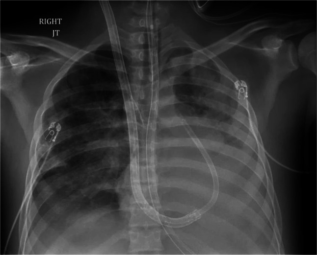
RVAD ECMO via a spectrum dual lumen single cannula (with extra drainage side holes in RV as well as RA), providing oxygenation, ventilation, as well as RV support — outflow/PA port is 15 Fr; for the 31-Fr cannula, smaller sizes can be flow limiting. Legend: RVAD, right ventricular assist device; RV, right ventricle; RA, right atrium
Central cannulation RVAD ECMO
Central cannulation consists of RA drainage with return to the main PA. This technique is infrequently utilized but may be considered if there is difficulty maintaining sufficient venous drainage, unfavorable peripheral venous anatomy, or if the percutaneous configuration does not allow safe mobility.
Conclusion
ECMO could be lifesaving in critically ill patients with respiratory or cardiac failure or combined cardiopulmonary failure. Cannulation is one of the most common causes of complications; therefore, precision during cannulation and ensuring hemostasis is key. ECMO is not an on/off therapy, and tailoring the cannulation strategy, ECMO configuration and ECMO flows to patient-specific pathology and degree of LV/RV dysfunction are essential to avoid complications. Patients are more likely to survive an ECMO run in a well-established ECMO program with more extensive resources. ECMO is very resource intensive, and patient selection is of utmost importance. In borderline situations with starting end-organ dysfunction, early initiation could improve end-organ function before it is irreversible.
Author contribution
AAA contributed to the conception or design of the work. MNF and SSR contributed to drafting the article. AAA, JH, and PT contributed to the critical revision of the article. All authors contributed to the final approval of the version to be published.
Funding
None.
Declarations
Ethics committee approval
Not applicable as it is a review article and no new research was carried out.
Informed consent
All appropriate consent was obtained for inclusion of patient data in this review.
Research involving human participants and/or animals
Research involving human and/or animal participants was not conducted.
Conflict of interest
All authors report no conflict of interest.
Footnotes
Meganne Nichole Ferrel and Syed Sikandar Raza are co-first authors.
Publisher's note
Springer Nature remains neutral with regard to jurisdictional claims in published maps and institutional affiliations.
References
- 1.Lafç G, Budak AB, Yener AÜ, et al. Use of extracorporeal membrane oxygenation in adults. Heart Lung Circ. 2014;23:10–23. doi: 10.1016/j.hlc.2013.08.009. [DOI] [PubMed] [Google Scholar]
- 2.Schmidt M, Burrell A, Roberts L, Bailey M, Sheldrake J, Rycus PT, et al. Predicting survival after ECMO for refractory cardiogenic shock: the Survival After Veno-Arterial-ECMO (SAVE)-score. Eur Heart J. 2015;36:2246–2256. doi: 10.1093/eurheartj/ehv194. [DOI] [PubMed] [Google Scholar]
- 3.Fletcher-Sandersjöö A, Frenckner B, Broman M. A single-center experience of 900 interhospital transports on extracorporeal membrane oxygenation. Ann Thorac Surg. 2019;107:119–127. doi: 10.1016/j.athoracsur.2018.07.040. [DOI] [PubMed] [Google Scholar]
- 4.Naidu SS, Baran DA, Jentzer JC, Hollenberg SM, van Diepen S, Basir MB, et al. SCAI SHOCK Stage Classification Expert Consensus update: a review and incorporation of validation studies: This statement was endorsed by the American College of Cardiology (ACC), American College of Emergency Physicians (ACEP), American Heart Association (AHA), European Society of Cardiology (ESC) Association for Acute Cardiovascular Care (ACVC), International Society for Heart and Lung Transplantation (ISHLT), Society of Critical Care Medicine (SCCM), and Society of Thoracic Surgeons (STS) in December 2021. J Am Coll Cardiol. 2022;79:933–946. doi: 10.1016/j.jacc.2022.01.018. [DOI] [PubMed] [Google Scholar]
- 5.Baran DA, Grines CL, Bailey S, Burkhoff D, Naidu SS, et al. SCAI clinical expert consensus statement on the classification of cardiogenic shock. Catheter Cardiovasc Interv. 2019;94:29–37. doi: 10.1002/ccd.28329. [DOI] [PubMed] [Google Scholar]
- 6.Telukuntla KS, Estep JD. Acute mechanical circulatory support for cardiogenic shock. Methodist Debakey Cardiovasc J. 2020;16:27–35. 10.14797/mdcj-16-1-27. [DOI] [PMC free article] [PubMed]
- 7.Abrams D, Combes A, Brodie D. Extracorporeal membrane oxygenation in cardiopulmonary disease in adults. J Am Coll Cardiol. 2014;63:2769–78. [DOI] [PubMed]
- 8.van Diepen S, Katz JN, Albert NM, Henry TD, Jacobs AK, Kapur NK, et al. Contemporary management of cardiogenic shock: a scientific statement from the American Heart Association. Circulation. 2017;136:e232–68. [DOI] [PubMed]
- 9.ELSO Adult Cardiac Failure Supplement to the ELSO General Guidelines Extracorporeal Life Support Organization (ELSO) Guidelines for Adult Cardiac Failure.; 2013. https://www.elso.org/Portals/0/IGD/Archive/FileManager/e76ef78eabcusersshyerdocumentselsoguidelinesforadultcardiacfailure1.3.pdf. Accessed 26 Aug 2018.
- 10.Extracorporeal Life Support Organization - ECMO and ECLS >about us >overview. https://www.elso.org/AboutUs/Overview.aspx. Accessed 21 Aug 2017.
- 11.Feldman D, Pamboukian SV, Teuteberg JJ, et al. The 2013 International Society for Heart and Lung Transplantation Guidelines for mechanical circulatory support: executive summary. J Heart Lung Transplant. 2013;32:157–187. doi: 10.1016/j.healun.2012.09.013. [DOI] [PubMed] [Google Scholar]
- 12.ELSO ECPR Supplement to the ELSO General Guidelines Extracorporeal Life Support Organization (ELSO) Guidelines for ECPR Cases.;2013. https://www.elso.org/Portals/0/IGD/Archive/FileManager/6713186745cusersshyerdocumentselsoguidelinesforecprcases1.3.pdf. Accessed 26 Aug 2018.
- 13.Soleimani B, Pae WE. Management of left ventricular distension during peripheral extracorporeal membrane oxygenation for cardiogenic shock. Perfusion. 2012;27:326–331. doi: 10.1177/0267659112443722. [DOI] [PubMed] [Google Scholar]
- 14.Peek GJ, Mugford M, Tiruvoipati R, Wilson A, Allen E, Thalanany MM, et al; CESAR trial collaboration. Efficacy and economic assessment of conventional ventilatory support versus extracorporeal membrane oxygenation for severe adult respiratory failure (CESAR): a multicentre randomized controlled trial. Lancet. 2009;374:1351–63. [DOI] [PubMed]
- 15.Davies A, Jones D, Bailey M, Beca J, Bellomo R, Blackwell N, et al. Extracorporeal membrane oxygenation for 2009 influenza A (H1N1) acute respiratory distress syndrome. JAMA. 2009;302:1888–95. [DOI] [PubMed]
- 16.Peek GJ, Clemens F, Elbourne D, Firmin R, Hardy P, Hibbert C, et al. CESAR: conventional ventilatory support vs extracorporeal membrane oxygenation for severe adult respiratory failure. BMC Health Serv Res. 2006;6:163. doi: 10.1186/1472-6963-6-163. [DOI] [PMC free article] [PubMed] [Google Scholar]
- 17.Eckman PM, Katz JN, El Banayosy A, Bohula EA, Sun B, van Diepen S. Veno-arterial extracorporeal membrane oxygenation for cardiogenic shock. An introduction for the busy clinician. Circulation. 2019;140:2019–37. [DOI] [PubMed]