Abstract
Estrogens play important roles in uterine growth and homeostasis through estrogen receptors (ESR1 and ESR2). To address the role of ESR1-mediated tissue events in the murine uterus, we analyzed mice with a mesenchymal tissue-specific knockout of Esr1. Isl1-driven Cre expression generated Esr1 deletion in the uterine stroma and endometrium (Isl-Esr1KO). We showed that overall structure of the Isl1-Esr1KO mouse uterus developed normally, but estrogen responsiveness and subsequent growth were defective, suggesting that mesenchymal ESR1 is necessary for both epithelial and mesenchymal cell proliferation. Furthermore, RNA-seq analysis revealed that the majority of estrogen-induced genes were regulated by stromal ESR1. In control mice, E2 administration induced 9476 up-regulated differentially expressed genes (DEGs), whereas only 1801 up-regulated DEGs were induced by E2 in Isl1-Esr1KO mice. We further showed that stromal ESR1-regulated genes in the mouse uterus included several growth factors and cytokines, which are potential factors that regulate epithelial and stromal tissue interaction, and also genes involved in lipid homeostasis. Therefore, we infer that stromal ESR1 expression is indispensable for most estrogen actions in the mouse uterus and the current results provide new insights into estrogen-mediated homeostasis in female reproductive organs.
Subject terms: Endocrinology, Reproductive biology
Introduction
Estrogens play important roles in vertebrate reproductive biology and their biological effects are principally mediated through estrogen receptors (ESRs), which are ligand-dependent transcription factors. Two ESRs (ESR1/ERα and ESR2/ERβ) have been identified from a variety of vertebrate species and the actions of estrogens on the uterus have been analyzed extensively. The uterus is comprised primarily of an epithelium, mesenchyme-derived stroma and smooth muscle and all of these tissues express ESR1. ESR1 is the predominant ESR subtype mediating the proliferative and differentiative effects of estrogens. Administration of estrogens increases uterine weight and promotes cell proliferation and differentiation and these effects were ablated in Esr1 KO mice1,2.
Epithelial-stromal interactions play pivotal roles in organogenesis, tissue differentiation and homeostasis in variety of organs. In the uterus, epithelial-stromal interactions have been demonstrated using ex vivo tissue recombination experiments with Esr1 KO- and wild type-derived epithelium and stroma. According to such experiments, effects of estrogens on uterine epithelial cell proliferation are mediated primarily via stromally-expressed ESR13. Thus, estrogen-induced growth factors secreted from the stroma were postulated to promote epithelial cell proliferation in a paracrine manner. IGF1 is a potential paracrine mediator for estrogen-induced uterine epithelial cell proliferation. Estrogen administration induced IGF1 expressed in the stroma and activated IGF1-IGF receptor signal in the epithelium4,5; however, IGF1 treatment alone cannot substitute for estrogen action6,7. In addition to mitogenic effects, the expression of various genes is regulated by paracrine signaling. Progesterone receptor (PGR) is expressed in the epithelial cells but not in the stromal cells in the absence of estrogen stimulation, whereas estrogen stimulation shifted PGR expression, decreasing epithelial PGR expression and up-regulated PGR expression in the stroma. The regulation of steroid hormone receptor expression and the subsequent control of cessation and activation of cell proliferation is required for pregnancy. Inhibition of epithelial PGR expression by estrogens does not occur in the absence of stromal ESR1 in tissue recombination experiments8. Epithelial ESR1 is dispensable for these events, although epithelial ESR1 expression is required for epithelial functionalization, such as lactoferrin (Ltf) gene expression and protein secretion9.
Subsequent in vivo mouse analyses supported such epithelial-stromal interactions observed in tissue recombination experiments10,11. Experiments using epithelial cell-specific KO of Esr1 driven by Wnt7a-Cre mouse line revealed that estrogen could induce proliferation of uterine epithelial cells despite the absence of epithelial ESR110. Similar approaches were conducted using an anti-Müllerian hormone receptor (Amhr)-Cre mouse line to investigate stromal ESR1 function11. In this mouse model, expression of ESR1 remained intact in the mesometrial side whereas Amhr-driven ablation of Esr1 was limited to the stromal cells of the anti-mesometrial side, with variable degrees of Esr1-deletion in individual animals. This study demonstrated that proliferative activity was reduced in the epithelial cells adjacent to stromal cells in which ESR1 was not expressed. However, due to a limited number of KO cells, the functional requirements of stromal ESR1 during estrogen-induced events in the uterus are not fully understood.
Epithelial-stromal interactions mediating estrogen action are commonly observed in other organs including mammary gland, oviduct, and vagina, and epithelial ESR1 functions in each organ were reported12–14. Until now, no mouse model for complete KO of uterine stromal Esr1 has been developed. Here we generated whole stromal ESR1-deletion in the mouse uterus by crossing an Isl1-Cre mouse line with Esr1-floxed mice to provide new insights into potential tissue-specific function of ESR1. We demonstrated that deletion of stromal ESR1 in the mouse uterus resulted in overall loss of estrogen action, including epithelial cell proliferation. Taking advantage of this in vivo mouse model, we evaluated gene expression by RNA-seq, and showed a large reduction in the number of estrogen-induced genes. Thus, stromal ESR1 controls a majority of the estrogen response and is indispensable for homeostasis in mouse uterus.
Results
Phenotypes of uterine mesenchyme-specific Esr1 Knockout Mice
ESR1 protein is expressed in all epithelial cells, most stromal and smooth muscle cells in the wild-type uterus (Fig. 1A,B). We generated a uterine stromal cell-specific Esr1 knockout mouse model (Isl1-Esr1KO) by crossing Esr1-floxed mice with Isl1-Cre knockin lines expressing Cre recombinase in caudal mesenchymal tissues15. ESR1 protein was detected in the epithelium but not in the stroma or smooth muscles (myometrium), demonstrating the mesenchyme-specificity of ESR1 loss-of-function in the Isl1-Esr1KO mouse uterus (Figs. 1C,D, and S1). Uterine stromal ESR1 has been lost at the neonatal stage in the Isl1-Esr1KO mice (Fig. S3). We note that Isl1-Cre is also active in a broad range of tissues16,17, and other tissues might be affected but were not studied here. Female reproductive tracts in the Isl1-Esr1KO mice were hypoplastic and ovaries lacked corpora lutea at 8 weeks of age. Histologically, the ovaries had few mature follicles and hemorrhagic cysts (Fig. S3), which resembles the ovary of conventional Esr1KO mice1,2. Thus, in the current study, we investigated histology and gene expression in the Isl1-Esr1KO mouse uterus using ovariectomized (OVX) mice to avoid any confounding effects of hypothalamus-pituitary–gonadal axis, and to simplify analysis of hormonal effects.
Figure 1.
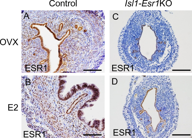
Stomal cell-specific ESR1 deletion in the Isl1-Esr1KO mice. ESR1 protein expression is evaluated by immunohistochemistry in control (A, B) and Isl1-Esr1KO (C, D) mice treated with OVX only (A, C) or E2 for three consecutive days (B, D), indicating successful deletion of Esr1 specifically in the stromal cells in the Isl1-Esr1KO mice. Scale bars, 100 μm.
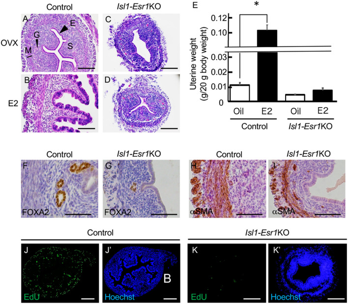
The uterus of 8-week-old control OVX mice was composed of a single layer of low columnar epithelial cells with relatively involuted stroma (Fig. 2A), and 17β-estradiol (E2) administration induced epithelial hypertrophy with water imbibition (Fig. 2B). The uterus of Isl1-Esr1KO OVX mice possess the definitive compartments, the epithelium, stroma and myometrium, however, stroma was less organized and hypotrophic compared with that of the controls (Figs. 2C and S1). Whole uterine weights were approximately half between control and Isl1-Esr1KO OVX mice in oil control injections (Fig. 2E). E2 administration for three consecutive days induced 10-folds increase in uterine wet weight in controls, but has no significant effects on uterine growth and weight in Isl1-Esr1KO mice (Figs. 2D,E, S1, and S4A). Luminal and glandular epithelial cells appeared normal but consistently low columnar morphology and lacked tall columnar structure in the E2-treated Isl1-Esr1KO mouse uterus. Isl1-Esr1KO uterus expressed Forkhead box A2 (FOXA2), an uterine gland marker, but a sparse distribution of uterine glands compared with those of controls (Figs. 2F,G, and S4B). Alpha-smooth muscle actin (αSMA) was normally expressed, suggesting normal differentiation of muscle tissue, but was somewhat disorganized (Fig. 2H,I). Thus, overall structure of Isl1-Esr1KO mouse uterus was reasonably normal but estrogen responsiveness and subsequent growth were impaired.
Figure 2.
Effects of stomal cell-specific ESR1 deletion in mouse uterus. The uterus of OVX control (A) and Isl1-Esr1KO (C) mice exhibit hypoplastic phenotypes. E2 treatment induces uterine organ growth and epithelial cell hyperplasia in the control (B), but fails to such phenotypes in the uterus of Isl1-Esr1KO mice (D). Uterine organ weight increases by E2 in controls, but not changed in Isl1-Esr1KO mice (E). More than 5 animals were analyzed. Error bars represent SEM. * indicates significant difference compared with OVX group assessed by student’s t-test (p < 0.05). Expression pattern of marker proteins FOXA2 for uterine gland (F, G) and αSMA for smooth muscle (H, I) in 8-week-old OVX mice uterus. Even in the absence of stromal ESR1, uterine glands are developed and smooth muscle cells ware differentiated. EdU-incorporation is detected in the control (J) and Isl1-Esr1KO (K) mouse uterus treated with E2 for three consecutive days, and representative images are shown. Blue fluorescent signal (J’, K’) indicates Hoechst staining in the same image of EdU staining (J, K). Scale bars, 200 µm (J), 100 μm (A–D, K) or 50 µm (F-I).
Epithelial-stromal tissue interaction in mouse uterus
Ex vivo tissue recombination experiments demonstrated that stromal ESR1 is required for paracrine regulation of epithelial cell proliferation and expression of several genes3,8. Incorporation of the deoxy-thymidine analog 5-ethynyl-2-deoxyuridine (EdU) showed that E2 administration induced cell proliferation in both epithelial and stromal cells in control mice (Fig. 2J). Note that stomal cell proliferation was not restricted in ESR1-expressing cells (Fig. S5). By contrast, proliferation of stromal and luminal epithelial cells of Isl1-Esr1KO mice uterus were not increased even after E2 administration (Fig. 2K). Accordingly, cyclin-dependent kinase inhibitor 1A (Cdkn1a) gene expression, a cell proliferation marker, was not induced by E2 administration in the Isl1-Esr1KO mice uterus (Fig. S6).
The uteri of OVX control and Isl1-Esr1KO mice expressed PGR in the epithelial cells but not in the stromal cells (Fig. 3A,C). Upon E2 administration, epithelial PGR was downregulated whereas stromal PGR was upregulated in controls (Fig. 3B). However, the Isl1-Esr1KO mouse uterus consistently expressed PGR in the epithelial cells but not in the stromal cells (Fig. 3D), suggesting that downregulation of PGR in the epithelial cells depends on stromal ESR1, and PGR expression in stromal cell depends on stromal ESR1.
Figure 3.
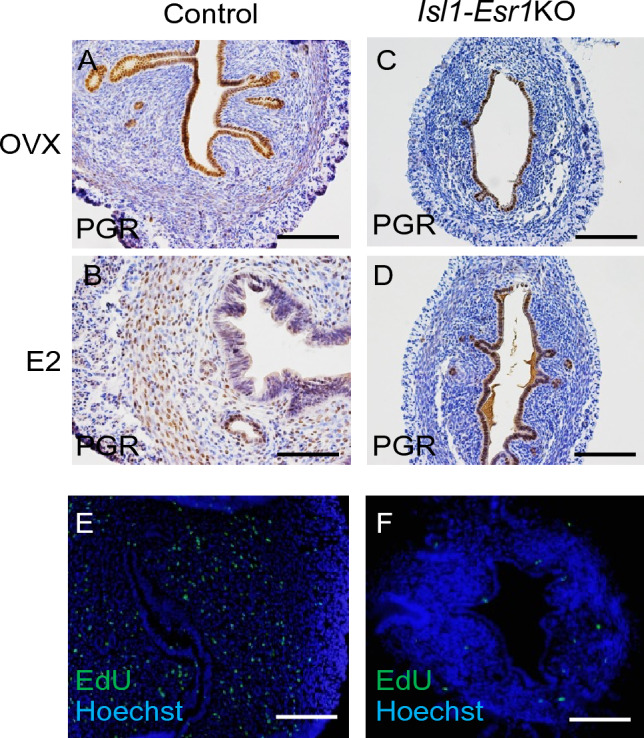
Regulation of PGR expression by in mouse uterus. Immunohistochemistry of PGR in control (A, B) and Isl1-Esr1KO (C) mice treated with OVX only (A, C) or E2 (B, D). PGR expression is detected in uterine epithelium (A), but E2 administration downregulates PGR expression in the epithelium, while upregulated in stroma (B). The shift of PGR expression is not observed in the Isl1-Esr1KO mice (C, D). Proliferation of the uterine stromal cells after a series of E2 and P4 treatments is detected EdU-incorporation (E, F). Isl1-Esr1KO mouse uterus fails to increase of stromal cell proliferation (F). Blue fluorescent signal indicates Hoechst staining. Scale bars, 100 μm.
During embryo implantation in normal mice, estrogen and progesterone cooperatively regulate the cessation of uterine epithelial cell proliferation and subsequent stromal cell proliferation18. We used hormonal regimens to mimic the hormonal profile during embryo implantation, in which 3 days of progesterone (P4) injection permits the uterine luminal epithelia to differentiate into a pre-receptive state, and the combination of P4 and E2 on the fourth day induced the receptive state19. In control mice, cell proliferation in the epithelial cells was not observed but was augmented in the stromal cells (Fig. 3E). However, in Isl1-Esr1KO mice, both epithelial and stromal cell proliferation was decreased (Fig. 3F).
Evaluation of E2-specific responses in mouse uterus
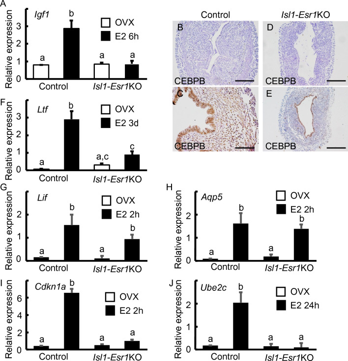
We next investigated the expression of representative estrogen-regulated genes involved in cell proliferation such as Igf1 and CCAAT/enhancer-binding protein β (Cebpb)10,20. Igf1 is a stromally expressed gene and E2 administration significantly upregulated its expression at 6 h in controls (Fig. 4A). By contrast, the expression levels of Igf1 in Isl1-Esr1KO mouse uterus were not significantly different (Fig. 4A). Expression of CEBPB protein was induced by E2 administration in the epithelial and stromal cells of control mouse uteri (Fig. 4B,C). Epithelial CEBPB immunoreactivity was found in the epithelium but not in the stroma of Isl1-Esr1KO uteri (Fig. 4D,E).
Figure 4.
Expression patterns of estrogen-regulated genes in the mouse uterus. Igf1 gene expression at 6 h after E2 administration is induced in control uteri, but not changed in the Isl1-Esr1KO mouse uteri (A). CEBPB protein is induced in both epithelial and stromal cells (B, C), but only epithelial cells express CEBPB after E2 administration in the Isl1-Esr1KO mouse uterus (D, E). Control mouse uteri increased Ltf gene expression but lost some estrogen response in the Isl1-Esr1KO mouse uterus (F). Lif (G) and Aqp5 (H) genes expression is increased in both E2 treated control and Isl1-Esr1KO mouse uterus. Isl1-Esr1KO mouse uterus. Loss of Cdkn1a (I) and ube2c (J) genes expression in the E2 treated Isl1-Esr1KO mouse uterus. Results are mean ± SEM. A two-way ANOVA followed by a Tukey–Kramer test was used and p < 0.05 was considered as significantly different.
LTF is a secreted protein regulated by E2 in mammalian uterine epithelium10. Control mouse uteri increased Ltf gene expression but lost some estrogen response in the Isl1-Esr1KO mouse (Fig. 4F), suggesting that full Ltf expression is required for both epithelial and stromal ESR1 as previously reported9. Remarkably, Ltf expression was augmented in the Isl1-Esr1KO mice compared to controls in the absence of estrogen. Other estrogen-regulated genes were also evaluated. Expression of early estrogen responsive genes, leukemia inhibitory factor (Lif), Cdkn1a, and aquaporin (Aqp5) was upregulated at 2 h after E2 administration in the control mice uterus (Fig. 4G–I). Of those, Lif and Aqp5 expression were similarly increased in the Isl1-Esr1KO mouse uterus, but Cdkn1a was not (Fig. 4G–I). The late estrogen responsive gene, ubiquitin-conjugating enzyme E2C (Ube2c) was not induced at 24 h in the Isl1-Esr1KO mice (Fig. 4J).
Gene expression analysis by RNA-seq
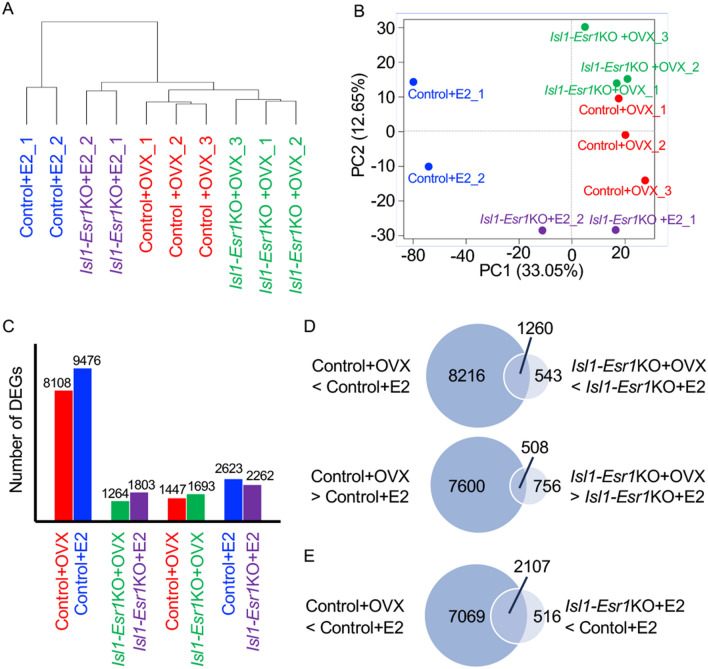
The response of the OVX mouse uterus to E2 was apparent within 6 h. These included metabolic responses in the form of increased water imbibition, vascular permeability and hyperemia, prostaglandin release, glucose metabolism, eosinophil infiltration, RNA polymerase and chromatin activity, lipid and protein synthesis21,22. Thus, we conducted RNA-seq analysis of uteri from control and Isl-Esr1KO mice treated with vehicle (Control + OVX or Isl1-Esr1KO + OVX) or E2 (Control + E or Isl1-Esr1KO + E) at 6 h (Fig. 5).
Figure 5.
RNA-seq analysis of uterine transcripts from control and Isl1-Esr1KO mice treated with vehicle and E2 at 6 h. Cluster dendrogram (A) and principal component analysis (PCA) plot showing the first and second principal components of all replicants (B) shows control E2 group (CON + E2) is differentially clustered from other group. Number of differentially expressed genes (DEGs) among uteri from control and Isl-Esr1KO mice treated with vehicle (Control + OVX or Isl1-Esr1KO + OVX) or E2 (Control + E2 or Isl1-Esr1KO + E2) at 6 h (C). Venn diagram of number of DEGs by E2 administration between control and Isl1-Esr1KO mouse uterus (D), and genes implicating stromal ESR1-induced genes (E).
The cluster dendrogram and principal component analysis (PCA) of all replicates showed that the Control + E2 group is differentially clustered from other groups (Fig. 5A,B). In control mice, E2 administration induced 9476 up-regulated differentially expressed genes (DEGs) and 8108 down-regulated DEGs, whereas, in Isl1-Esr1KO mouse, 1803 up-regulated DEGs and 1264 down-regulated DEGs (Fig. 5C; DEGs are given in Supplemental Tables S2-S5). Thus, approximately 90% of estrogen-induced genes were regulated by stromal ESR1 (Fig. 5D). We found 1447 Control + OVX biased genes and 1693 Isl1-Esr1KO + OVX biased genes between Control + OVX and Isl1-Esr1KO + OVX groups (Fig. 5C), indicating that the Isl1-Esr1KO mouse uterus was strongly affected by loss of stromal ESR1 function even in the absence of E2. When comparing E2-administered control and Isl1-Esr1KO mouse uterus, the number of DEGs are increased, and 2623 DEGs were control-E2 biased while 2262 DEGs were Isl1-Esr1KO-E2 biased (Fig. 5C).
The genes commonly found as DEGs between Control + OVX and Control + E2, and between Isl1-Esr1KO + OVX and Isl1-Esr1KO + E2 were postulated as epithelial expressed ESR1-madiating genes. We found 1260 up-regulated and 508 down-regulated genes were identified, which were satisfied such criteria (Fig. 5D and Supplement Table S6). Gene ontology (GO) analysis for biological process revealed that the ribosome biology and RNA processing are enriched (Supplement Table S7).
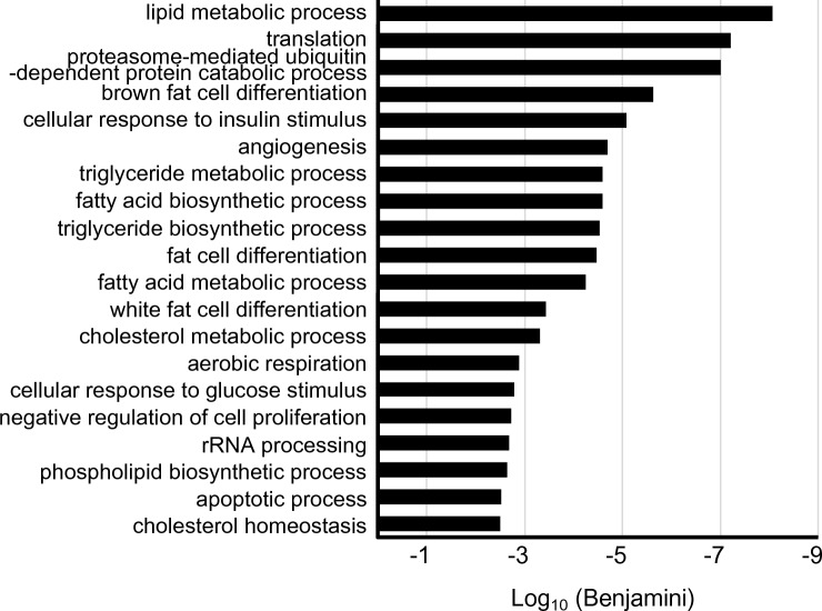
We next focused on estrogen-induced genes in the stromal cells. We investigated the upregulated genes by E2 administration in controls (Control + OVX < Control + E2) and the augmented genes in control E2 uterus rather than those of Isl1-Esr1KO E2 uterus (Isl1-Esr1KO + E2 < Control + E2). We identified 2107 genes that satisfied this criterion (Fig. 5E and Supplement Table S8). Expression of these genes was not induced without stromal ESR1. Secreted growth factors that could be candidates for mediating epithelial and stromal tissue interaction were identified. These included Igf1 and several Wnt ligands (Wnt4, Wnt7b, Wnt9a, Wnt9b), fibroblast growth factors (Fgf1, Fgf21), neuregulins (Nrg2, Nrg4), transforming growth factor beta (Tgf-β) superfamily member [Tgfb2, bone morphogenetic proteins (Bmp1, Bmp6), growth differentiation factors (Gdf6, Gdf15), inhibin beta-B (Inhbb)], and other cytokines such as chemokine (C–C motif) ligands (Cxcl6, Cxcl7, Cxcl9, Cxcl11, Cxcl12), interleukin (Il11), vascular endothelial growth factor member [Vegfa, placental growth factor (Pgf)] (Table 1). Further, GO analysis for biological process revealed that terms related to lipid, triglyceride, fatty acid metabolism, and fat cell differentiation were enriched (Fig. 6 and Supplement Table S9). These included important lipid metabolism regulatory genes such as Cebpa, Cebpb, sterol regulatory element binding transcription factor 1 (Srebf1), Kruppel-like factor 4 (Klf4), activating transcription factor 5(Atf5) (Table 2).
Table 1.
Expression profiles for selected secreted growth factor and cytokine genes.
| Id | Gene | log2 FoldChange Control + OVX < Control + E |
padj | log2 FoldChange Control + E > Isl1-Esr1KO + E |
padj |
|---|---|---|---|---|---|
| ENSMUST00000115713 | Nrg2 | 3.124 | 1.93E−57 | − 3.058 | 8.46E−22 |
| ENSMUST00000108783 | Wnt9a | 2.165 | 1.61E−52 | − 2.612 | 4.15E−16 |
| ENSMUST00000004913 | Pgf | 4.955 | 6.91E−40 | − 3.948 | 3.85E−53 |
| ENSMUST00000095360 | Igf1 | 3.082 | 1.51E−34 | − 3.31 | 4.83E−24 |
| ENSMUST00000071648 | Vegfa | 2.32 | 9.77E−32 | − 2.162 | 7.89E−07 |
| ENSMUST00000110103 | Gdf15 | 8.042 | 1.93E−31 | − 4.036 | 8.42E−05 |
| ENSMUST00000073043 | Cxcl12 | 1.564 | 2.32E−29 | − 1.279 | 4.26E−07 |
| ENSMUST00000045747 | Wnt4 | 2.968 | 4.11E−27 | − 2.882 | 7.88E−06 |
| ENSMUST00000038765 | Inhbb | 4.596 | 2.18E−22 | − 4.738 | 2.32E−18 |
| ENSMUST00000040750 | Lif | 4.271 | 2.29E−20 | − 2.731 | 0.0307 |
| ENSMUST00000109424 | Wnt7b | 3.556 | 5.53E−14 | − 2.334 | 0.0090 |
| ENSMUST00000019266 | Cxcl9 | 2.489 | 2.53E−10 | − 2.162 | 0.0002 |
| ENSMUST00000019071 | Cxcl6 | 2.853 | 9.22E−10 | − 2.401 | 0.0000 |
| ENSMUST00000033099 | Fgf21 | 8.565 | 1.40E−09 | − 7.836 | 8.05E−05 |
| ENSMUST00000000194 | Cxcl12 | 4.058 | 1.61E−09 | − 2.481 | 0.0029 |
| ENSMUST00000021011 | Cxcl7 | 4.799 | 6.80E−09 | − 3.361 | 0.0066 |
| ENSMUST00000045288 | Tgfb2 | 1.867 | 3.14E−06 | − 1.446 | 0.0015 |
| ENSMUST00000171970 | Bmp6 | 2.348 | 9.59E−05 | − 2.259 | 0.0165 |
| ENSMUST00000094892 | Il11 | 3.802 | 0.0002 | − 3.092 | 0.0039 |
| ENSMUST00000057613 | Gdf6 | 3.181 | 0.0003 | − 3.957 | 0.0070 |
| ENSMUST00000000342 | Cxcl11 | 1.699 | 0.0005 | − 1.166 | 0.0214 |
| ENSMUST00000040647 | Fgf1 | 0.81 | 0.0058 | − 1.259 | 0.0442 |
| ENSMUST00000018630 | Wnt9b | 2.422 | 0.0102 | − 5.767 | 0.0439 |
| ENSMUST00000126368 | Nrg4 | 4.135 | 0.0346 | − 7.795 | 0.0159 |
| ENSMUST00000022693 | Bmp1 | 0.464 | 0.0390 | − 0.858 | 0.0406 |
Figure 6.
Top 20 biological process gene ontology (GO) terms mapped to the candidate stromal ESR1-mediated events in mouse uterus. The terms related lipid, triglyceride, and fatty acid metabolism, and fat cell differentiation are enriched.
Table 2.
Expression profiles for selected transcriptional factor for regulating lipid metabolism.
| Id | Gene | log2 FoldChange Control + OVX < Control + E |
padj | log2 FoldChange Control + E > Isl1− Esr1KO + E |
padj |
|---|---|---|---|---|---|
| ENSMUST00000070642 | Cebpb | 2.591 | 5.26E−96 | 1.765 | 3.32E−09 |
| ENSMUST00000047356 | Atf5 | 2.653 | 2.85E−47 | 1.989 | 4.67E−08 |
| ENSMUST00000042985 | Cebp1 | 4.089 | 2.64E−15 | 3.086 | 2.82E−06 |
| ENSMUST00000107619 | Klf4 | 2.716 | 2.25E−14 | 1.745 | 5.00E−05 |
| ENSMUST00000020846 | Srebf1 | 1.65 | 3.24E−10 | 1.339 | 0.00286 |
| ENSMUST00000171644 | Pparg | 3.871 | 2.09E−07 | 3.259 | 8.26E−05 |
Discussion
Epithelial-stromal interactions are essential for regulating organogenesis, tissue/cell differentiation and functions throughout the body. Cell proliferation and differentiation in female reproductive organs have been studied extensively as an excellent model to analyze such tissue interactions. Tissue recombination experiments23 suggested that epithelial cell proliferation in female reproductive organs, including uterus, vagina and mammary gland, is mediated by stromal ESR1 in a paracrine manner3,24,25. Therefore, epithelial ESR1 may be dispensable for epithelial mitogenic response to estrogens. Subsequent genetic studies using an epithelial cell-specific Esr1KO mouse model demonstrated that epithelial ESR1 is neither necessary nor sufficient for uterine cell proliferation in female reproductive organs10,13,26. Winuthayanon et al.11 reported that epithelial cells failed to proliferate without ESR1 in neighboring stromal cells. However, the function of ESR1 has not been fully investigated, because of a lack of efficient Cre mouse lines for stromal cell-specific KO of Esr1. To clarify and extend those observations, we used an Isl1-Cre mouse line and Esr1-floxed mouse, to create stromal-specific knockout of ESR1, which allowed us to investigate the roles of stromal ESR1 in mediating the effects of E2 in the mouse uterus. We found that stromal ESR1 is necessary for epithelial cell proliferation, which is consistent with the previous tissue recombination experiments, but our findings also demonstrated that stromal ESR1 is indispensable for organ growth and for the majority of estrogen-induced actions in mouse uterus.
Phenotypes and response to E2 in the Isl1-Esr1KO mouse uterus
The uterus is derived from the Müllerian duct and consists of an epithelium and mesenchyme during early development. During neonatal development, the mesenchyme further differentiates into stoma and smooth muscle cells (an outer longitudinal and an inner circular smooth muscle layer). Previous reports using a conventional Esr1KO mouse line showed that ESR1 is not required for uterine tissue differentiation1,2. Similarly, Isl1-Esr1KO mouse uterus did not response to E2 for cell proliferation in both epithelium and stroma, resulting in hypoplastic phenotypes.
It is postulated that a stroma-derived secreted growth factor mediates estrogen-induced uterine epithelial cell proliferation in a paracrine manner. Several growth factors were proposed to fill this role and IGF1 is considered as a plausible candidate; Igf1 is expressed predominantly in the stroma upon estrogen stimulation, accompanied by phosphorylation pf IGF1 receptor in the epithelium5,27. Moreover, IGF1 administration can elicit epithelial cell proliferation in vivo6,10,28. By contrast, tissue grafting experiments using Igf1 KO mouse uteri showed that systemic but not local IGF1 is required for E2-induced uterine epithelial cell proliferation7. Thus, complementary or combination of other growth factors will be required for paracrine induction of uterine epithelial cell proliferation.
In the current RNA-seq analysis of control and Isl1-Esr1KO mouse uterus, we provided candidates for such paracrine factors that fulfill the following two conditions for stromal ESR1-regulated genes. These are 1) “genes that are upregulated by E2 in control mouse uterus” and 2) “highly expressed genes in E2-treated controls compared with E2-treated Isl1-Esr1KO mouse uterus”. We found several secreted growth factors and related genes, including Igf1, that fulfill these criteria. Fibroblast growth factors (FGFs) are expressed in the stroma in the presence of E2 and activate FGF receptor signaling in the uterine epithelium in a paracrine manner, leading to subsequent cell proliferation via MAPK activation29. Wnts play multiple roles in uterine physiology and diseases30 and contribute to stem cell-like characteristics in the uterus31. Several Tgfβ superfamily member genes were identified as stromal ESR1-mediating secreted factors where they were suggested to be possible regulators of cell proliferation and differentiation in the uterus during pregnancy and carcinogenesis32–34.
Regulation of cell proliferation in the uterine epithelium and stroma is important for implantation, and the establishment and maintenance of pregnancy. An important mechanism underlying this response is mediated by the expression of PGR and CEBPB, which could regulate cell proliferation in the stroma20,35. The Isl1-Esr1KO mouse uterus did not show stromal expression of PGR and CEBPB. In normal mice, stromal cell proliferation is independent of ESR1 expression, suggesting paracrine or juxtacrine regulation of stromal cell proliferation (Fig. S4). Therefore, it remains unknown whether regulation of Pgr and Cebpb gene expression is directly mediated through ESR1 in the uterine stroma. Epithelial expression of PGR and CEBPB is differentially regulated by estrogens. In control mice, CEBPB was induced by estrogens in both epithelium and stroma. By contrast, PGR was expressed in epithelial cells in the absence of E2 while E2 administration down-regulated PGR expression. In the Isl1-Esr1KO mouse uterus, epithelial CEBPB expression was probably mediated by epithelial ESR1 while epithelial downregulation of PGR failed to occur, although the mediating factor(s) remain to be elucidated. We were unable to evaluate whether implantation could be successful in the Isl1-Esr1KO model due to anovulation, which was likely due to deletion of ESR1 in ovary and/or hypothalamus-pituitary axis.
LTF is an epithelial secreted protein and a primary marker for estrogen actions in mouse uterine epithelium. Previous tissue recombination experiments suggested that both stromal and epithelial ESR1 were required for the production of E2-dependent epithelial LTF9. The current results supported this conclusion and the idea that overall estrogen action in the uterus is via stromal ESR1. Intriguingly, Ltf expression was augmented in the Isl1-Esr1KO OVX mice compared to OVX controls. Additionally, Mucin 1 (MUC1), also regulated by estrogen and secreted at the epithelial cell surface36, was increased in Isl1-Esr1KO mice compared to controls in the absence of estrogen (Table S5). Therefore, epithelial cells in Isl1-Esr1KO exhibit secretory characteristics normally seen after estrogen stimulation by stromal Esr1 deletion.
Gene expression in response to E2 in the Isl1-Esr1KO mouse uterus
The current RNA-seq analysis revealed that DEGs elicited by E2 at 6 h were decreased in the Isl1-Esr1KO mice compared with those of controls. This indicated that the majority of transcripts induced by E2 in the mouse uterus through ESR1 occurred in the stroma rather than the epithelium. This is consistent with previous RNA-seq analyses conducted using epithelial cell-specific Esr1KO mouse uterus in the early phase of estrogenic response37. We also evaluated gene expression with qRT-PCR analysis, and showed that expression of early estrogen responsive genes, Lif and Aqp5 was upregulated at 2 h after E2 administration in both control and Isl1-Esr1KO mouse uterus. Expression of Lif and aqp genes were not increased in epithelial Esr1KO mouse uterus10; therefore, these genes are probably induced directly by the luminal and glandular epithelial ESR1. On the other hand, some genes, such as Cdkn1a, were not induced in either epithelial-specific or stromal-specific Esr1 KO mice10 and the current study.
GO analysis was performed on the stromal ESR1-induced genes. In addition to cell proliferation-related genes, we found that “lipid metabolism” was one of the most enriched biological process terms. Most genes were biased in the E2-treated control group. Thus, stromal ESR1 contributes to metabolic regulation, which is definitely required for subsequent uterine physiological events during the very early phase of estrogen stimulation. Lipid metabolism is intriguing because a conditional deletion of Ctnnb1/β-catenin in mouse uterus transformed myometrial cells to adipocytes38. CTNNB1 is an effector molecule for Wnt signaling, suggesting that metabolic regulation by an ESR1-Wnt axis maybe important for tissue homeostasis in the uterus. Furthermore, treatment with E2 and the peroxisome proliferator activated receptor gamma (PPARγ)-specific agonist, rosiglitazone, induced abnormal uterine glands and atypical endometrial hyperplasia39. The direct PPARγ target gene, fatty acid-binding protein 4 (Fabp4) is expressed in the epithelium and is involved in embryonic implantation40. Therefore, stromal ESR1 regulates a variety of physiological events in the uterus, in part through regulation of lipid metabolism-related genes.
In the human uterus, endometrial cell proliferation is controlled by estrogen levels in the body during the menstrual cycle. Nevertheless, the mechanisms of cell proliferation at the tissue level in the normal uterine epithelium are still not well understood. Estrogen is strongly associated with the development of cancers and thus aberrant regulation of uterine cell homeostasis is involved in endometrial cancer and infertility. In this study, we used mice in which Esr1 was knocked out in the entire uterine stroma to elucidate estrogen-mediated tissue interactions and regulation of estrogen actions. Overall, an improved understanding of the distinct roles of epithelial and stromal ESR1 will shed light on the mechanisms of estrogen-mediated homeostasis underlying disorders in female reproductive organs.
Methods
Mouse and treatment
C57BL/6J (Sankyo, Tokyo, Japan), Isl1-Cre41, Esr1-null and Esr1-floxed1 mice were maintained under 12 h light/12 h dark at 23–25 °C, and fed laboratory chow (MR Standard; Sankyo) and tap water ad libitum. To obtain uterine stromal cell-specific Esr1KO mice (Isl1Cre/ + ;Esr1flox/−), Isl1Cre/ + ;Esr1+/− male were crossed with Esr1flox/flox female mice. For control mice, Cre-negative-Esr1flox/+ siblings were used. In most experiments, mice were ovariectomized under combination of anesthetic with midazolam (0.3 mg/kg body weight), medetomidine (4 mg/kg body weight) and butorphanol (5 mg/kg body weight) at 6 weeks of age and sacrificed at 8 weeks of age. For examining effects of estrogen, a single injection of 100 ng E2 (Sigma, St. Louis, MO, USA) was given to OVX mice and sacrificed 2, 6, 12, and 24 h after the injection. Some mice were given a single daily injection of 100 ng E2 for 3 days and sacrificed 24 h after the last injection. For examining effects of progesterone, OVX mice were primed with 100 ng E2 for 2 days. After resting for another 2 days, four daily injections of 1 mg progesterone (Sigma) with one injection of 50 ng E2 with the last injection of P4 on the fourth day.
All experiments involving animals and their care were conducted in compliance with ARRIVE guidelines. All experiments were performed in accordance with relevant guidelines and regulations. The present study was approved by the Animal Care and Use Committee at the Tokyo University of Science (No. K19013, K20013, K21011, K22012).
Histology and immunohistochemistry
Hematoxylin and eosin staining and immunohistochemistry were performed as previously described13,42. For immunohistochemistry, paraformaldehyde-fixed, paraffin-embedded sections were incubated with the following primary antibodies: ESR1 (sc-8005), PGR (sc-538), CEBPB (sc-150), FOXA2 (sc-6554), αSMA (sc-53142, Santa Cruz, Santa Cruz, CA, USA). The sections were stained with the Vectastain ABC Kit (Vector Laboratories, Burlingame, CA, USA). Immunofluorescence analysis was performed with Alexa Fluor protein-conjugated secondary antibodies (Thermo Fisher Scientific, Waltham, MA, USA) and counterstained with Hoechst 33342 (Sigma).
For EdU-immunostaining, mice were injected with EdU at 50 mg/kg body weight. One hour after the injection, animals were euthanized and tissues collected. EdU-incorporated cells were detected using Click-iT EdU Imaging Kits (Themo Fisher Scientific) as described in the manufacture’s protocol. In some samples, ESR1 (sc-8005) was detected with Alexa Fluor protein-conjugated secondary antibodies (Thermo Fisher Scientific) and immunofluorescent imaging. More than 3 animals were analyzed, and representative pictures are shown.
Quantitative reverse transcription-polymerase chain reaction (qRT-PCR)
Total RNA was isolated from each group using ISOGEN II reagent (Nippon Gene, Tokyo, Japan) then reverse transcribed with PrimeScript RT reagent Kit (Takara, Kusatsu, Japan). qRT-PCR was performed with a StepOnePlus Real-Time PCR system (Thermo fisher Scientific) with TB Green Premix Ex Taq II (Takara). The reaction profile consisted of 2 min 50 °C and 5 min at 95 °C followed by 40 cycles at 95 °C for 15 s and 60 °C for 1 min. The expression levels of the target genes were normalized against the expression level of the ribosomal protein L7 (Rpl7). Sequences of the specific primers are given in Supplemental Table S1. At least three samples were run in triplicate to determine sample reproducibility. A two-way ANOVA followed by a Tukey–Kramer test was used to analyze differences in gene expression. p < 0.05 was considered as significantly different.
RNA sequence (RNA-seq)
RNA was extracted from the uteri of three mice to make one sample and three biological replicates (n = 3) from each group were analyzed. However, due to quality issues during the data analysis, control E2 and Isl1-Esr1KO E2 group were analyzed with N = 2. Total RNA was isolated from whole uteri using the ISOGEN II reagent and purified with RNeasy micro kit (Qiagen, Hilden, Germany) according to the manufacturer’s instructions. Total RNAs extracted from the uteri of three mice were combined into one sample, and three samples from each group were processed for RNAseq analysis at Macrogen Japan (Tokyo, Japan) using the NovaSeq6000 platform with the Truseq stranded mRNA library constructed for paired-end 100 bp applications, according to Macrogen’s protocol. The quality of output sequences was inspected using the FastQC program (version 0.11.2, available online at: http://www.bioinformatics.babraham.ac.uk/projects/fastqc). The reads from each biological replicate were mapped to the mouse genome (GRCm38.p6) for quantification by Salmon (version 1.2.1). Differentially expressed genes (DEGs) were calculated using DESeq2 package (version 1.22.2) in the SARTools package (version 1.6.6)43 with R (version 3.5.3)44. Gene ontology enrichment analyses were conducted using DAVID web service (version 6.8)45.
Supplementary Information
Acknowledgements
We would like to thank Dr. Sylvia Evans (University of California at San Diego, USA) and Dr. Pierre Chambon (Institute for Genetics and Cellular and Molecular Biology, France) for providing Isl1-Cre and Esr1-floxed mice, respectively. We also thank Dr. Tomomi Sato (Yokohama City University, Japan), Dr. Gen Yamada (Wakayama Medical University, Japan), and Dr. Yu Hirano (Wakayama Medical University, Japan) for their invaluable support. We are grateful to Dr. Bruce Blumberg (University of California at Irvine) for his critical readings of the manuscript. This work was supported by Grants-in-Aid for Scientific Research from the Ministry of Education, Culture, Sports, Science and Technology of Japan (17H06432, 20H006301, 21H02522; S.Miy.).
Author contributions
S.Miy. and T.I. conceived and designed the study. K.F., S.Min. and E.S. performed majority of experiment and data collection. T.G., S.Y. and M.S. contributed to some experiments, and mouse preparation and sampling. K.T. performed data analysis for RNA-seq. All authors investigated and discussed the results and the data interpretation. S.Miy. wrote the original draft, and all authors contributed to review, comment, and edit the manuscript.
Data availability
Sequencing data have been deposited in DDBJ under the accession code DRA016091 (https://ddbj.nig.ac.jp/search?query=%22DRA016091%22).
Competing interests
The authors declare no competing interests.
Footnotes
Publisher's note
Springer Nature remains neutral with regard to jurisdictional claims in published maps and institutional affiliations.
These authors contributed equally: Keita Furuminato and Saki Minatoya.
Supplementary Information
The online version contains supplementary material available at 10.1038/s41598-023-39474-y.
References
- 1.Dupont S, et al. Effect of single and compound knockouts of estrogen receptors alpha (ERalpha) and beta (ERbeta) on mouse reproductive phenotypes. Development. 2000;127:4277–4291. doi: 10.1242/dev.127.19.4277. [DOI] [PubMed] [Google Scholar]
- 2.Lubahn DB, et al. Alteration of reproductive function but not prenatal sexual development after insertional disruption of the mouse estrogen receptor gene. Proc. Natl. Acad. Sci. U. S. A. 1993;90:11162–11166. doi: 10.1073/pnas.90.23.11162. [DOI] [PMC free article] [PubMed] [Google Scholar]
- 3.Cooke PS, et al. Stromal estrogen receptors mediate mitogenic effects of estradiol on uterine epithelium. Proc. Natl. Acad. Sci. U. S. A. 1997;94:6535–6540. doi: 10.1073/pnas.94.12.6535. [DOI] [PMC free article] [PubMed] [Google Scholar]
- 4.Kahlert S, et al. Estrogen receptor alpha rapidly activates the IGF-1 receptor pathway. J. Biol. Chem. 2000;275:18447–18453. doi: 10.1074/jbc.M910345199. [DOI] [PubMed] [Google Scholar]
- 5.Zhu L, Pollard JW. Estradiol-17beta regulates mouse uterine epithelial cell proliferation through insulin-like growth factor 1 signaling. Proc. Natl. Acad. Sci. U. S. A. 2007;104:15847–15851. doi: 10.1073/pnas.0705749104. [DOI] [PMC free article] [PubMed] [Google Scholar]
- 6.Klotz DM, et al. Requirement of estrogen receptor-alpha in insulin-like growth factor-1 (IGF-1)-induced uterine responses and in vivo evidence for IGF-1/estrogen receptor cross-talk. J. Biol. Chem. 2002;277:8531–8537. doi: 10.1074/jbc.M109592200. [DOI] [PubMed] [Google Scholar]
- 7.Sato T, et al. Role of systemic and local IGF-I in the effects of estrogen on growth and epithelial proliferation of mouse uterus. Endocrinology. 2002;143:2673–2679. doi: 10.1210/endo.143.7.8878. [DOI] [PubMed] [Google Scholar]
- 8.Kurita T, et al. Paracrine regulation of epithelial progesterone receptor by estradiol in the mouse female reproductive tract. Biol. Reprod. 2000;62:821–830. doi: 10.1093/biolreprod/62.4.821. [DOI] [PubMed] [Google Scholar]
- 9.Buchanan DL, et al. Tissue compartment-specific estrogen receptor-alpha participation in the mouse uterine epithelial secretory response. Endocrinology. 1999;140:484–491. doi: 10.1210/endo.140.1.6448. [DOI] [PubMed] [Google Scholar]
- 10.Winuthayanon W, Hewitt SC, Orvis GD, Behringer RR, Korach KS. Uterine epithelial estrogen receptor α is dispensable for proliferation but essential for complete biological and biochemical responses. Proc. Natl. Acad. Sci. U. S. A. 2010;107:19272–19277. doi: 10.1073/pnas.1013226107. [DOI] [PMC free article] [PubMed] [Google Scholar]
- 11.Winuthayanon W, et al. Juxtacrine activity of estrogen receptor α in uterine stromal cells is necessary for estrogen-induced epithelial cell proliferation. Sci. Rep. 2017;7:8377. doi: 10.1038/s41598-017-07728-1. [DOI] [PMC free article] [PubMed] [Google Scholar]
- 12.Feng Y, Manka D, Wagner KU, Khan SA. Estrogen receptor-alpha expression in the mammary epithelium is required for ductal and alveolar morphogenesis in mice. Proc. Natl. Acad. Sci. U. S. A. 2007;104:14718–14723. doi: 10.1073/pnas.0706933104. [DOI] [PMC free article] [PubMed] [Google Scholar]
- 13.Miyagawa S, Iguchi T. Epithelial estrogen receptor 1 intrinsically mediates squamous differentiation in the mouse vagina. Proc. Natl. Acad. Sci. U. S. A. 2015;112:12986–12991. doi: 10.1073/pnas.1513550112. [DOI] [PMC free article] [PubMed] [Google Scholar]
- 14.Winuthayanon W, et al. Oviductal estrogen receptor α signaling prevents protease-mediated embryo death. Elife. 2015;4:e10453. doi: 10.7554/eLife.10453. [DOI] [PMC free article] [PubMed] [Google Scholar]
- 15.Miyagawa S, et al. Dosage-dependent hedgehog signals integrated with Wnt/beta-catenin signaling regulate external genitalia formation as an appendicular program. Development. 2009;136:3969–3978. doi: 10.1242/dev.039438. [DOI] [PMC free article] [PubMed] [Google Scholar]
- 16.Ching ST, et al. Isl1 mediates mesenchymal expansion in the developing external genitalia via regulation of Bmp4, Fgf10 and Wnt5a. Hum. Mol. Genet. 2018;27:107–119. doi: 10.1093/hmg/ddx388. [DOI] [PMC free article] [PubMed] [Google Scholar]
- 17.Kaku Y, et al. Islet1 deletion causes kidney agenesis and hydroureter resembling CAKUT. J. Am. Soc. Nephrol. 2013;24:1242–1249. doi: 10.1681/asn.2012050528. [DOI] [PMC free article] [PubMed] [Google Scholar]
- 18.Daikoku T, et al. Conditional deletion of Msx homeobox genes in the uterus inhibits blastocyst implantation by altering uterine receptivity. Dev. Cell. 2011;21:1014–1025. doi: 10.1016/j.devcel.2011.09.010. [DOI] [PMC free article] [PubMed] [Google Scholar]
- 19.Pan H, Zhu L, Deng Y, Pollard JW. Microarray analysis of uterine epithelial gene expression during the implantation window in the mouse. Endocrinology. 2006;147:4904–4916. doi: 10.1210/en.2006-0140. [DOI] [PubMed] [Google Scholar]
- 20.Mantena SR, et al. C/EBPbeta is a critical mediator of steroid hormone-regulated cell proliferation and differentiation in the uterine epithelium and stroma. Proc. Natl. Acad. Sci. U. S. A. 2006;103:1870–1875. doi: 10.1073/pnas.0507261103. [DOI] [PMC free article] [PubMed] [Google Scholar]
- 21.Clark J, Markaverich B. In: The Physiology of Reproduction. Knobil E, editor. Raven Press; 1988. pp. 675–724. [Google Scholar]
- 22.Couse JF, Korach KS. Estrogen receptor null mice: What have we learned and where will they lead us? Endocr. Rev. 1999;20:358–417. doi: 10.1210/edrv.20.3.0370. [DOI] [PubMed] [Google Scholar]
- 23.Cunha GR, Cooke PS, Kurita T. Role of stromal-epithelial interactions in hormonal responses. Arch. Histol. Cytol. 2004;67:417–434. doi: 10.1679/aohc.67.417. [DOI] [PubMed] [Google Scholar]
- 24.Cunha GR, et al. Elucidation of a role for stromal steroid hormone receptors in mammary gland growth and development using tissue recombinants. J. Mammary Gland Biol. Neoplasia. 1997;2:393–402. doi: 10.1023/a:1026303630843. [DOI] [PubMed] [Google Scholar]
- 25.Buchanan DL, et al. Role of stromal and epithelial estrogen receptors in vaginal epithelial proliferation, stratification, and cornification. Endocrinology. 1998;139:4345–4352. doi: 10.1210/endo.139.10.6241. [DOI] [PubMed] [Google Scholar]
- 26.Mueller SO, Clark JA, Myers PH, Korach KS. Mammary gland development in adult mice requires epithelial and stromal estrogen receptor alpha. Endocrinology. 2002;143:2357–2365. doi: 10.1210/endo.143.6.8836. [DOI] [PubMed] [Google Scholar]
- 27.Murphy LJ, Ghahary A. Uterine insulin-like growth factor-1: Regulation of expression and its role in estrogen-induced uterine proliferation. Endocr. Rev. 1990;11:443–453. doi: 10.1210/edrv-11-3-443. [DOI] [PubMed] [Google Scholar]
- 28.Hewitt SC, et al. Role of ERα in mediating female uterine transcriptional responses to IGF1. Endocrinology. 2017;158:2427–2435. doi: 10.1210/en.2017-00349. [DOI] [PMC free article] [PubMed] [Google Scholar]
- 29.Li Q, et al. The antiproliferative action of progesterone in uterine epithelium is mediated by Hand2. Science. 2011;331:912–916. doi: 10.1126/science.1197454. [DOI] [PMC free article] [PubMed] [Google Scholar]
- 30.van der Horst PH, Wang Y, van der Zee M, Burger CW, Blok LJ. Interaction between sex hormones and WNT/β-catenin signal transduction in endometrial physiology and disease. Mol. Cell Endocrinol. 2012;358:176–184. doi: 10.1016/j.mce.2011.06.010. [DOI] [PubMed] [Google Scholar]
- 31.Syed SM, et al. Endometrial axin2(+) cells drive epithelial homeostasis, regeneration, and cancer following oncogenic transformation. Cell Stem Cell. 2020;26:64–80.e13. doi: 10.1016/j.stem.2019.11.012. [DOI] [PubMed] [Google Scholar]
- 32.Monsivais D, et al. Uterine ALK3 is essential during the window of implantation. Proc. Natl. Acad. Sci. U. S. A. 2016;113:E387–395. doi: 10.1073/pnas.1523758113. [DOI] [PMC free article] [PubMed] [Google Scholar]
- 33.Monsivais D, Peng J, Kang Y, Matzuk MM. Activin-like kinase 5 (ALK5) inactivation in the mouse uterus results in metastatic endometrial carcinoma. Proc. Natl. Acad. Sci. U. S. A. 2019;116:3883–3892. doi: 10.1073/pnas.1806838116. [DOI] [PMC free article] [PubMed] [Google Scholar]
- 34.Ni N, Li Q. TGFβ superfamily signaling and uterine decidualization. Reprod. Biol. Endocrinol. 2017;15:84. doi: 10.1186/s12958-017-0303-0. [DOI] [PMC free article] [PubMed] [Google Scholar]
- 35.Wang W, Li Q, Bagchi IC, Bagchi MK. The CCAAT/enhancer binding protein beta is a critical regulator of steroid-induced mitotic expansion of uterine stromal cells during decidualization. Endocrinology. 2010;151:3929–3940. doi: 10.1210/en.2009-1437. [DOI] [PMC free article] [PubMed] [Google Scholar]
- 36.Braga VM, Gendler SJ. Modulation of Muc-1 mucin expression in the mouse uterus during the estrus cycle, early pregnancy and placentation. J. Cell Sci. 1993;105(Pt 2):397–405. doi: 10.1242/jcs.105.2.397. [DOI] [PubMed] [Google Scholar]
- 37.Winuthayanon W, Hewitt SC, Korach KS. Uterine epithelial cell estrogen receptor alpha-dependent and -independent genomic profiles that underlie estrogen responses in mice. Biol. Reprod. 2014;91:110. doi: 10.1095/biolreprod.114.120170. [DOI] [PMC free article] [PubMed] [Google Scholar]
- 38.Arango NA, et al. Conditional deletion of beta-catenin in the mesenchyme of the developing mouse uterus results in a switch to adipogenesis in the myometrium. Dev. Biol. 2005;288:276–283. doi: 10.1016/j.ydbio.2005.09.045. [DOI] [PubMed] [Google Scholar]
- 39.Gunin AG, Bitter AD, Demakov AB, Vasilieva EN, Suslonova NV. Effects of peroxisome proliferator activated receptors-alpha and -gamma agonists on estradiol-induced proliferation and hyperplasia formation in the mouse uterus. J. Endocrinol. 2004;182:229–239. doi: 10.1677/joe.0.1820229. [DOI] [PubMed] [Google Scholar]
- 40.Wang P, et al. Fatty acid-binding protein 4 in endometrial epithelium is involved in embryonic implantation. Cell Physiol. Biochem. 2017;41:501–509. doi: 10.1159/000456886. [DOI] [PubMed] [Google Scholar]
- 41.Yang L, et al. Isl1Cre reveals a common Bmp pathway in heart and limb development. Development. 2006;133:1575–1585. doi: 10.1242/dev.02322. [DOI] [PMC free article] [PubMed] [Google Scholar]
- 42.Miyagawa S, Sato M, Sudo T, Yamada G, Iguchi T. Unique roles of estrogen-dependent Pten control in epithelial cell homeostasis of mouse vagina. Oncogene. 2015;34:1035–1043. doi: 10.1038/onc.2014.62. [DOI] [PubMed] [Google Scholar]
- 43.Varet H, Brillet-Guéguen L, Coppée JY, Dillies MA. SARTools: A DESeq2- and EdgeR-based R pipeline for comprehensive differential analysis of RNA-seq data. PLoS One. 2016;11:e0157022. doi: 10.1371/journal.pone.0157022. [DOI] [PMC free article] [PubMed] [Google Scholar]
- 44.Team, R. C. in R: A Language and Environment for Statistical Computing, version 3.0. 2. 2013 (R Foundation for Statistical Computing, 2019).
- 45.Sherman BT, et al. DAVID: A web server for functional enrichment analysis and functional annotation of gene lists (2021 update) Nucleic Acids Res. 2022;50:W216–W221. doi: 10.1093/nar/gkac194. [DOI] [PMC free article] [PubMed] [Google Scholar]
Associated Data
This section collects any data citations, data availability statements, or supplementary materials included in this article.
Supplementary Materials
Data Availability Statement
Sequencing data have been deposited in DDBJ under the accession code DRA016091 (https://ddbj.nig.ac.jp/search?query=%22DRA016091%22).


