Abstract
Background
Diabetic kidney disease (DKD) has become a major cause of chronic kidney disease. However, early diagnosis of DKD is challenging. Trimethylamine oxide (TMAO) is an intestinal microbial metabolite which might be associated with diabetes complications. The aim of this study was to investigate the correlation between TMAO and DKD.
Methods
A cross-sectional study was conducted. A total of 108 T2DM patients and 33 healthy subjects were enrolled in this study. Multiple logistic regression analyses and area under receiver operating characteristic curves (AUROC) were performed to evaluate the correlation between serum TMAO and DKD.
Results
Serum TMAO levels were significantly higher in DKD patients than healthy control group and the NDKD (T2DM without combined DKD) group (P < 0.05). TMAO levels were negatively correlated with eGFR and positively correlated with urea nitrogen, ACR and DKD (P < 0.05). Logistic regression analysis indicated that serum TMAO was one of the independent risk factors for DKD patients (P < 0.05). In the diagnostic model, the AUROC of TMAO for the diagnosis of DKD was 0.691.
Conclusion
Elevated levels of serum TMAO levels were positively associated with the risk of DKD in T2DM patients, which might be a potential biomarker for DKD.
Keywords: trimethylamine N-oxide, type 2 diabetes, diabetic kidney disease
Introduction
Diabetic kidney disease (DKD) is one of the most common and important microvascular complications of diabetes, with more than 25–40% of diabetic patients developing nephropathy 20–35 years after the onset of the disease (1). DKD has become a major cause of chronic kidney disease (2), which is a serious threat to human health. On this basis, early diagnosis of DKD is particularly important.
In recent years, the relationship between intestinal flora metabolites and chronic metabolic diseases has received increasing attention. Trimethylamine oxide (TMAO) is an intestinal microbial metabolite that is generated from choline and l-carnitine in food by the action of intestinal microorganisms to trimethylamine (TMA), which is catalyzed in the liver by flavin monooxygenase 3 to TMAO (3, 4). Previous studies have confirmed that TMAO was a new predictor of cardiovascular disease and was associated with major adverse cardiovascular events and all-cause mortality (5, 6, 7). TMAO levels were associated with insulin resistance, impaired glucose tolerance, and a positive association with risk of diabetes (8, 9). Recent clinical studies suggested that TMAO levels were not only associated with the development of diabetes but also diabetic complications. Signe et al (10) reported that elevated plasma TMAO concentrations were associated with impaired renal function in type 1 diabetes patients; however, the correlation became uncorrelated after adjustment for baseline glomerular filtration rate, indicating that TMAO might be a biomarker of renal function or as a risk factor for macro- and microvascular complications, especially impaired renal function. Another cross-sectional survey study showed that elevated plasma TMAO levels in type 2 diabetic patients (T2DM) correlated with the incidence and severity of diabetic retinopathy (11).
In this study, we aimed to investigate the correlation between serum TMAO levels and DKD in T2DM patients and then tried to explore that TMAO might be a promising biomarker for diabetic microangiopathy.
Materials and methods
Study population
A total of 108 patients with T2DM from May 2021 to October 2021 were enrolled. Inclusion criteria: the diagnostic criteria for diabetes mellitus by the World Health Organization in 1999. Exclusion criteria are as follows: (i) patients with combined acute complications of diabetes mellitus; (ii) patients with severe infections, surgery, trauma, and other stressful conditions within the last 1 month; (iii) patients with malignant tumors; (iv) long-term vegetarians; (v) patients with systematic use of antibiotics or probiotics within 3 months; (vi) patients with autoimmune diseases; (vii) patients with severe heart, liver and other organ diseases; and (viii) patients with primary glomerular diseases and other patients with secondary glomerular diseases. Thirty-three healthy individuals who were examined at our hospital during the same period were also included as a control group. All studies in this study were conducted with the informed consent of the study subjects and were approved by the ethics committee of the Second Affiliated Hospital of Fujian Medical University (Ethics No. 2022-81).
Clinical data and biochemical analyses
Clinical data including gender, age, body mass index (BMI, in kg/m²), duration of diabetes (in years), and history of hypertension were collected. Laboratory parameters included fasting blood glucose (mmol/L), urea nitrogen (BUN in mmol/L), blood creatinine (SCR in μmol/L), blood uric acid (UA in μmol/L), glycated hemoglobin (HbA1c in %), urinary microalbumin (mg/dL), urinary creatinine (μmol/L), and calculate urinary albumin to creatinine ratio (ACR in mg/g) and glomerular filtration rate (eGFR in mL/(min·1.73 m2)) with the following formulas:
 |
 |
Quantitative detection of serum TMAO concentration
The quantitative detection of serum TMAO concentration was performed by isotope dilution high-performance liquid chromatography-tandem mass spectrometry with ultraperformance liquid chromatography-triple quadrupole tandem mass spectrometry (Shimadzu LC-MS 8050CL).
Statistical analyses
Statistical analysis was conducted using SPSS 26.0 statistical software. Data are presented as frequencies (percentages) or means ± s.d. We assessed differences between groups with Student’s t-test, Kruskal–Wallis H-test, chi-square test, and Mann–Whitney U test. Spearman's correlation analysis and multi-factor logistic regression analysis were used to explore the influencing factors. P < 0.05 was considered statistically significant. The diagnostic value of serum TMAO was assessed by the area under the subject's operating characteristic curve (AUROC).
Results
Clinical characteristics and serum TMAO levels of study subjects
A total of 108 patients with T2DM and 33 non-diabetic patients (control group) were included in this study. T2DM patients were divided into two groups according to ACR, NDKD group (ACR<30 mg/g, n = 73) and DKD group (ACR >30 mg/g, n = 35). The differences in age and gender among the three groups were not statistically significant, suggesting that the groups were comparable.
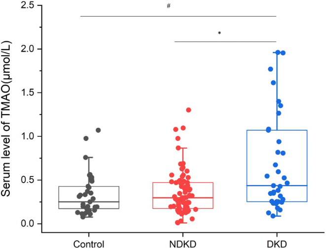
Serum TMAO levels in the control group, NKD group, and DKD group increased sequentially (Fig. 1). Serum TMAO levels in the DKD group were significantly higher than those in the control and NDKD groups (P < 0.05). Serum TMAO levels in the NDKD group were higher than those in the control group, without statistically significance (P > 0.05) (Table 1).
Figure 1.

Comparation of serum TMAO concentrations in NDKD and DKD group vs control group, # P < 0.05, vs NDKD group,*P < 0.05.
Table 1.
Clinical characteristics and serum TMAO levels.
| control group (n = 33) | NDKD group (n = 73) | DKD group (n = 35) | F/X²/Z | P | |
|---|---|---|---|---|---|
| Age (year) | 52.00 ± 12.69 | 51.58 ± 12.52 | 55.86 ± 14.17 | 1.356 | 0.261 |
| Male | 60.6% (20/33) | 65.8% (48/73) | 77.1% (27/35) | 2.294 | 0.318 |
| BMI (kg/m²) | 22.18 ± 2.83 | 25.31 ± 3.42a | 23.86 ± 3.99 | 9.651 | 0.000d |
| Diabetes duration (year) | NA | 5.0 (1.0, 10) | 10 (4.25,16.0)b | −2.752 | 0.006d |
| Hypertension (%) | NA | 32.9% (24/73) | 51.4% (18/35)b | 4.210 | 0.040b |
| BUN (mmol/L) | 4.6 (3.93, 5.55) | 5.11 (4.24, 6.07) | 5.83 (4.93, 7.30)a,b | 9.892 | 0.007e |
| UA (μmol/L) | 313 (282, 413.5) | 305 (235, 358.5) | 370 (309, 407)b | 8.525 | 0.014b |
| FPG (mmol/L) | 5.39 (5.10, 5.80) | 8.0 (6.68, 10.13)a | 7.85 (6.52, 9.55)a | 46.249 | 0.000d |
| HbA1c (%) | NA | 9.2 (7.0, 11.05) | 9.3 (7.0,10.3) | -0.420 | 0.674 |
| SCR (μmol/L) | 70.0 (56.95, 78.1) | 66 (55.1, 76.0) | 75.9 (63.0, 96.0)b | 9.690 | 0.008c |
| eGFR (mL/min•1.73 m²) | 102.32 ± 22.31 | 113.14 ± 27.38 | 95.99 ± 41.64b | 4.10 | 0.029b |
| TMAO (μmol/L) | 0.25 (0.16, 0.46) | 0.30 (0.17, 0.48) | 0.44 (0.25, 1.07)a,b | 11.789 | 0.003c |
vs control group, aP < 0.05, vs NDKD group,bP < 0.05 vs NDKD group, cP < 0.01, vs NDKD group dP < 0.001.
Clinical characteristics of the high- and low-level groups of TMAO
First, all subjects were divided into two groups according to the median serum TMAO level (0.3126 μmol/L): low TMAO level group (n = 70); and high TMAO level group (n = 71). High TMAO level group showed higher age, BUN, and lower eGFR (P < 0.05) (Supplementary Table 1, see section on supplementary materials given at the end of this article).
Further, we analyzed the clinical characteristics in T2DM patients. 108 T2DM patients were divided into two groups according to the median TMAO level (0.322 μmol/L): low TMAO level group (n = 54) and high TMAO level group (n = 54). Age, duration of diabetes, BUN, SCR, ACR, and DKD prevalence were significantly higher in the high TMAO level group, while eGFR was significantly lower (P < 0.05) (Table 2).
Table 2.
Comparison of clinical characteristics between the high-level and low-level TMAO groups in T2DM patients.
| Low TMAO level group (n = 54) | High TMAO level group (n = 54) | t/X²/Z | P | |
|---|---|---|---|---|
| Age (year) | 49.13 ± 12.55 | 56.80 ± 12.74 | −3.150 | 0.002b |
| Male | 72.2% (39/54) | 66.7% (36/54) | 0.393 | 0.531 |
| BMI (kg/m²) | 25.27 ± 3.99 | 24.41 ± 3.28 | 1.220 | 0.225 |
| Diabetes duration (year) | 4 (1, 10) | 10 (3, 15) | −2.693 | 0.007b |
| Hypertension (%) | 42.6% (23/54) | 35.2% (19/54) | 0.623 | 0.43 |
| BUN (mmol/L) | 4.78 (3.91, 5.86) | 5.74 (4.95, 7.06) | −3.017 | 0.003b |
| UA (μmol/L) | 324.5 (233.25, 384.25) | 331.0 (288.75, 383.75) | −0.839 | 0.402 |
| FPG (mmol/L) | 7.78 (6.47, 9.25) | 8.08 (6.72, 10.35) | −1.085 | 0.278 |
| HbA1c (%) | 8.6 (6.9, 10.75) | 9.5 (7.15, 11.28) | −0.562 | 0.574 |
| SCR (μmol/L) | 65.5 (54.98, 76.0) | 72.4 (60.58, 83.50) | −2.095 | 0.036a |
| eGFR (mL/min•1.73m²) | 116.85 ± 24.68 | 98.32 ± 38.44 | 2.981 | 0.004b |
| ACR (mg/g) | 13.35 (7.98, 24.83) | 17.22 (11.11, 179.04) | −2.366 | 0.018a |
| DKD (%) | 22.2% (12/54) | 42.6% (23/55) | 5.115 | 0.024a |
aP < 0.05, bP < 0.01.
Linear correlation analysis between serum TMAO and kidney function
First, we analyzed the correlation between serum TMAO and kidney function in all subjects. Spearman correlation analysis showed that TMAO was negatively correlated with eGFR (r = −0.370, P < 0.001) and positively correlated with age, BUN, and SCR (r = 0.304, 0.440, 0.308, P < 0.001) (Supplementary Table 2).
Further, we analyzed the correlation between serum TMAO and kidney function in T2DM patients. Spearman correlation analysis showed that TMAO was negatively correlated with eGFR (r = −0.445, P < 0.001) and positively correlated with age, disease duration, BUN, SCR, ACR, and DKD prevalence (r = 0.326, 0.242, 0.423, 0.407, 0.231, 0.293, P ≤ 0.01) (Table 3).
Table 3.
Linear correlation analysis of serum TMAO with clinical characteristics in T2DM patients.
| Variables | TMAO | |
|---|---|---|
| r | P | |
| Age (year) | 0.326 | 0.001b |
| Male | −0.047 | 0.626 |
| BMI (kg/m²) | −0.117 | 0.229 |
| Diabetes duration (year) | 0.242 | 0.012a |
| BUN (mmol/L) | 0.423 | 0.000c |
| UA (μmol/L) | 0.181 | 0.061 |
| FPG (mmol/L) | 0.038 | 0.693 |
| HbA1c (%) | 0.065 | 0.507 |
| SCR (μmol/L) | 0.407 | 0.000c |
| eGFR (mL/min/1.73m²) | −0.445 | 0.000c |
| ACR (mg/g) | 0.231 | 0.016a |
| Hypertension (%) | 0.000 | 1.000 |
| DKD (%) | 0.293 | 0.002b |
aP < 0.05, bP < 0.01, cP < 0.001.
Regression analysis of DKD risk factors
Multiple logistic regression analysis showed that serum TMAO (odds ratio (OR) = 7.880,95% CI 1.993–31.152, P = 0.003) was independent risk factor for DKD patients after adjusted for eGFR, BUN, and UA (Table 4).
Table 4.
Regression analysis of DKD risk factors
| b | Wald | P | OR | OR (95% CI) | |
|---|---|---|---|---|---|
| TMAO | 2.064 | 8.664 | 0.003b | 7.880 | (1.993, 31.152) |
| eGFR | 0.008 | 0.812 | 0.368 | 1.008 | (0.991, 1.025) |
| BUN | 0.174 | 1.273 | 0.259 | 1.190 | (0.880, 1.609) |
| UA | 0.003 | 1.953 | 0.162 | 1.003 | (0.999, 1.007) |
| Hypertension | 1.335 | 8.232 | 0.004b | 3.799 | (1.526, 9.457) |
b P < 0.01.
Serum TMAO in DKD diagnosis
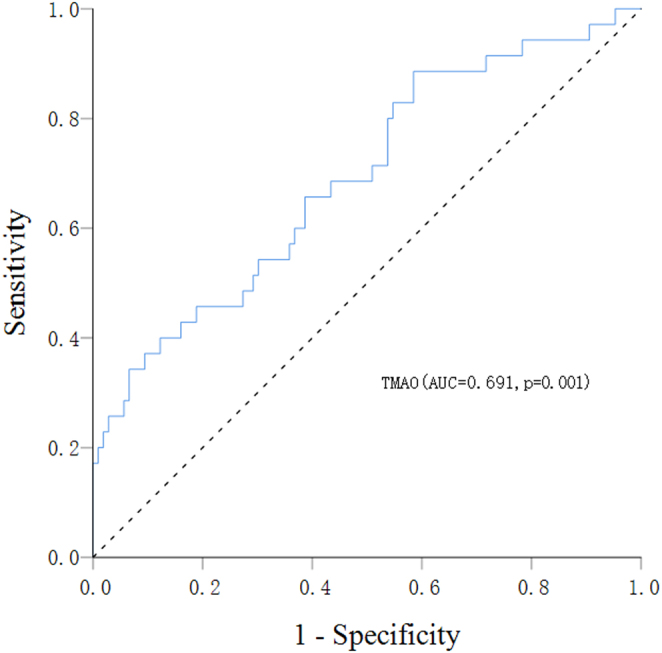
As shown in Fig. 2, the AUROC of TMAO was 0.691 (95% CI 0.588–0.795; P = 0.001), with a sensitivity of 88.6% and specificity of 58.5% at an optimal cut value of 0.227 mol/L.
Figure 2.

AUROC of serum TMAO level for the diagnosis of DKD.
Discussion
In the present study, we found a novel relationship between serum TMAO and DKD. TMAO was significantly elevated in DKD patients compared with control group and NDKD patients. Besides, serum TMAO was independent risk factor for DKD patients.
Studies showed gut microbiota-dependent TMAO may be associated with the development of T2DM. The addition of TMAO to the diet has been reported to increase fasting insulin levels and insulin resistance in mice, exacerbating impaired glucose tolerance (9). TMAO levels were significantly higher in patients with T2DM than in controls in a case–control study (12), which confirmed that elevated TMAO levels were associated with an increased risk of diabetes, in line with the results of a meta-analysis (8). However, a recent cohort study (13) showed that plasma TMAO levels were associated with insulin resistance in the elderly and did not significantly correlate with T2DM. In the present study, our results showed that serum TMAO levels were higher in the diabetes group than the control group, but the difference was not statistically significant, which might be related to the small sample size.
Although the difference in serum TMAO levels between the NDKD and control groups was not statistically significant, our study showed that the DKD group had significantly elevated TMAO levels. Besides, the serum TMAO levels in the control, NDKD and DKD groups increased sequentially. The serum TMAO levels in the DKD group were significantly higher than in the other two groups. Previous studies also suggested the relationship between TMAO and renal function. Another study (14) showed significantly higher TMAO levels in patients with T2DM and advanced CKD than in the control group.
In our study, we found a positive correlation between serum TMAO levels and BUN and SCR levels, and a negative correlation with eGFR, which was consistent with Bell's observations in patients with chronic renal failure (15). We also found that serum TMAO levels were positively correlated with ACR levels. In addition, patients in the high-level group had significantly lower eGFR and higher ACR values, as well as a higher proportion of combined DKD, compared with patients in the low TMAO level group, suggesting that diabetic patients with high circulating TMAO levels have worse renal function. Further, multifactorial logistic regression analysis confirmed that elevated serum TMAO levels were an independent risk factor for the development of DKD. In a cohort study that included patients with end-stage renal disease, patients had significantly higher circulating TMAO levels than normal controls, and TMAO levels were positively correlated with serum BUN and SCR levels (15), which was in consist with our study. As we know, the kidney is the main route of TMAO clearance (16). The cause of the significant increase in circulating TMAO in CKD patients has been suggested to be related to the disruption of gut microbial homeostasis. Mohammed et al. showed a significant increase in the number of bacteria that metabolize choline and carnitine and produce TMA in the gut microbiome of T2DM patients with combined chronic kidney disease (14). It has also been suggested that it may be related to increased FMO enzyme-mediated TMAO formation. Alexander et al. (17) performed FMO enzyme activity experiments using liver microsomes from experimental CKD rats and control rats and showed for the first time that metabolic activation of FMO enzymes by uremic solutes may contribute to the elevated TMAO levels in CKD. Further basic studies at the ex vivo cellular and animal level could help to reveal the causes of elevated TMAO levels in DKD patients.
However, several limitations should be considered. First, our sample size is small. In future studies, we will expand the sample size to provide a better understanding of the changes in TMAO concentrations with DKD. . In addition, our study did not address the underlying mechanisms of TMAO in DKD, which needs to be further explored in the future. It has been shown that TMAO activates the production of NLRP3 (Pyrin structural domain-3) inflammatory vesicles and nuclear factor κB signaling, which is involved in the process of DKD by promoting vascular inflammation and oxidative stress (18, 19). Animal studies have shown that increased dietary TMAO promotes phosphorylation of Smad3, a pro-fibrotic regulator in kidney disease, and increases plasma cystatin C and renal injury marker-1 levels, which are sensitive indicators of impaired kidney function (20, 21).
Inconclusion, our study demonstrated that elevated serum TMAO levels are closely associated with the risk of DKD in patients with T2DM and are one of the independent risk factors for the development of DKD.
Supplementary Materials
Declaration of interest
The authors declare no conflict of interest.
Funding
This work was supported by National Natural Science Foundation of China (NSFC 82200871); Natural Science Foundation of Fujian Province (2020J01221); Fujian Provincial Health Technology Project (2020GGA057).
Availability of data and material
The data presented in this study are available on request from the corresponding author.
Ethics approval and consent to participate
The research proposal was approved by the ethics committee of the Second Affiliated Hospital to Fujian Medical University (ethical approval ID:2022-81). All participants signed an informed consent form.
Consent for publication
All authors consent for publication.
Author contribution statement
YH and ZZ contributed equally to this manuscript. YH and ZZ contributed in data analysis and manuscript writing. JZ conceptualized and designed these studies, performed them, and supervise the programme. ZH contributed in data analysis. All authors contributed to manuscript revision and read and approved the submitted version.
Acknowledgements
The authors would like to acknowledge all the subjects participating in this study.
References
- 1.Remuzzi G Schieppati A & Ruggenenti P. Clinical practice. Nephropathy in patients with type 2 diabetes. New England Journal of Medicine 20023461145–1151. ( 10.1056/NEJMcp011773) [DOI] [PubMed] [Google Scholar]
- 2.Zhang LX, Zhao MH, Zuo L, Wang Y, Yu F, Zhang H, Wang HB, CK-NET Work Group. Kidney C. & Disease Network. Annual data report. Kidney International Supplements 20159e1–e81. [DOI] [PMC free article] [PubMed] [Google Scholar]
- 3.Craciun S & Balskus EP. Microbial conversion of choline to trimethylamine requires a glycyl radical enzyme. PNAS 201210921307–21312. ( 10.1073/pnas.1215689109) [DOI] [PMC free article] [PubMed] [Google Scholar]
- 4.Fennema D Phillips IR & Shephard EA. Trimethylamine and trimethylamine N-oxide, a flavin-containing monooxygenase 3(FMO3)-mediated host-microbiome metabolic axis implicated in health and disease. Drug Metabolism and Disposition 2016441839–1850. ( 10.1124/dmd.116.070615) [DOI] [PMC free article] [PubMed] [Google Scholar]
- 5.Tang WH Wang Z Levison BS Koeth RA Britt EB Fu XM Wu YP & Hazen SL. Intestinal microbial metabolism of phosphatidylcholine and cardiovascular risk. New England Journal of Medicine 20133681575–1584. ( 10.1056/NEJMoa1109400) [DOI] [PMC free article] [PubMed] [Google Scholar]
- 6.Schiattarella GG Sannino A Toscano E Giugliano G Gargiulo G Franzone A Trimarco B Esposito G & Perrino C. Gut microbe-generated metabolite trimethylamine-N-oxide as cardiovascular risk biomarker: a systematic review and dose-response meta-analysis. European Heart Journal 2017382948–2956. ( 10.1093/eurheartj/ehx342) [DOI] [PubMed] [Google Scholar]
- 7.Guasti L Galliazzo S Molaro M Visconti E Pennella B Gaudio GV Lupi A Grandi AM & Squizzato A. TMAO as a biomarker of cardiovascular events: a systematic review and meta-analysis. Internal and Emergency Medicine 202116201–207. ( 10.1007/s11739-020-02470-5) [DOI] [PubMed] [Google Scholar]
- 8.Zhuang R, Ge X, Han L, Yu P, Gong X, Meng Q, Zhang Y, Fan H, Zheng L, Liu Z, et al. Gut microbe-generated metabolite trimethylamine N-oxide and the risk of diabetes: A systematic review and dose-response meta-analysis. Obesity Reviews 201920883–894. ( 10.1111/obr.12843) [DOI] [PubMed] [Google Scholar]
- 9.Gao X Liu X Xu J Xue C Xue Y & Wang Y. Dietary trimethylamine N-oxide exacerbates impaired glucose tolerance in mice fed a high fat diet. Journal of Bioscience and Bioengineering 2014118476–481. ( 10.1016/j.jbiosc.2014.03.001) [DOI] [PubMed] [Google Scholar]
- 10.Winther SA, Øllgaard JC, Tofte N, Tarnow L, Wang Z, Ahluwalia TS, Jorsal A, Theilade S, Parving HH, Hansen TW, et al. Utility of plasma concentration of trimethylamine N-oxide in predicting cardiovascular and renal complications in individuals with Type 1 diabetes. Diabetes Care 2019421512–1520. ( 10.2337/dc19-0048) [DOI] [PMC free article] [PubMed] [Google Scholar]
- 11.Liu W Wang C Xia Y Xia W Liu G Ren C Gu Y Li X & Lu P. Elevated plasma trimethylamine-N-oxide levels are associated with diabetic retinopathy. Acta Diabetologica 202158221–229. ( 10.1007/s00592-020-01610-9) [DOI] [PMC free article] [PubMed] [Google Scholar]
- 12.Shan Z, Sun T, Huang H, Chen S, Chen L, Luo C, Yang W, Yang X, Yao P, Cheng J, et al. Association between microbiota-dependent metabolite trimethylamine-N-oxide and type 2 diabetes. American Journal of Clinical Nutrition 2017106888–894. ( 10.3945/ajcn.117.157107) [DOI] [PubMed] [Google Scholar]
- 13.Lemaitre RN, Jensen PN, Wang Z, Fretts AM, McKnight B, Nemet I, Biggs ML, Sotoodehnia N, de Oliveira Otto MC, Psaty BM, et al. Association of trimethylamine N-oxide and related metabolites in plasma and incident Type 2 diabetes: the cardiovascular health study. JAMA Network Open 20214 e2122844. ( 10.1001/jamanetworkopen.2021.22844) [DOI] [PMC free article] [PubMed] [Google Scholar]
- 14.Al-Obaide MAI Singh R Datta P Rewers-Felkins KA Salguero MV Al-Obaidi I Kottapalli KR & Vasylyeva TL. Gut microbiota-dependent trimethylamine-N-oxide and serum biomarkers in patients with T2DM and advanced CKD. Journal of Clinical Medicine 20176. ( 10.3390/jcm6090086) [DOI] [PMC free article] [PubMed] [Google Scholar]
- 15.Bell JD Lee JA Lee HA Sadler PJ Wilkie DR & Woodham RH. Nuclear magnetic resonance studies of blood plasma and urine from subjects with chronic renal failure: identification of trimethylamine-N-oxide. Biochimica et Biophysica Acta 19911096101–107. ( 10.1016/0925-4439(9190046-c) [DOI] [PubMed] [Google Scholar]
- 16.Al-Waiz M Mitchell SC Idle JR & Smith RL. The metabolism of 14C-labelled trimethylamine and its N-oxide in man. Xenobiotica; the Fate of Foreign Compounds in Biological Systems 198717551–558. ( 10.3109/00498258709043962) [DOI] [PubMed] [Google Scholar]
- 17.Prokopienko AJ West RE Schrum DP Stubbs JR Leblond FA Pichette V & Nolin TD. Metabolic activation of flavin monooxygenase-mediated trimethylamine-N-oxide formation in experimental kidney disease. Scientific Reports 20199 15901. ( 10.1038/s41598-019-52032-9) [DOI] [PMC free article] [PubMed] [Google Scholar]
- 18.Chen ML Zhu XH Ran L Lang HD Yi L & Mi MT. Trimethylamine-N-oxide induces vascular inflammation by activating the NLRP3 inflammasome through the SIRT3-SOD2-mtROS signaling pathway. Journal of the American Heart Association 20176. ( 10.1161/JAHA.117.006347) [DOI] [PMC free article] [PubMed] [Google Scholar]
- 19.Zhang X, Li Y, Yang P, Liu X, Lu L, Chen Y, Zhong X, Li Z, Liu H, Ou C, et al. Trimethylamine-N-oxide promotes vascular calcification through activation of NLRP3 (nucleotide-binding domain, leucine-rich-containing family, pyrin domain-Containing-3) inflammasome and NF-κB (nuclear factor κB) signals. Arteriosclerosis, Thrombosis, and Vascular Biology 202040751–765. ( 10.1161/ATVBAHA.119.313414) [DOI] [PubMed] [Google Scholar]
- 20.Runyan CE Schnaper HW & Poncelet AC. Smad3 and PKCdelta mediate TGF-beta1-induced collagen I expression in human mesangial cells. American Journal of Physiology. Renal Physiology 2003285F413–F422. ( 10.1152/ajprenal.00082.2003) [DOI] [PubMed] [Google Scholar]
- 21.Tang WH Wang Z Kennedy DJ Wu Y Buffa JA Agatisa-Boyle B Li XS Levison BS & Hazen SL. Gut microbiota-dependent trimethylamine N-oxide (TMAO) pathway contributes to both development of renal insufficiency and mortality risk in chronic kidney disease. Circulation Research 2015116448–455. ( 10.1161/CIRCRESAHA.116.305360) [DOI] [PMC free article] [PubMed] [Google Scholar]
Associated Data
This section collects any data citations, data availability statements, or supplementary materials included in this article.
Supplementary Materials
Data Availability Statement
The data presented in this study are available on request from the corresponding author.


 This work is licensed under a