Main text
(iScience 26, 105835; January 20, 2023)
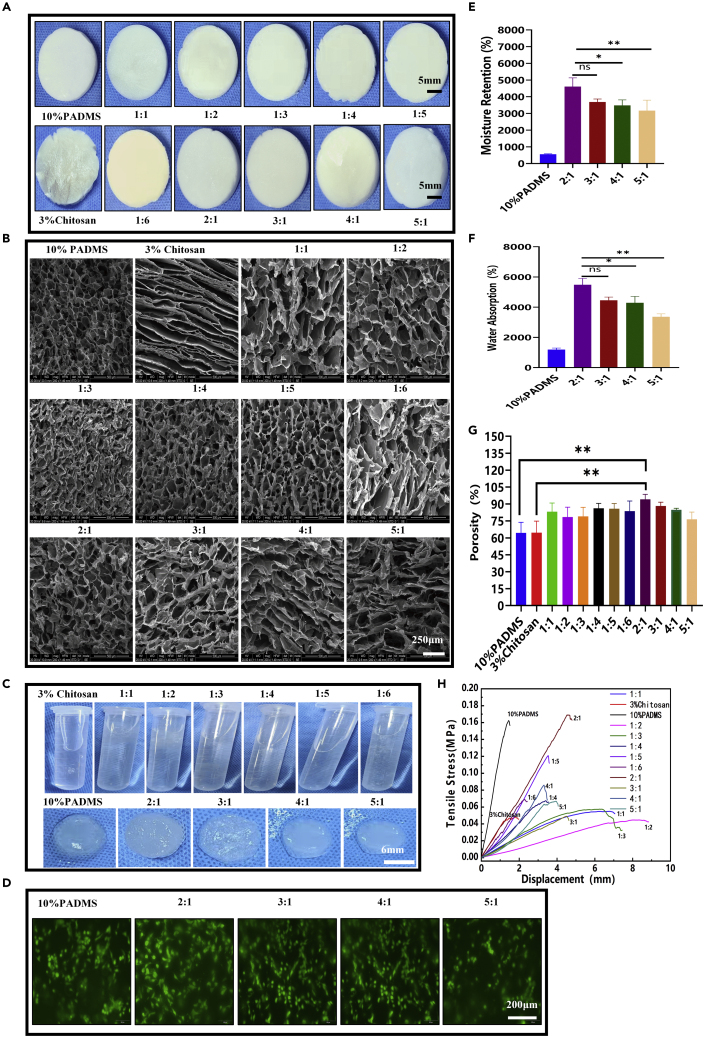
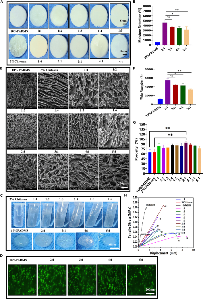
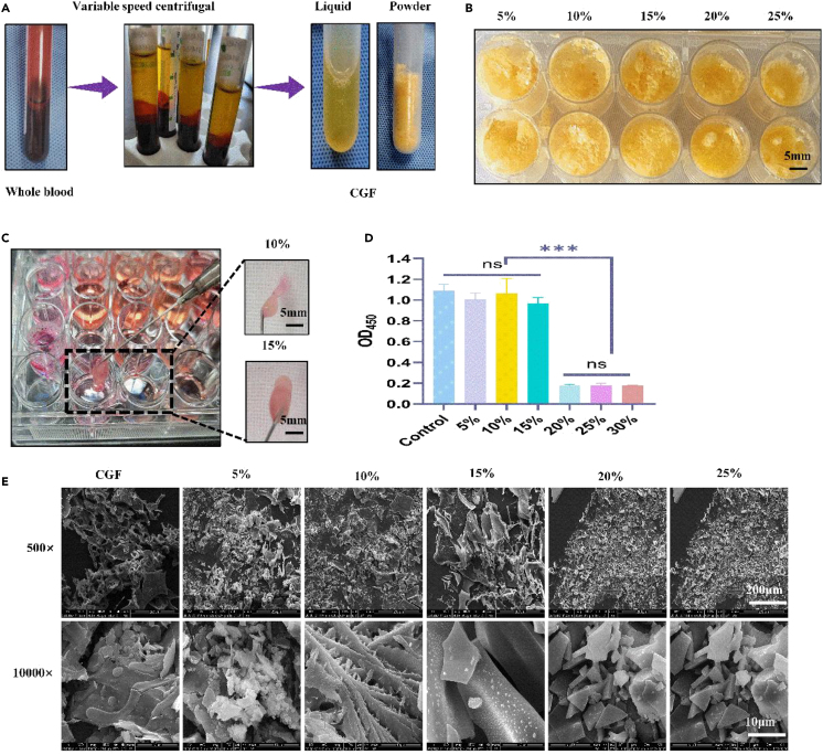
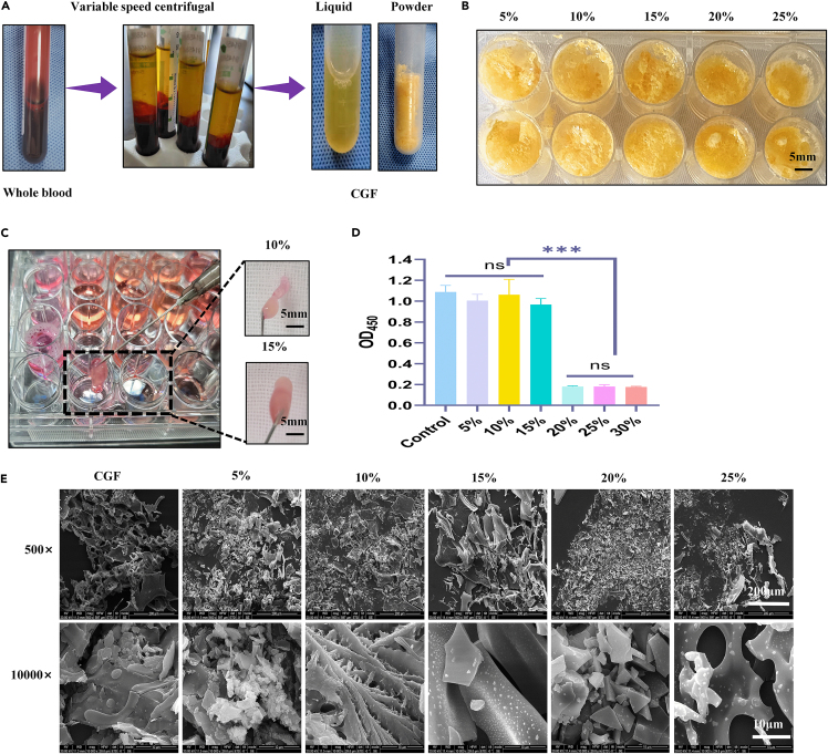
Recently, after careful retrospective examination, our team found that. In the staining of live and dead cells in Figure 3D, the fluorescence staining pictures of living and dead cells in groups 3:1 and 4:1 were repeated. In Figure 3C morphology pictures soaked in deionized water, group 4:1 and group 5:1 showed repetition. From the microstructural images of CGF scans at different concentrations in Figure S2E. The SEM images at 20% and 25% concentrations were repeated. Our team immediately checked the raw data. After investigation, we believe that the team members were negligent in the final picture typesetting, and inappropriate picture import occurred, which resulted in the repetition of two adjacent pictures. However, it does not involve quantitative data analysis and does not affect this study's final results and conclusions. (1) We corrected the 3:1 image immersed in deionized water in Figure 3C. (2) We have corrected the 4:1 image of the live and dead cells stained in Figure 3D. (3) We have corrected the SEM picture of the 25% concentration in Figure S2E.These corrections do not significantly affect the overall findings and conclusions of the paper, and we want to assure the reader that the corrected values and labels do not alter the interpretation or validity of the study. We sincerely apologize for any confusion these errors may have caused and appreciate the opportunity to correct them. We thank the editors and the readers for their understanding.
Figure 3. Preparation and characterization of ADM-CS composite sponge scaffolds (original)
Figure 3. Preparation and characterization of ADM-CS composite sponge scaffolds (corrected)
Figure S2. Preparation and characterization of potent growth factor (CGF), related to STAR Methods (original)
Figure S2. Preparation and characterization of potent growth factor (CGF), related to STAR Methods (corrected)
Contributor Information
Yanbin Gao, Email: gaoyanbin6@yeah.net.
Jun Ma, Email: nfyy_majun@163.com.
Lei Yang, Email: yuanyang@smu.edu.cn.


