Abstract
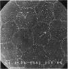
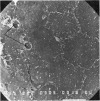
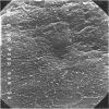
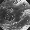
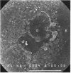
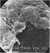
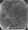
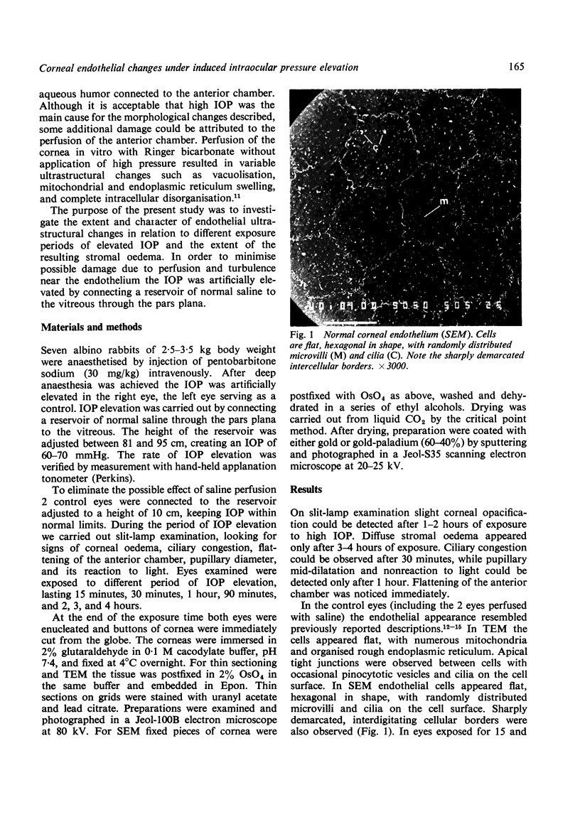
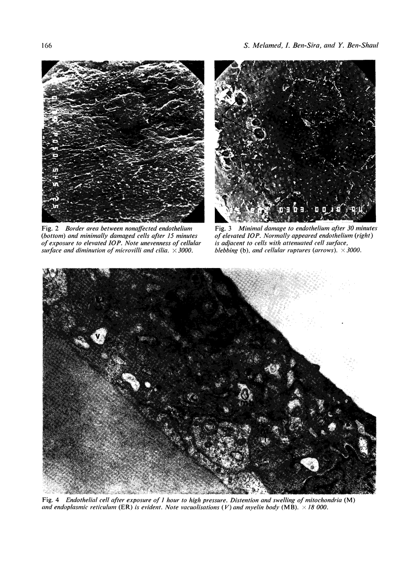
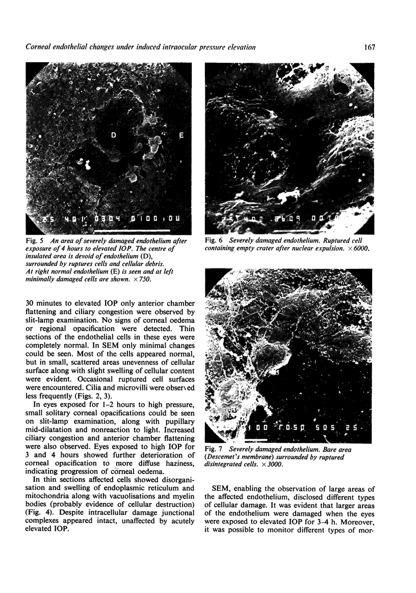
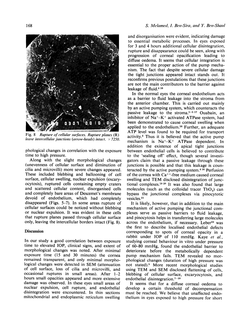
Intraocular pressure was artificially raised to 60--70 mmHg in 7 albino rabbits for periods of 15 minutes to 4 hours. The corneal endothelium of these eyes was studied by transmission and scanning electron microscopy. A correlation between exposure time to elevated IOP, clinical signs observed by slit-lamp examination, and extent of morphological damage is clearly shown. In eyes exposed to high pressure for 15 and 30 minutes corneas remained transparent and only minimal changes could be detected by SEM, which consisted of small areas of cell with unevenness of their surface, occasional cellular ruptures, and diminution of cilia and microvilli. After 1--2 hours of exposure small, solitary corneal opacifications appeared. In these eyes more severe morphological changes affecting larger areas were observed, with additional cellular blebbing, excariocytosis, cellular rupture, disintegration, and disappearance seen in SEM. Thin sections revealed swelling of mitochondria, disorganisation of endoplasmic reticulum, and the existence of myelin bodies. In eyes exposed for 3 and 4 hours to high IOP corneal haziness, implying stromal oedema, appeared. In these eyes the areas affected were larger, the extent of damage being more severe. Many areas were bare of endothelium, surrounded by scattered cellular debris, and showed cells with ballooning surfaces and multiple ruptures. Even in severe cellular damage cellular junctions appeared intact. It is assumed that endothelial cells are more sensitive to IOP elevation than the cellular junctions and that injury to the active pump system due to morphological damage is responsible for the resultant corneal oedema.
Full text
PDF
Images in this article
Selected References
These references are in PubMed. This may not be the complete list of references from this article.
- Blümcke S., Morgenroth K., Jr The stereo ultrastructure of the external and internal surface of the cornea. J Ultrastruct Res. 1967 Jun;18(5):502–518. doi: 10.1016/s0022-5320(67)80200-4. [DOI] [PubMed] [Google Scholar]
- Donn A. Cornea and sclera. Arch Ophthalmol. 1966 Feb;75(2):261–288. doi: 10.1001/archopht.1966.00970050263021. [DOI] [PubMed] [Google Scholar]
- Doughman D. J., Van Horn D., Harris J. E., Miller G. E., Lindstrom R., Good R. A. The ultrastructure of human organ-cultured cornea. I. Endothelium. Arch Ophthalmol. 1974 Dec;92(6):516–523. doi: 10.1001/archopht.1974.01010010530015. [DOI] [PubMed] [Google Scholar]
- Fischbarg J. Active and passive properties of the rabbit corneal endothelium. Exp Eye Res. 1973 May 10;15(5):615–638. doi: 10.1016/0014-4835(73)90071-7. [DOI] [PubMed] [Google Scholar]
- IRVINE A. R., Jr The role of the endothelium in bullous keratopathy. AMA Arch Ophthalmol. 1956 Sep;56(3):338–351. doi: 10.1001/archopht.1956.00930040346003. [DOI] [PubMed] [Google Scholar]
- KAYE G. I., PAPPAS G. D., DONN A., MALLETT N. Studies on the cornea. II. The uptake and transport of colloidal particles by the living rabbit cornea in vitro. J Cell Biol. 1962 Mar;12:481–501. doi: 10.1083/jcb.12.3.481. [DOI] [PMC free article] [PubMed] [Google Scholar]
- Kaye G. I., Hoefle F. B., Donn A. Studies on the cornea. 8. Reversibility of the effects of in vitro perfusion of the rabbit corneal endothelium with calcium-free medium. Invest Ophthalmol. 1973 Feb;12(2):98–113. [PubMed] [Google Scholar]
- Kaye G. I., Mishima S., Cole J. D., Kaye N. W. Studies on the cornea. VII. Effects of perfusion with a Ca++-free medium on the corneal endothelium. Invest Ophthalmol. 1968 Feb;7(1):53–66. [PubMed] [Google Scholar]
- Kaye G. I., Sibley R. C., Hoefle F. B. Recent studies on the nature and function of the corneal endothelial barrier. Exp Eye Res. 1973 May 10;15(5):585–613. doi: 10.1016/0014-4835(73)90070-5. [DOI] [PubMed] [Google Scholar]
- McCarey B. E., Edelhauser H. F., Van Horn D. L. Functional and structural changes in the corneal edothelium during in vitro perfusion. Invest Ophthalmol. 1973 Jun;12(6):410–417. [PubMed] [Google Scholar]
- Svedbergh B., Bill A. Scanning electron microscopic studies of the corneal endothelium in man and monkeys. Acta Ophthalmol (Copenh) 1972;50(3):321–336. doi: 10.1111/j.1755-3768.1972.tb05955.x. [DOI] [PubMed] [Google Scholar]
- Trenberth S. M., Mishima S. The effect of ouabain on the rabbit corneal endothelium. Invest Ophthalmol. 1968 Feb;7(1):44–52. [PubMed] [Google Scholar]
- YTTEBORG J., DOHLMAN C. CORNEAL EDEMA AND INTRAOCULAR PRESSURE. 1. ANIMAL EXPERIMENTS. Arch Ophthalmol. 1965 Sep;74:375–381. doi: 10.1001/archopht.1965.00970040377018. [DOI] [PubMed] [Google Scholar]
- Ytteborg J., Dohlman C. H. Corneal edema and intraocular pressure. II. Clinical results. Arch Ophthalmol. 1965 Oct;74(4):477–484. doi: 10.1001/archopht.1965.00970040479008. [DOI] [PubMed] [Google Scholar]


