Pneumonia is one of the commonest differential diagnoses of a homogeneous opacity on chest X-ray. In the absence of clinical and laboratory evidence of infection, it is important to investigate for pathologies which may mimic consolidation on chest X-ray but may be related to extrathoracic abnormalities.
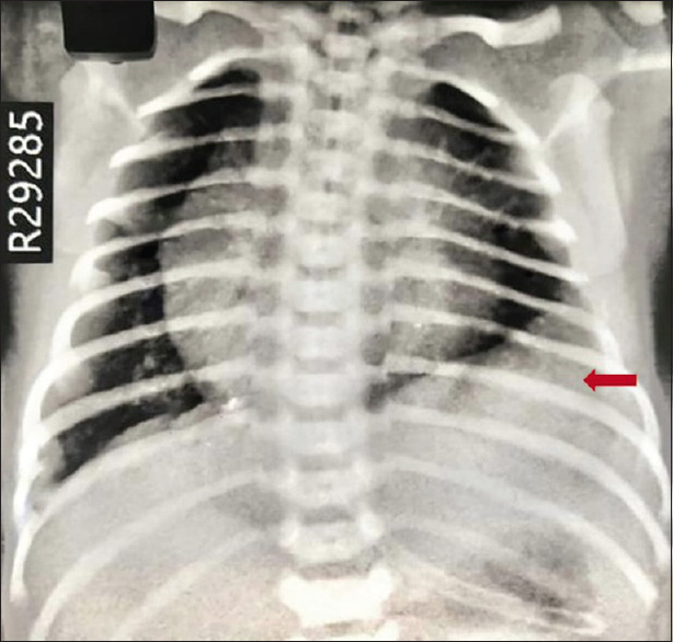
A full-term male child was born by emergency cesarean section in view of fetal distress. His Apgar scores were 8 and 9 at 1 and 5 min, respectively. He had mild respiratory distress with an oxygen saturation of 99% on room air. On respiratory examination, he had reduced air entry on the lower side of the left chest. Chest X-ray [Figure 1] revealed a homogeneous, well-defined opacity in the lower lobe of the left lung with blunting of the costophrenic angle, suggestive of left lower lobe consolidation with pleural effusion. However, his sepsis screen was negative, and the neonate was clinically stable and feeding well.
Figure 1.

Chest X-ray showing a well-defined, homogenous opacity in the lower lobe of the left lung with blunting of the left costophrenic angle (arrow)
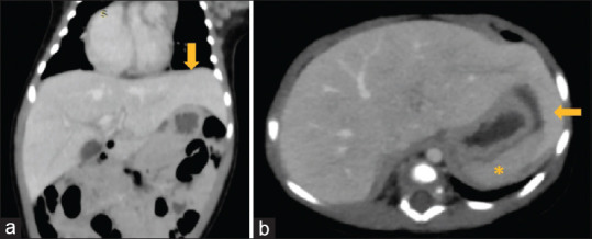
An ultrasonography of the thorax and abdomen showed an enlarged left lobe of the liver. A computed tomography (CT) of the abdomen showed an elongated left lobe of the liver reaching up to the left hypochondrium and roofing the spleen [Figure 2a and b]. The liver parenchyma was otherwise unremarkable. This abnormal morphology of liver was identified as beaver tail liver. Interestingly, on review of chest X-ray, this enlarged left lobe of liver mimicked left lower lobe consolidation. The neonate had uneventful hospital stay and was discharged.
Figure 2.

CT abdomen showing (a) enlarged left lobe of the liver reaching up to the left upper quadrant of the abdomen (arrow); (b) enlarged left liver lobe (arrow) encircling the spleen (asterisk)
Beaver tail liver, also known as a sliver of liver, is a variant of normal hepatic morphology with an elongated left liver lobe extending laterally up to the left hypochondrium and often surrounding the spleen.[1] The term was coined because of its resemblance to a beaver's tail. Identification of this variant is clinically important in trauma patients, as the left upper quadrant abdominal trauma can cause injury to the left lobe of the liver and can be misdiagnosed as subcapsular splenic hematoma or perisplenic fluid collection. Additionally, this variation may prove fatal during invasive procedures of the abdomen.[2,3,4]
To our knowledge, this is the first reported case of a beaver tail variant of liver in a neonate. It may mimic a lung pathology on chest X-ray as seen in the present case. Clinical percussion in older children and adults will reveal a dull note in the left lower chest, as it is occupied by the beaver tail variant of liver. A lower lobe pneumonia will also have dullness on percussion, but it can be differentiated from the liver by performing vocal resonance or tactile vocal fremitus. However, as this clinical examination is challenging in a neonate, a simple bedside ultrasonography can help reach the diagnosis. Thus, a differential diagnosis of beaver tail variant should be considered in radiologically suspected left lower lobe pathologies in appropriate clinical setting.
Declaration of patient consent
The authors certify that appropriate patient consent was obtained.
Financial support and sponsorship
Nil.
Conflicts of interest
There are no conflicts of interest.
References
- 1.Atalar MH, Karakus K. Beaver tail liver. Abdom Radiol. 2018;43:1851–2. doi: 10.1007/s00261-017-1395-x. [DOI] [PubMed] [Google Scholar]
- 2.Glennison M, Salloum C, Lim C, Lacaze L, Malek A, Enriquez A, et al. Accessory liver lobes: Anatomical description and clinical implications. J Visc Surg. 2014;151:451–6. doi: 10.1016/j.jviscsurg.2014.09.013. [DOI] [PubMed] [Google Scholar]
- 3.Jones R, Tabbut M, Gramer D. Elongated left lobe of the liver mimicking a subcapsular hematoma of the spleen on the focused assessment with sonography for trauma exam. Am J Emerg Med. 2014;32:814–e3. doi: 10.1016/j.ajem.2013.12.050. [DOI] [PubMed] [Google Scholar]
- 4.Crivello MS, Peterson IM, Austin RM. Left lobe of the liver mimicking perisplenic collections. J Clin Ultrasound. 1986;14:697–701. doi: 10.1002/jcu.1870140906. [DOI] [PubMed] [Google Scholar]


