Abstract
Neuroblastoma (NB), the most frequent solid extracranial tumor in children, is not always cured by current aggressive therapies that have notable adverse effects; therefore, novel treatments are necessary. Phosphoinositide 3-kinase (PI3K) and fibroblast growth factor receptor inhibitors exhibit synergistic effect in NB cell lines. In the present study, mono- and combination therapy of the United States Food and Drug Administration-approved PI3K, cyclin-dependent kinase-4/6 (CDK4/6), poly-ADP-ribose-polymerase (PARP) and WEE1 G2 checkpoint kinase (WEE1) inhibitors (BYL719, PD-0332991, BMN673 and MK-1775, respectively), were used to treat NB cell lines SK-N-AS, SK-N-BE(2)-C, SK-N-DZ, SK-N-FI and SK-N-SH and viability (assessed by WST-1 assay), proliferation (incucyte analysis) and cell cycle (FACS) changes were assessed. Treatments with all single drugs presented dose-dependent responses with decreased viability and proliferation and combining BYL719 with PD-0332991 or BMN673 with MK-1775 resulted in additive or synergistic effects in most cell lines., except for SK-N-SH for the former and for SK-N-AS for the latter. Moreover, combining MK-1775 and BMN673 decreased the numbers of cells in S phase to a greater extent than either drug alone, while when combining PD-0332991 and BYL719 the observed effect was close to that of PD-0332991 alone. To summarize, PI3K and CDK4/6 or PARP and WEE1 exhibited synergistic anti-NB effects and lower doses of the inhibitors could be utilized, thereby potentially reducing adverse side effects.
Keywords: neuroblastoma, targeted therapy, WEE1 inhibitor, poly-ADP-ribose-polymerase inhibitor, PI3K inhibitor, CDK4/6 inhibitor
Introduction
Neuroblastoma (NB) is the most frequently diagnosed cancer during the first year of life and has genetic, morphological and clinical heterogeneity and can spontaneously regress or progress aggressively, with many patients succumbing to recurrent/refractory metastatic disease (1–6). For children with low- and intermediate-risk disease, survival is >90%, while for those with high-risk disease, 5-year survival is <50% despite multimodal application of chemotherapy, stem cell transplantation, operative treatment, irradiation, retinoid therapy and different types of immunotherapy (1,6). NB does not respond well to conventional chemotherapy and irradiation, and current treatments accepted for NB have narrow targeted specificity (1,7,8). To improve individualized therapy for NB, analysis of the molecular mechanisms underlying the etiology and pathogenesis of NB is essential and new therapeutic targets have been disclosed (9–13).
The PI3K/Akt signaling pathway has been shown to be involved in the control of many key cellular responses, such as proliferation, survival, metabolism and migration (9,14,15). Moreover, PI3K/Akt is activated in several tumor types, including NB (16,17). Clinical trials with PI3K inhibitor monotherapy have been performed for NB; for example, a phase I clinical trial with SF1126 (PI3K/mTOR inhibitor) for relapsed/refractory NB (trial no. NCT02337309) (18) has been conducted by the New Approaches to Neuroblastoma Therapy Consortium.
Furthermore, our previous studies assessed in vitro effects of the United States Food and Drug Administration (FDA)-approved PI3K and fibroblast growth factor receptor (FGFR) inhibitors BYL719 and JNJ- 42756493, respectively, as single or combined treatment, with or without chemotherapy in NB and medulloblastoma (MB) cell lines (10–13). Combined use of PI3K and FGFR inhibitors resulted in synergistic effects, suggesting possible clinical utility for NB and MB, while their joint effects with chemotherapy drugs were less clear and need further investigation. The present study aimed to assess combinations of targeted therapies.
The contribution of regulators of the cell cycle such as cyclin-dependent kinase (CDK) 4/6 and WEE1 G2 checkpoint kinase (WEE1) in NB implies that deregulation of the normal cell cycle could be important in promoting NB tumorigenesis (19,20). CDK4 and 6 are enzymes that act with D-type cyclins to promote progression of the cell cycle from G1 to S phase (21–23). When the cyclin D/CDK4/6/inhibitor of CDK4/retinoblastoma pathway is dysregulated, proliferation is increased in many types of cancer such as glioblastoma, melanoma, liposarcoma and NB (24–26). Therefore, CDK4/6 inhibitors may serve as therapeutic candidates in malignant cells. Currently there are three FDA-approved CDK4/6 inhibitors, palbociclib, ribociclib and abemaciclib, which are primarily used for breast cancer (27). Moreover, several studies have shown that PI3K/Akt inhibitors combined with CDK4/6 inhibitors are effective in preclinical cancer models and exhibit synergism (28–31).
Other FDA approved inhibitors include poly ADP-ribose polymerase (PARP) inhibitor olaparib and the WEE1 inhibitor IMP7068 (32–37). Olaparib is used in patients with recurrent ovarian, early breast, metastatic castration-resistant prostate and metastatic pancreatic cancer, while IMP7068 indirectly targets non-functional p53 (present in some cases of high-risk NB) and has potential synergy with PARP inhibitors (32–37).
To explore options for NB therapy, the present study investigated PI3K, CDK4/6, PARP and WEE1 inhibitors in different combinations to identify potential synergistic effects in NB cell lines with different mutational profiles. NB cell lines, SK-N-AS, SK-N-BE(2)-C, SK-N-DZ, SK-N-FI and SK-N-SH, were assessed for their sensitivity to single and combined treatments with FDA-approved PI3K, CDK4/6, PARP inhibitors BYL719, PD-0332991 and BMN673, respectively, as well as the non-FDA-approved WEE1 inhibitor MK-1775.
Materials and methods
Cell lines and culture
Non-MYCN proto-oncogene, bHLH transcription factor (MYCN)-amplified SK-N-AS, SK-N-FI (both mutant p53) and SK-N-SH (wild-type p53) as well as MYCN-amplified SK-N-DZ (deletion of PIK3 catalytic subunit type 2γ) and SK-N-BE(2)-C (both mutant p53) were a kind gift from Professor Per Kogner, Karolinska Institute (Stockholm, Sweden) and their identities were verified previously (10). Cells were all grown in RPMI)1640 medium including 10% fetal bovine serum (both Gibco; Thermo Fisher Scientific, Inc.), 1% L-glutamine, 100 U/ml penicillin and 100 µg/ml streptomycin at 37°C in a humidified incubator with 5% CO2.
Inhibitors
Stock solutions of the FDA-approved PI3K, CDK4/6 and PARP inhibitors BYL719, PD-0332991 and BMN673 and non-FDA-approved WEE1 inhibitor MK-1775 were all purchased from Selleck Chemicals, prepared in DMSO and then diluted in PBS. Titration experiments were performed using 0.01–50.0 MK-1775, 0.001–50.0 BMN673, 1.0–20.0 PD-0332991 and 0.25–20.0 µM BYL719) in all cell lines (data not shown). Subsequently, 1.0–10.0 µM for BYL719 and PD-0332991 and 0.5–1.0 µM for BMN673 and MK-1775 were tested to identify additive or synergistic effects.
WST-1 viability assay
To assess viability, SK-N-AS, SK-N-BE(2)-C, SK-N-DZ, SK-N-FI and SK-N-SH cells (5,000 cells/well were cultured in 90 µl RPMI-1640 medium in 96-well plates at 37°C, 5% CO2 for 24 h followed by treatment with inhibitors. Viability was assessed after 24, 48 and 72 h exposure to inhibitors by adding 10 µl cell proliferation WST-1 reagent (Roche Diagnostics GmbH, Solna, Sweden) per each well as previously described (10,11). The plates were incubated at 37°C, 5% CO2 for 1 h and the absorbance was measured at 450 nm using the VersaMax microplate reader (Molecular Devices, LLC). Moreover, half-maximal inhibitory concentration (IC50) was estimated from log concentration-effect curves with non-linear regression analysis in GraphPad Prism Software version 8 (GraphPad Software, Inc.). All graphs represent at least three experiments and absorbance values after treatment were compared with PBS controls.
Confluence, cytotoxicity and apoptosis assay
To estimate confluence, cytotoxicity and apoptosis, SK-N-AS, SK-N-BE(2)-C, SK-N-DZ, SK-N-FI and SK-N-SH cells (5,000 cells/well) were cultured in 200 µl RPMI-1640 medium in 96-well plates in an IncuCyte S3 Live® Cell Analysis System (Essen Bioscience, Welwyn Garden City, UK) at 37°C. After 24 h, the medium was replaced with fresh RPMI-1640 medium that contained Incucyte Cytotox Red Reagent to assess cytotoxicity or IncuCyte Caspase-3/7 Green Apoptosis Assay Reagent [both (Essen Bioscience, Welwyn Garden City, UK)] to assess apoptosis for 72 h at 37°C. Cell confluence was assessed by image-based measurements of cell growth based on area using IncuCyte Analysis Software version 2021A, Sartorius. Apoptosis was determined when IncuCyte Caspase-3/7 Green Apoptosis Assay Reagent crossed cell membrane and was cleaved by active caspase-3/7. DNA intercalating dye was released leading to the fluorescent staining of nuclear DNA. By using IncuCyte Analysis Software (Sartorius), fluorescent objects were quantified. Images were captured with IncuCyte S3 Live® Cell Analysis System (Essen Bioscience) every 2 h with PBS used as negative control.
FACS analysis
To follow the effects of the drugs on the cell cycle a FACS analysis was performed. All procedures were performed according to the protocol of the manufacturer (Invitrogen; Thermo Fisher Scientific, Inc.), SK-N-AS, SK-N-BE(2)-C, SK-N-DZ, SK-N-FI and SK-N-SH cells were collected 48 h after treatment with 0.5 and 1.0 MK-1775, 1.0 BMN673, 5.0 and 10.0 PD-0332991 and 5.0 and 10.0 µM BYL719, resuspended in cold PBS in 15-ml tubes and cold 70% ethanol was included dropwise while vortexing at a speed of 1,800 rpm for 10 sec at room temperature. Controls were made with PBS and DMSO at the tested concentrations. Subsequently, tubes were stored at 4°C until they were utilized. A total of 1×10⁶ cells was analyzed per sample. Following centrifugation at 204 g for 10 min at 4°C, the pellet was dissolved in 0.5 ml FxCyclePI/RNAse solution according to the manufacturer's instructions (Invitrogen; Thermo Fisher Scientific, Inc. Stockholm, Sweden) at room temperature (without light) for 30 min before analysis using FACS Novocyte, Agilent and Flowjo_v10.8.1 software (Sapio Sciences, London, UK).
Statistical analysis
The efficacy of the joint therapies was analyzed by highest single agent effect-based approach and median effect dose-effect-based approach (based on Loewe additivity) (38,39). To validate the effect of single drugs or using them in combination in comparison to a negative control, multiple unpaired t tests followed by post hoc Holm-Sidak correction was performed (11). All the experiments were performed at least three times and are presented as mean and SD. Data were analyzed using Graphpad Prism 9.4.1. Median-effect equation to calculate a median-effect value D (equivalent to IC50) and slope, as described previously (10). Goodness-of-fit was analyzed with linear correlation coefficient; r>0.85 was set as a threshold for subsequent analysis. The interaction degree of the drugs was assessed using the combination index (CI) for mutually exclusive drugs as follows: CI=d1/D1 + d2/D2, where D1 and D2 represent the doses of drug 1 and 2 alone needed to obtain a certain effect and d1 and d2 represent the doses of drug 1 and 2 needed to obtain the same effect in combination. CI<0.75 was considered to indicate synergy and CI<1.45; values between the two were considered assessed to indicate additive effects, in concordance with the recommendations of the ComboSyn (combosyn.com/). One-way ANOVA with Bonferroni's post hoc test was used to calculate the difference in mean between two single drugs and their combined treatment. P<0.05 was considered to indicate a statistically significant difference.
Results
Viability of the SK-N-AS, SK-N-BE(2)-C, SK-N-DZ, SK-N-FI and SK-N-SH NB cell lines treated with BYL719, PD-0332991, BMN673 and MK-1775 is affected in a dose-dependent manner
The effects of BYL719, PD-0332991, BMN673 and MK-1775 on SK-N-AS, SK-N-BE(2)-C, SK-N-DZ, SK-N-FI and SK-N-SH viability were assessed by WST-1 assay for 72 h following treatment. When combining PD-0332991 with BYL719 or MK-1775 with BMN673 only low doses of the drugs exposing possible synergistic effects were utilized.
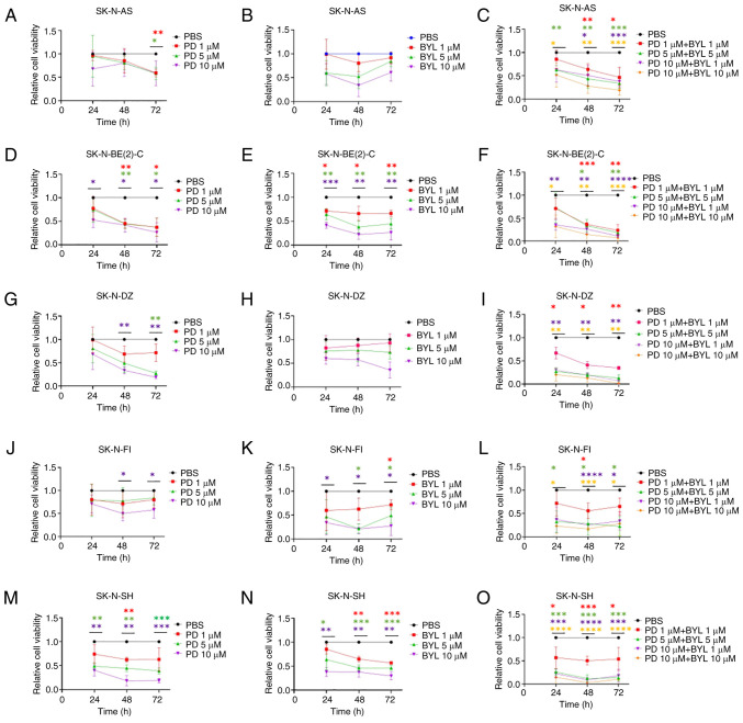
All NB cell lines presented dose-dependent responses following treatment with 1–10 µM PD-0332991. The highest PD-0332991 dose (10 µM) caused a notable decrease in absorbance at 48 and 72 h after treatment compared with PBS in all lines except for SK-N-AS (Fig. 1A, D, G, J, M). A similar response was observed with 5 µM for all cell lines except SK-N-FI and 1 µM PD-0332991 at 72 h for SK-N-BE(2)-C and SK-N-AS.
Figure 1.
WST-1 viability assay in NB cell lines following treatment with CDK4/6 inhibitor PD and PI3K inhibitor BYL. Viability of SK-N-AS treated with (A) PD, BYL (B) and combination of PD and BYL (C), viability of SK-N-BE(2)-C treated with PD (D), BYL (E) and combination of PD and BYL (F), viability of SK-N-DZ treated with PD (G), BYL (H) and combination of PD and BYL (I), viability of SK-N-FI treated with PD (J), BYL (K) and combination of PD and BYL (L) and viability of SK-N-SH treated with PD (M), BYL (N) and combination of PD and BYL (O) was measured by absorbance. *P<0.05, **P<0.01, ***P<0.001, ****P<0.0001 treatments compared with PBS each time point. NB, neuroblastoma; PD, PD-0332991; BYL, BYL719.
All NB lines exhibited dose-dependent responses to 1–10 µM BYL719 (Fig. 1B, E, H, K, N). The highest dose (10 µM) gave a notable decrease in absorbance at 48 and 72 h after treatment compared with PBS in all lines except for SK-N-AS and SK-N-DZI. A similar response with 1 and 5 µM BYL719 was observed at 72 h for all NB lines except SK-N-AS and SK-N-DZ.
NB cell lines presented a significant drop in absorbance following treatment with almost all PD-0332991 + BYL719 combinations at 48 and 72 h (Fig. 1C, F, I, L, O).
SK-N-BE(2)-C and SK-N-SH were more sensitive to single treatment with PD-0332991 and BYL719, while the other cell lines were more resistant, but upon combining the drugs this was not the case for any of the NB lines.
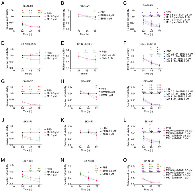
All NB cell lines presented dose-dependent responses with 0.5 and 1 µM MK-1775; SK-N-AS and SK-N-DZ were the most and SK-N-BE(2)-C the least sensitive (Fig. 2A, D, G, J, M). The higher MK-1775 dose (1 µM) resulted in a notable absorbance decrease at 48 and 72 h compared with PBS in all NB lines except SK-N-BE(2)-C, while the lower dose (0.5 µM) showed a significant effect at all timepoints only in SK-N-AS and SK-N-DZ.
Figure 2.
WST-1 viability assay in NB cell lines after treatment with WEE1 inhibitor MK and PARP inhibitor BMN. Viability of SK-N-AS treated with MK (A), BMN (B) and combination of MK and BMN (C), viability of SK-N-BE(2)-C treated with MK (D), BMN (E) and combination of MK and BMN (F), viability of SK-N-DZ treated with MK (G), BMN (H) and combination of MK and BMN (I), viability of SK-N-FI treated with MK (J), BMN (K) and combination of MK and BMN (L) and viability of SK-N-SH treated with MK (M), BMN (N) and combination of MK and BMN (O) was measured by absorbance 24, 48 and 72 h. *P<0.05, **P<0.01, ***P<0.001, ****P<0.0001 vs. PBS control at each time point. NB, neuroblastoma; BMN, BMN673; MK, MK-1775; WEE1, WEE1 G2 checkpoint kinase; PARP, poly-ADP-ribose-polymerase.
BMN673 was less potent when administered alone compared with MK-1775, although dose-dependent responses were observed in all the NB cell lines (Fig. 2B, E, H, K, N). The higher BMN673 dose (1 µM) significantly decreased absorbance 72 h after treatment in all cell lines except SK-N-FI, while the lower dose (0.5 µM) showed a notable effect after 72 h only in SK-N-SH and SK-N-DZ cells.
MK-1775 + BMN673 (both 0.5–1.0 µM) resulted in a notable decrease in absorbance for all cell lines at 72 h after treatment; SK-N-SH and SK-N-DZ showed significant decreases in absorbance with almost all combinations and timepoints (Fig. 2C, F, I, L, O).
To summarize, all NB lines except SK-N-BE(2)-C were sensitive to MK-1775, while only SK-N-SH showed pronounced sensitivity to BMN673; when combining MK-1775 + BMN673, all cell lines showed a consistent sensitivity especially after 72 h.
IC50 of PD-0332991, BMN673 and MK-1775
IC50 values for PD-0332991, BMN673 and MK-1775 for SK-N-AS, SK-N-BE(2)-C, SK-N-DZ, SK-N-FI and SK-N-SH at 24–72 h after treatment are presented in Table I. BYL719 IC50 values have been reported previously (12). PD-0332991 IC50 values were 0.05–1532.00 µM. SK-N-BE(2)-C was the most sensitive cell line, followed by SK-N-SH and SK-N-DZ, while SK-N-FI and SK-N-AS were less sensitive. IC50 values for MK-1775 were 0.14–6.61 µM. SK-N-DZ and SK-N-AS were the most sensitive lines, while SK-N-BE(2)-C, SK-N-FI and SK-N-SH were less sensitive. For BMN673, IC50 values were 0.99–621.20 µM. SK-N-SH was the most and SK-N-FI the least sensitive NB cell line.
Table I.
IC50 based on WST-1 viability assay following treatment with CDK4/6 PD-0332991, WEE1 G2 checkpoint kinase MK-1775 and poly-ADP-ribose-polymerase inhibitor BMN-673.
| A, PD-0332991 | |||
|---|---|---|---|
|
| |||
| IC50, µM | |||
|
|
|||
| Cell line | 24 h | 48 h | 72 h |
| SK-N-AS | 12.7a | 24.5a | 11.5a |
| SK-N-BE(2)-C | 19.8a | 0.1a | 0.1a |
| SK-N-DZ | 18.8a | 3.9 | 2.3 |
| SK-N-FI | 1532a | 26.4a | 34.9a |
| SK-N-SH | 5.0 | 2.3 | 2.1 |
|
| |||
| B, MK-1775 | |||
|
| |||
| IC50, µM | |||
|
|
|||
| Cell line | 24 h | 48 h | 72 h |
|
| |||
| SK-N-AS | 1.5 | 0.3 | 0.3 |
| SK-N-BE(2)-C | 1.3 | 1.0 | 1.0 |
| SK-N-DZ | 0.4 | 0.2 | 0.1 |
| SK-N-FI | 6.6 | 1.8 | 0.6 |
| SK-N-SH | 4.9 | 2.5 | 1.0 |
|
| |||
| C, BMN673 | |||
|
| |||
| IC50, µM | |||
|
|
|||
| Cell line | 24 h | 48 h | 72 h |
|
| |||
| SK-N-AS | 109.8a | 174.5a | 63.6 |
| SK-N-BE(2)-C | 152.1a | 20.4 | 7.1 |
| SK-N-DZ | 109.6a | 621.2a | 13.2 |
| SK-N-FI | 195.4a | - | - |
| SK-N-SH | 83.0 | 1.0 | 0.1 |
IC50 was determined from log concentrations effect curves in GraphPad Prism using non-linear regression analysis.
Extrapolated value outside the tested concentration range. IC50, half-maximal inhibitory concentration; -, not determined.
To summarize, most cell lines were more sensitive to MK-1775 than BMN673 but response to PD-0332991 varied.
Highest single agent and median effect following combined inhibitor treatment
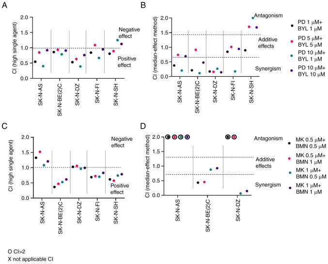
CIs of PD-0332991 + BYL719 and MK-1775 + BMN673 were measured using highest single agent and dose-effect-based median effect (38,39). To summarize, when using the highest single agent, CI<1 indicates a positive effect, while a CI>1 suggests a negative effect. When using the dose-effect-based median effect, CI<0.75 indicates synergism, 0.75≤CI<1.45 indicates an additive effect, CI=1.45 indicates a neutral effect and CI>1.45 suggests an antagonistic effect. CI was measured 24 (data not shown), 48 and 72 h (data not shown) following drug administration for all NB lines; CI at 48 h is presented in Fig. 3.
Figure 3.
Effect of combined PI3K and CDK4/6 inhibitors WEE1 G2 checkpoint kinase and PARP inhibitors on neuroblastoma cell lines. Upon combination of BYL and PD, CIs were calculated by (A) highest single agent and (B) median effect method after 48 h. When combining MK together with BMN, CIs were calculated by (C) highest single agent and (D) median effect method after 48 h. CI>1.45 suggests antagonism, 0.75<CI>1.45 additive, and CI<0.75 synergistic effects. o indicates CI>2, which implies a negative combination effect. CI, combination index; PD, PD-0332991; BYL, BYL719; BMN, BMN673; MK, MK-1775; PARP, poly-ADP-ribose-polymerase.
The lowest doses (1 and 5 µM) of PD-0332991 + BYL719 yielded synergistic or neutral combinational effects (CI<1) 48 h after treatment in all NB lines except SK-N-SH, where a slightly negative effect was noted (Fig. 3A). In addition, combining 10 PD-0332991 + 1 or 10 µM BYL719 resulted in positive or neutral combinational effects in all NB lines except SK-N-SH.
PD-0332991 + BYL719 presented synergistic or neutral combinational effects in all NB lines with the exception of SK-N-SH, where some antagonistic effects were observed (Fig. 3A and B).
Low doses (0.5 and 1.0 µM) of MK-1775 + BMN673 analyzed according to the highest single agent approach showed mainly positive or neutral effects (CI<1) 48 h after treatment for all NB cell lines except SK-N-AS, where negative effects were observed (Fig. 3C). Using the dose-effect-based median effect approach, synergistic effects were observed in SK-N-BE(2)-C and SK-N-DZ, while for SK-N-FI and SK-N-SH they could not be analyzed (Fig. 3D).
To summarize, MK-1775 + BMN673 exhibited positive and neutral effects for NB cell lines except SK-N-AS, but it was not possible to calculate the dose-effect-based median effect in all cases.
Cell confluence after single and combined treatments with PD-0332991, BYL719, BMN673 and MK-1775
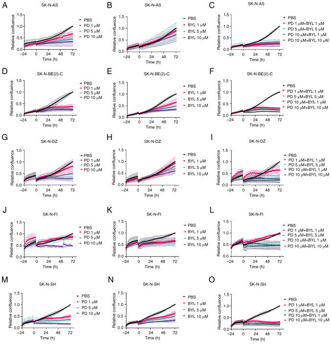
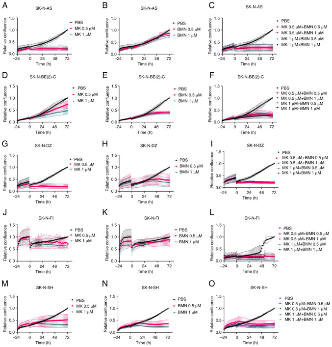
Confluence as a measure of proliferation of NB cell lines following single and combined treatment with PD-0332991, BYL719, BMN673 and MK-1775 was analyzed using the IncuCyte S3 Live-Cell Analysis System every 2 h for 72 h.
Monotherapy with 5–10 µM PD-0332991 decreased confluence in all NB cell lines, while 1 µM PD-0332991 decreased confluence only in SK-N-BE(2)-C and SK-N-SH cells with only marginal effects on the other cell lines (Fig. 4A, D, G, J, M).
Figure 4.
Effect of PD and BYL on confluence of neuroblastoma cell lines. Confluence of SK-N-AS cells treated with PD (A), BYL (B) and combination of PD and BYL (C), confluence of SK-N-BE(2)-C treated with PD (D), BYL (E) and combination of PD and BYL (F), confluence of SK-N-DZ treated with PD (G), BYL (H) and combination of PD and BYL (I), confluence of SK-N-FI treated with PD (J), BYL (K) and combination of PD and BYL (L) and confluence of SK-N-SH treated with PD (M), BYL (N) and combination of PD and BYL (O) was measured every 2 h up to 72 h after the treatment. Confluence indicates proliferation. BYL, BYL719; PD, PD-0332991; CDK4/6, cyclin-dependent kinase-4/6; PI3K, phosphoinositide 3-kinase.
Monotherapy with 1–10 µM BYL719 showed decreases in confluence in the NB cell lines, with SK-N-AS and SK-N-DZ exhibiting the least difference (Fig. 4B, E, H, K, N).
PD-0332991 + BYL719 (both 1–10 µM) decreased confluence for all NB lines, except SK-N-DZ and SK-N-FI at the lowest dose combinations (Fig. 4C, F, I, L, O).
Low doses (0.5 and 1.0 µM) of MK-1775 alone decreased cell confluence in all NB cell lines 72 h after treatment compared with PBS control (Fig. 5A, D, G, J, M).
Figure 5.
Confluence of neuroblastoma cell lines following administration of MK and BMN. Confluence of SK-N-AS treated with (A) MK (A), BMN (B) and combination of MK and BMN (C), confluence of SK-N-BE(2)-C treated with (D) MK, BMN (E) and combination of MK and BMN (F), confluence of SK-N-DZ treated with MK (G), BMN (H) and combination of MK and BMN (I), confluence of SK-N-FI treated with MK (J), BMN (K) and combination of MK and BMN (L) and confluence of SK-N-SH treated with MK (M), BMN (N) and combination of MK and BMN (O) was measured every 2 h up to 72 h after the treatment. Confluence denotes proliferation. BMN, BMN673; MK, MK-1775; PARP, poly-ADP-ribose-polymerase.
Low doses (0.5 and 1.0 µM) of BMN673 tended to decrease confluence, especially in SK-N-BE(2)-C and SK-N-SH compared with PBS control (Fig. 5B, E, H, K, N).
Combining 0.5 and 1.0 µM MK-1775 + BMN673 decreased confluence for all NB cell lines compared with PBS control (Fig. 5C, F, I, L, O).
To summarize, combining PD-0332991 + BYL719 or MK-1775 + BMN673 tended to decrease cell confluence at lower doses compared with monotherapy.
Cytotoxic effects of single and combined treatments of PD-0332991, BYL719, BMN673 and MK-1775
The cytotoxic effects of the drugs on SK-N-AS, SK-N-BE(2)-C, SK-N-DZ, SK-N-FI and SK-N-SH were analyzed utilizing the Cytotox Red Reagent and IncuCyte S3 Live-Cell Analysis System (Figs. S1 and S2).
Marked cytotoxicity was not observed in the NB lines after exposure to 1–10 µM PD-0332991 or BYL719, however, treatment with 10 µM PD-0332991 induced slight cytotoxic effects in SK-N-FI and SK-N-SH cells (Fig. S1A, B, D, E, G, H, J, K, M, N).
When combining 10 µM PD-0332991 + BYL719, cytotoxicity was observed in almost all NB cell lines. Moreover, cytotoxicity was observed for SK-N-FI with all combinations of 1.0–10.0 µM PD-0332991 + BYL719 (Fig. S1C, F, I, L, O).
Cytotoxic effects were observed with 0.5–1.0 µM MK-1775 in SK-N-AS, S-K-N-FI and SK-N-SH, but not SK-N-BE(2)-C and SK-N-DZ cells (Fig. S2A, D, G, J, M).
Cytotoxicity was observed with 0.5–1.0 µM BMN673 in SK-N-SH but not any of the other cell lines (Fig. S2B, E, H, K, N).
Combined 0.5–1.0 µM MK-1775 + BMN673 tended to provide similar cytotoxic effects on the NB lines as 0.5–1.0 µM MK-1775 alone with moderate effects on SK-N-AS, S-K-N-FI and SK-N-SH (Fig. S2C, F, I, L, O).
To summarize, marked cytotoxic effects were shown only when using high doses of PD-0332991 or MK-1775. Moreover, although combining PD-0332991 with BYL719 gave additive positive effects while combining MK-1775 + BMN673 did not increase the cytotoxic effects compared with MK-1775 alone.
Apoptotic effects on SK-N-AS, SK-N-BE(2)-C, SK-N-DZ, SK-N-FI and SK-N-SH after single and combined treatments of PD-0332991, BYL719, BMN673 and MK-1775
Apoptotic effects on SK-N-AS, SK-N-BE(2)-C, SK-N-DZ, SK-N-FI and SK-N-SH was analyzed using the Caspase-3/7 Green Reagent in the IncuCyte S3 Live-Cell Analysis System (Figs. S3 and S4). Slight apoptosis was observed only with 10 µM PD-0332991 in SK-N-DZ and SK-N-SH (Fig. S3A, D, G, J, M).
Marked apoptosis was not observed with 1–10 µM BYL719 in any of the NB lines (Fig. S3B, E, H, K, N).
When combining 1–10 µM PD-0332991 + BYL719, apoptosis was observed in SK-N-DZ and 10 µM PD-0332991 + BYL719 caused apoptosis in SK-N-FI and SK-N-BE(2)-C cells (Fig. S3C, F, I, L, O).
Apoptosis was observed in all NB lines at the highest dose (1 µM) of MK-1775 (Fig. A, D, G, J, M). Apoptosis was also observed with 0.5–1.0 µM BMN673 in SK-N-BE(2)-C and SK-N-SH (Fig. S4B, E, H, K, N).
Combining 0.5–1.0 µM MK-1775 + BMN673 did not enhance the apoptotic effects compared with 0.5–1.0 µM MK-1775 or BMN673 alone (Fig. S4C, F, I, L, O).
To summarize, MK-1775 induced apoptosis in almost all NB lines, as did BMN673 in SK-N-BE(2)-C and SK-N-SH cells. PD-0332991 induced apoptosis only at high doses, whereas BYL719 did not induce apoptotic effects. Moreover, combining MK-1775 + BMN673 did not increase the apoptotic effects compared with MK-1775 or BMN673 alone. However, PD-0332991 + BYL719 increased apoptosis in SK-N-BE(2)-C, SK-N-DZ and SK-N-FI cells.
Cell cycle distribution after single and combined treatments with CDK4/6 and PI3K inhibitors PD-0332991 and BYL719 on SK-N-DZ and SK-N-SH and PARP and WEE1 inhibitors MK-1775 and BMN673 on SK-N-BE(2)-C and SK-N-AS
Effects on cell cycle distribution were examined in selected samples using FACS Novocyte machine and FlowJo software. Effects of single and combined PD-0332991 and BYL719 were analyzed for one sensitive and one resistant cell line, SK-N-DZ and SK-N-SH, respectively. Likewise, effects of single and combined MK-1775 and BMN673 were analyzed for one sensitive and one resistant cell line, SK-N-BE(2)-C and SK-N-AS, respectively.
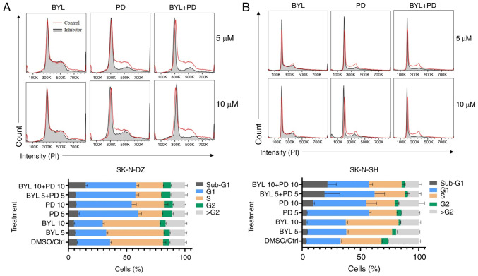
Using either 5 or 10 µM PD-0332991 increased the number of cells in G1 and decreased the numbers of cells in S phase for SK-N-DZ and SK-N-SH, while 5 and 10 µM BYL719 had no effect on the cell cycle of these NB lines (Fig. 6A and B). In addition, combining PD-0332991 + BYL719 did not result in any notable changes in the cell cycle distribution compared with PD-0332991 alone.
Figure 6.
Cell cycle profiles of SK-N-DZ and SK-N-SH obtained by FACS analysis. Cell cycle distribution of (A) SK-N-DZ and (B) SK-N-SH NB cell lines treated with CDK4/6 and PI3K inhibitors PD and BYL for 48 h. BYL, BYL719; PD, PD-0332991; CDK4/6, cyclin-dependent kinase-4/6; PI3K, phosphoinositide 3-kinase.
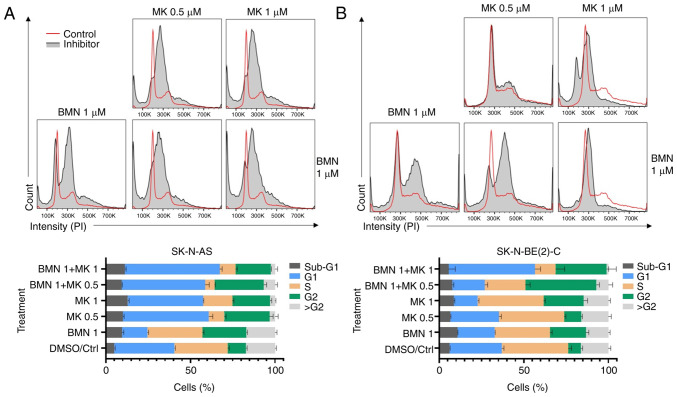
Treatment with 0.5 and 1 µM MK-1775 increased the number of cells in G1 and G2 and decreased the numbers of cells in S phase for SK-N-AS (Fig. 7A and B). For SK-N-BE(2)-C, 1 µM MK-1775 notably decreased number of cells in G1 and increased number of cells in G2. In addition, 1 µM BMN673 alone decreased the number of cells in G1 and increased the number of cells in G2 in both cell lines. Combining 1 µM MK-1775 + BMN673 induced a notable increase in the number of cells in both G1 and G2 and a notable decrease in number of cells in S phase in both cell lines.
Figure 7.
Cell cycle profiles of SK-N-AS and SK-N-BE(2)-C obtained by FACS analysis. Cell cycle distribution of (A) SK-N-AS and (B) SK-N-BE(2)-C neuroblastoma cell lines treated with WEE1 inhibitor MK-1775 and PARP inhibitor BMN673 for 48 h. BMN, BMN673; MK, MK-1775; WEE1, WEE1 G2 checkpoint kinase; PARP, poly-ADP-ribose-polymerase.
To summarize, upon combining MK-1775 with BMN673 additive effects were observed for both SK-N-BE(2)-C and SK-N-AS cells, with large increases in the number of cells in G1 and G2 and a notable decrease in the numbers of cells in S phase. By contrast, following treatment with PD-0332991 + BYL719, the effect was similar to using PD-0332991 alone with a rise in the number of cells in G1 and a decrease in the number of cells in S phase.
Discussion
Here, the effects of CDK4/6, PI3K, WEE1 and PARP inhibitors PD-0332991, BYL719, MK-1775 and BMN673 alone or combined on NB cell lines SK-N-AS, SK-N-BE(2)-C, SK-N-DZ, SK-N-FI and SK-N-SH were assessed. Briefly, all single inhibitor applications induced dose-dependent decreases in viability and confluence in all NB lines and combining PD-0332991 + BYL719 or BMN673 + MK-1775 resulted in additive and synergistic effects in most NB lines. A slight cytotoxic effect was induced by high doses of PD-0332991 or MK-1775 and apoptosis was induced in most NB cell lines by MK-1775; apoptosis induced by PD-0332991, BYL719 and BMN673 was cell line-specific. In addition, effects on the cell cycle were examined in SK-N-DZ and SK-N-SH after single and combined PD-0332991 and BYL719 applications and SK-N-BE(2)-C and SK-N-AS after single and combined MK-1775 and BMN673 treatment. Notably, while combining PD-0332991 + BYL719 did not result in any notable effects on cell cycle, combining MK-1775 + BMN673 showed a notable decrease in the number of cells in S phase for both cell lines.
As expected, single inhibitor applications induced dose-dependent responses (12,13). Similarly, when combining CDK4/6 inhibitor PD-0332991 + PI3K inhibitor BYL719, positive and synergistic effects on viability and confluence were observed in almost all NB lines, consistent with pre-clinical models using other types of tumor (28,29,40,41). However, SK-N-SH cell line showed mainly antagonistic, despite decreases in viability and cell confluence. This may be because SK-N-SH cell line was sensitive and lacked mutated p53 and it was difficult to observe further decreases in viability upon combination treatment, especially with doses that induced effects in more resistant cell lines. Our previous study showed that when combining PI3K and FGFR inhibitors in NB, MB and head and neck cancer cell lines, effects in cell lines that were particularly sensitive to one of the drugs were not observed (10–13,40,42). It is unknown why combining PD-0332991 + BYL719 resulted in enhanced inhibition in SK-N-AS, SK-N-BE(2)-C, SK-N-DZ and SK-N-FI, however all four cell lines have mutated p53. Moreover, SK-N-BE(2)-C and SK-N-DZ have amplified MYCN. On the other hand, SK-N-SH with wild-type p53 was also sensitive to treatments with both PD-0332991 and BYL719 alone.
WEE1 inhibitor MK-1775 + PARP inhibitor BMN673 induced some positive and synergistic effects on viability and confluence in SK-N-BE(2)-C, SK-N-DZ, SK-N-FI, SK-N-SH cells, similar to what has been shown for advanced prostate and ovarian cancer (36,37). SK-N-AS and SK-N-DZ cells exhibited antagonistic effects; this could be because they were both sensitive to MK-1775 but resistant to BMN673.
Cytotoxicity and apoptosis were assessed. For PD-0332991, only high dose treatment induced cytotoxicity and apoptosis in NB cell lines, while for BYL719 cytotoxic effects were not observed and combining the two did not markedly improve cytotoxic or apoptotic effects compared with PD-0332991 alone. However, when combining PD-0332991 + BYL719 in SK-N-BE(2)-C, SK-N-DZ and SK-N-FI cells, enhanced apoptosis was observed. These data are similar to our previous study on MB cell lines DAOY and UW228-3, where PD-0332991 exhibited a greater effect on both cytotoxicity and apoptosis (13).
Similarly, while MK-1775 induced both cytotoxicity and apoptosis in some NB cell lines, BMN673 did not show cytotoxic and apoptotic effects and when combining them their effects were not enhanced. MK-1775 induced cytotoxic effects in SK-N-AS, SK-N-FI and SK-N-SH, but these effects were not enhanced by the addition of BMN673 in any cell lines. Likewise, when analyzing apoptosis, no additive effect was observed upon combining MK-1775 + BMN673 compared with single MK-1775 treatment. To conclude, there were no additive effects on either cytotoxicity or apoptosis when combining MK-1775 + BMN673.
PD-0332991 increased the proportion of cells in G1 and limited the proportion of cells in S phase for SK-N-DZ and SK-N-SH, while using BYL719 alone had no effect on the cell cycle distribution of either NB line. MK-1775 increased the proportion of cells in G1 and G2 and decreased the proportion of cells in S phase for SK-N-AS, effects on SK-N-BE(2)-C were less pronounced. All the effects of using BMN673 alone were limited in comparison to MK-1775, although an increase in numbers of cells in G2 was observed following treatment with BMN673 in both SK-N-AS and SK-N-BE(2)-C.
The observation that both PD-0332991 and MK-1775 increased the proportion of cells in G1 and limited the number of cells in S phase and that MK-1775 can have effects on the cell cycle and mitosis by increasing the number of cells arresting in G2 is in line with previous data (43,44). Likewise, BMN673 has also been shown to induce cell cycle arrest (42).
Notably, combining MK-1775 + BMN673 in SK-N-BE(2)-C and SK-N-AS cells resulted in a marked decrease in the number of cells in the S phase; higher doses resulted in a marked increase in the proportion of cells in G1 and G2. This may be due to the effects of BMN673, as previously described (45).
The present study had limitations. Lower doses in combination were not used when the NB lines were sensitive to one drug alone. However, if a cell line is sensitive to low doses of one inhibitor, combination therapies may not be needed. Furthermore, the present study did not investigate changes in pathways in relation to the molecular profile of each cell line. Another limitation of the present study was that all treatments were performed in vitro; studies on cells grown as spheroids and treatment in in vivo in mouse models will be performed in future.
To summarize, PD-0332991, BYL719, MK-1775 and BMN673 gave dose dependent effects on viability the NB cell lines. Notably however, using combinations of PD-0332991 with BYL719 and combinations of MK-1775 and BMN763 gave additive and synergistic effects on the inhibition of viability compared with each drug alone. Inhibition of cell confluence tended to be more prominent when combining two drugs especially for some of the cell lines which tended to be relatively resistant to one drug alone. Enhanced cytotoxicity or apoptosis was not observed when combining PD-0332991 + BYL719 or MK-1775 + BMN673. BYL719 did not affect PD-0332991-induced changes in the cell cycle distribution. Notably, MK-1775 + BMN673 resulted in a notable increase of cells in G1 and G2 and a marked decrease of cells in S phase.
Altogether, the aforementioned data indicated that NB, similar to other tumor types, may be sensitive to single and combined treatment with CDK4/6, PI3K, PARP and WEE1 inhibitors (40,41). It is unknown why some NB cell lines were more sensitive to some combinations.
To conclude, combined treatment with either CDK4/6 and PI3K inhibitor PD-0332991 + BYL719 or WEE1 and PARP inhibitors MK-1775 + BMN673 synergistically decreased viability and cell confluence in most NB cell lines. These combinations did not have any marked effects on cytotoxicity or apoptosis. MK-1775 + BMN673 resulted in marked effects on the cell cycle distribution compared with PD-0332991 + BYL719 combination. The present data suggest that in the future, combinations of MK-1775 + BMN673 or PD-0332991 and BYL719 could serve as a treatment of resilient or recurrent NB. Further studies utilizing NB cell lines with specific molecular characteristics grown as spheroid cultures or in vivo models are required to determine the best drug combination for each cell line based on its molecular profile.
Supplementary Material
Acknowledgements
The authors would like to thank Professor Per Kogner, Karolinska Institute (Stockholm, Sweden) for kindly providing neuroblastoma cell lines.
Funding Statement
The present study was supported by Swedish Childhood Cancer Foundation (grant no. TJ2022-0067), Swedish Cancer Foundation (grant no. 20 0704), the Stockholm Cancer Society (grant no. 201092), Stockholm City Council (grant no. 20180037), AnnaBrita o Bo Casters Minne Foundation (Lindhés Advokatbyrå) (grant no. LA2022-0070), Svenska Läkaresällskapet (grant no. SLS-934161), Åke Wiberg Foundation (grant no. M21-0012), Karolinska Institutet Sweden (grant no. 2022-01587), Tornspiran Foundation (grant no. 839), Mary Beves Foundation and Magnus Bergvalls Foundation (grant no. 2022-109).
Availability of data and materials
The datasets used and/or analyzed during the current study are available from the corresponding author on reasonable request.
Authors' contributions
ML and ONK performed experiments, interpreted the data and wrote the manuscript. SH and EP performed experiments. SH and ML performed experiments, analyzed data and wrote the manuscript. TM analyzed FACS data and constructed figures. TD and ONK conceived and designed the study, analyzed and interpreted data and wrote and revised the manuscript. ML and ONK confirm the authenticity of all the raw data. All authors have read and approved the final manuscript.
Ethics approval and consent to participate
Not applicable.
Patient consent for publication
Not applicable.
Competing interests
The authors declare that they have no competing interests.
References
- 1.Ward E, DeSantis C, Robbins A, Kohler B, Jemal A. Childhood and adolescent cancer statistics, 2014. CA Cancer J Clin. 2014;64:83–103. doi: 10.3322/caac.21219. [DOI] [PubMed] [Google Scholar]
- 2.D'Angio GJ, Evans AE, Koop CE. Special pattern of widespread neuroblastoma with a favourable prognosis. Lancet. 1971;1:1046–1049. doi: 10.1016/S0140-6736(71)91606-0. [DOI] [PubMed] [Google Scholar]
- 3.Nakagawara A, Li Y, Izumi H, Muramori K, Inada H, Nishi M. Neuroblastoma. Jpn J Clin Oncol. 2018;1:214–241. doi: 10.1093/jjco/hyx176. [DOI] [PubMed] [Google Scholar]
- 4.Bo Q, Matthay KK. Advancing therapy for neuroblastoma. Nat Rev Clin Oncol. 2022;19:515–533. doi: 10.1038/s41571-022-00643-z. [DOI] [PubMed] [Google Scholar]
- 5.Hero B, Simon T, Spitz R, Ernestus K, Gnekow AK, Scheel-Walter HG, Schwabe D, Schilling FH, Benz-Bohm G, Berthold F. Localized infant neuroblastomas often show spontaneous regression: Results of the prospective trials NB95-S and NB97. J Clin Oncol. 2008;26:1504–1510. doi: 10.1200/JCO.2007.12.3349. [DOI] [PubMed] [Google Scholar]
- 6.Matthay KK, Maris JM, Schleiermacher G, Nakagawara A, Mackall CL, Diller L, Weiss WA. Neuroblastoma. Nat Rev Dis Prim. 2016;2:16078. doi: 10.1038/nrdp.2016.78. [DOI] [PubMed] [Google Scholar]
- 7.London WB, Bagatell R, Weigel BJ, Fox E, Guo D, Van Ryn C, Naranjo A, Park JR. Historical time to disease progression and progression-free survival in patients with recurrent/refractory neuroblastoma treated in the modern era on children's oncology group early-phase trials. Cancer. 2017;123:4914–4923. doi: 10.1002/cncr.30934. [DOI] [PMC free article] [PubMed] [Google Scholar]
- 8.Johnsen JI, Dyberg C, Wickström M. Neuroblastoma-A neural crest derived embryonal malignancy. Front Mol Neurosci. 2019;12:9. doi: 10.3389/fnmol.2019.00009. [DOI] [PMC free article] [PubMed] [Google Scholar]
- 9.Hemmings BA, Restuccia DF. The PI3K-PKB/Akt pathway. Cold Spring Harb Perspect Biol. 2015;7:a026609. doi: 10.1101/cshperspect.a026609. [DOI] [PMC free article] [PubMed] [Google Scholar]
- 10.Kostopoulou ON, Holzhauser S, Lange BKA, Ohmayer A, Andonova T, Bersani C, Wickström M, Dalianis T. Analyses of FGFR3 and PIK3CA mutations in neuroblastomas and the effects of the corresponding inhibitors on neuroblastoma cell lines. Int J Oncol. 2019;55:1372–1384. doi: 10.3892/ijo.2019.4896. [DOI] [PubMed] [Google Scholar]
- 11.Holzhauser S, Lukoseviciute M, Andonova T, Ursu RG, Dalianis T, Wickström M, Kostopoulou ON. Targeting fibroblast growth factor receptor (FGFR) and Phosphoinositide 3-kinase (PI3K) signaling pathways in medulloblastoma cell lines. Anticancer Res. 2020;40:53–66. doi: 10.21873/anticanres.13925. [DOI] [PubMed] [Google Scholar]
- 12.Holzhauser S, Lukoseviciute M, Papachristofi C, Vasilopoulou C, Herold N, Wickström M, Kostopoulou ON, Dalianis T. Effects of PI3K and FGFR inhibitors alone and in combination, and with/without cytostatics in childhood neuroblastoma cell lines. Int J Oncol. 2021;58:211–225. doi: 10.3892/ijo.2021.5167. [DOI] [PMC free article] [PubMed] [Google Scholar]
- 13.Lukoseviciute M, Maier H, Poulou-Sidiropoulou E, Rosendahl E, Holzhauser S, Dalianis T, Kostopoulou ON. Targeting PI3K, FGFR, CDK4/6 signaling pathways together with cytostatics and radiotherapy in two medulloblastoma cell lines. Front Oncol. 2021;11:748657. doi: 10.3389/fonc.2021.748657. [DOI] [PMC free article] [PubMed] [Google Scholar]
- 14.Katso R, Okkenhaug K, Ahmadi K, White S, Timms J, Waterfield MD. Cellular function of phosphoinositide 3-kinases: Implications for development, homeostasis, and cancer. Annu Rev Cell Dev Biol. 2001;17:615–675. doi: 10.1146/annurev.cellbio.17.1.615. [DOI] [PubMed] [Google Scholar]
- 15.Engelman JA, Luo J, Cantley LC. The evolution of phosphatidylinositol 3-kinases as regulators of growth and metabolism. Nat Rev Genet. 2006;7:606–619. doi: 10.1038/nrg1879. [DOI] [PubMed] [Google Scholar]
- 16.King D, Yeomanson D, Bryant HE. PI3King the lock: Targeting the PI3K/Akt/mTOR pathway as a novel therapeutic strategy in neuroblastoma. J Pediatr Hematol Oncol. 2015;37:245–251. doi: 10.1097/MPH.0000000000000329. [DOI] [PubMed] [Google Scholar]
- 17.Khezri MR, Jafari R, Yousefi K, Zolbanin NM. The PI3K/AKT signaling pathway in cancer: Molecular mechanisms and possible therapeutic interventions. Exp Mol Pathol. 2022;127:104787. doi: 10.1016/j.yexmp.2022.104787. [DOI] [PubMed] [Google Scholar]
- 18.Zage PE. Novel therapies for relapsed and refractory neuroblastoma. Children (Basel) 2018;5:148. doi: 10.3390/children5110148. [DOI] [PMC free article] [PubMed] [Google Scholar]
- 19.Creevey L, Ryan J, Harvey H, Bray IM, Meehan M, Khan AR, Stallings RL. MicroRNA-497 increases apoptosis in MYCN amplified neuroblastoma cells by targeting the key cell cycle regulator WEE1. Mol Cancer. 2013;12:23. doi: 10.1186/1476-4598-12-23. [DOI] [PMC free article] [PubMed] [Google Scholar]
- 20.Rader J, Russell MR, Hart LS, Nakazawa MS, Belcastro LT, Martinez D, Li Y, Carpenter EL, Attiyeh EF, Diskin SJ, et al. Dual CDK4/CDK6 inhibition induces cell-cycle arrest and senescence in neuroblastoma. Clin Cancer Res. 2013;19:6173–6182. doi: 10.1158/1078-0432.CCR-13-1675. [DOI] [PMC free article] [PubMed] [Google Scholar]
- 21.Knudsen ES, Pruitt SC, Hershberger PA, Witkiewicz AK, Goodrich DW. Cell cycle and beyond: Exploiting new RB1 controlled mechanisms for cancer therapy. Trends Cancer. 2019;5:308–324. doi: 10.1016/j.trecan.2019.03.005. [DOI] [PMC free article] [PubMed] [Google Scholar]
- 22.Morgan DO. Cyclin-dependent kinases: Engines, clocks, and microprocessors. Annu Rev Cell Dev Biol. 1997;13:261–291. doi: 10.1146/annurev.cellbio.13.1.261. [DOI] [PubMed] [Google Scholar]
- 23.Sherr CJ, Roberts JM. Living with or without cyclins and cyclin-dependent kinases. Genes Dev. 2004;18:2699–2711. doi: 10.1101/gad.1256504. [DOI] [PubMed] [Google Scholar]
- 24.Hamilton E, Infante JR. Targeting CDK4/6 in patients with cancer. Cancer Treat Rev. 2016;45:129–138. doi: 10.1016/j.ctrv.2016.03.002. [DOI] [PubMed] [Google Scholar]
- 25.Ortega S, Malumbres M, Barbacid M. Cyclin D-dependent kinases, INK4 inhibitors and cancer. Biochim Biophys Acta. 2022;1602:73–87. doi: 10.1016/s0304-419x(02)00037-9. [DOI] [PubMed] [Google Scholar]
- 26.Braal CL, Jongbloed EM, Wilting SM, Mathijssen RHJ, Koolen SLW, Jager A. Inhibiting CDK4/6 in breast cancer with palbociclib, ribociclib, and abemaciclib: Similarities and differences. Drugs. 2021;81:317–331. doi: 10.1007/s40265-020-01461-2. [DOI] [PMC free article] [PubMed] [Google Scholar]
- 27.Schmidt EE, Ichimura K, Reifenberger G, Collins VP. CDKN2 (p16/MTS1) gene deletion or CDK4 amplification occurs in the majority of glioblastomas. Cancer Res. 1994;15:6321–6324. [PubMed] [Google Scholar]
- 28.Muranen T, Meric-Bernstam F, Mills GB. Promising rationally derived combination therapy with PI3K and CDK4/6 inhibitors. Cancer Cell. 2014;26:7–9. doi: 10.1016/j.ccr.2014.06.020. [DOI] [PubMed] [Google Scholar]
- 29.Bonelli MA, Digiacomo G, Fumarola C, Alfieri R, Quaini F, Falco A, Madeddu D, Monica SL, Cretella D, Ravelli A, et al. Combined inhibition of CDK4/6 and PI3K/AKT/mTOR pathways induces a synergistic anti-tumor effect in malignant pleural mesothelioma cells. Neoplasia. 2017;19:637–648. doi: 10.1016/j.neo.2017.05.003. [DOI] [PMC free article] [PubMed] [Google Scholar]
- 30.Loibl S, Furlanetto J. Integrating CDK4/6 inhibitors in the treatment of patients with early breast cancer. Breast. 2022;62:S70–S79. doi: 10.1016/j.breast.2021.12.008. [DOI] [PMC free article] [PubMed] [Google Scholar]
- 31.Vora SR, Juric D, Kim N, Mino-Kenudson M, Huynh T, Costa C, Lockerman EL, Pollack SF, Liu M, Li X, et al. CDK 4/6 inhibitors sensitize PIK3CA mutant breast cancer to PI3K inhibitors. Cancer Cell. 2014;26:136–149. doi: 10.1016/j.ccr.2014.05.020. [DOI] [PMC free article] [PubMed] [Google Scholar]
- 32.Paik J. Olaparib: A review as first-line maintenance therapy in advanced ovarian cancer. Target Oncol. 2021;16:847–856. doi: 10.1007/s11523-021-00842-1. [DOI] [PubMed] [Google Scholar]
- 33.Brown TJ, Reiss KA. PARP inhibitors in pancreatic cancer. Cancer J. 2021;27:465–475. doi: 10.1097/PPO.0000000000000554. [DOI] [PMC free article] [PubMed] [Google Scholar]
- 34.De Bono J, Mateo J, Fizazi K, Saad F, Shore N, Sandhu S, Chi KN, Sartor O, Agarwal N, Olmos D, et al. Olaparib for metastatic castration-resistant prostate cancer. N Engl J Med. 2022;382:2091–2102. doi: 10.1056/NEJMoa1911440. [DOI] [PubMed] [Google Scholar]
- 35.Banerjee S, Moore KN, Colombo N, Scambia G, Kim BG, Oaknin A, Friedlander M, Lisyanskaya A, Floquet A, Leary A, et al. Maintenance olaparib for patients with newly diagnosed advanced ovarian cancer and a BRCA mutation (SOLO1/GOG 3004): 5-year follow-up of a randomised, double-blind, placebo-controlled, phase 3 trial. Lancet Oncol. 2021;22:1721–1731. doi: 10.1016/S1470-2045(21)00531-3. [DOI] [PubMed] [Google Scholar]
- 36.Fang Y, McGrail DJ, Sun C, Labrie M, Chen X, Zhang D, Ju Z, Vellano CP, Lu Y, LI Y, et al. Sequential therapy with PARP and WEE1 inhibitors minimizes toxicity while maintaining efficacy. Cancer Cell. 2019;35:851–867.e857. doi: 10.1016/j.ccell.2019.05.001. [DOI] [PMC free article] [PubMed] [Google Scholar]
- 37.di Rorà AG, Cerchione C, Martinelli G, Simonetti G. A WEE1 family business: Regulation of mitosis, cancer progression, and therapeutic target. J Hematol Oncol. 2020;13:126. doi: 10.1186/s13045-020-00959-2. [DOI] [PMC free article] [PubMed] [Google Scholar]
- 38.Foucquier J, Guedj M. Analysis of drug combinations: Current methodological landscape. Pharmacol Res Perspect. 2015;3:e00149. doi: 10.1002/prp2.149. [DOI] [PMC free article] [PubMed] [Google Scholar]
- 39.Chou TC. Drug combination studies and their synergy quantification using the Chou-Talalay method. Cancer Res. 2010;70:440–446. doi: 10.1158/0008-5472.CAN-09-1947. [DOI] [PubMed] [Google Scholar]
- 40.Kostopoulou ON, Zupancic M, Pont M, Papin E, Lukoseviciute M, Mikelarena BA, Holzhauser S, Dalianis T. Targeted therapy of HPV positive and negative tonsillar squamous cell carcinoma cell lines reveals synergy between CDK4/6, PI3K and sometimes FGFR inhibitors, but rarely between PARP and WEE1 inhibitors. Viruses. 2022;14:1372. doi: 10.3390/v14071372. [DOI] [PMC free article] [PubMed] [Google Scholar]
- 41.Agostinetto E, Debien V, Marta GN, Lambertini M, Piccart-Gebhart M, de Azambuja E. CDK4/6 and PI3K inhibitors: A new promise for patients with HER2-positive breast cancer. Eur J Clin Invest. 2021;51:e13535. doi: 10.1111/eci.13535. [DOI] [PubMed] [Google Scholar]
- 42.Holzhauser S, Wild N, Zupancic M, Ursu RG, Bersani C, Näsman A, Kostopoulou Ourania N, Dalianis T. Targeted therapy with PI3K and FGFR inhibitors on human papillomavirus positive and negative tonsillar and base of tongue cancer lines with and without corresponding mutations. Front Oncol. 2021;11:640490. doi: 10.3389/fonc.2021.640490. [DOI] [PMC free article] [PubMed] [Google Scholar]
- 43.Rihani A, Vandesompele J, Speleman F, Van Maerken T. Inhibition of CDK4/6 as a novel therapeutic option for neuroblastoma. Cancer Cell Int. 2015;15:76. doi: 10.1186/s12935-015-0224-y. [DOI] [PMC free article] [PubMed] [Google Scholar]
- 44.Fry DW, Harvey PJ, Keller PR, Elliott WL, Meade M, Trachet E, Albassam M, Zheng X, Leopold WR, Pryer NK, Toogood PL. Specific inhibition of cyclin-dependent kinase 4/6 by PD 0332991 and associated antitumor activity in human tumor xenografts. Mol Cancer Ther. 2004;3:1427–1438. doi: 10.1158/1535-7163.1427.3.11. [DOI] [PubMed] [Google Scholar]
- 45.Challa S, Kraus WL. Two birds, one stone: Non-canonical therapeutic effects of the PARP inhibitor Talazoparib. Cell Chem Biol. 2022;29:171–173. doi: 10.1016/j.chembiol.2022.02.002. [DOI] [PubMed] [Google Scholar]
Associated Data
This section collects any data citations, data availability statements, or supplementary materials included in this article.
Supplementary Materials
Data Availability Statement
The datasets used and/or analyzed during the current study are available from the corresponding author on reasonable request.


