

Abstract
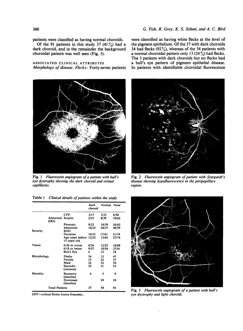
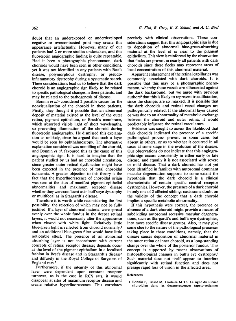
Many patients with heredomacular degeneration exhibit a peculiar fluorescein angiographic finding of absence of the normal background fluorescence (a dark choroid). The cause of this is unknown but may relate to the deposition of an abnormal material in the retinal pigment epithelial cells. The finding does not correlate with severity or duration of disease but is more frequent in patients with flecks. The finding may be useful in subdividing heredomacular degenerations into more specific disease groups.
Full text
PDF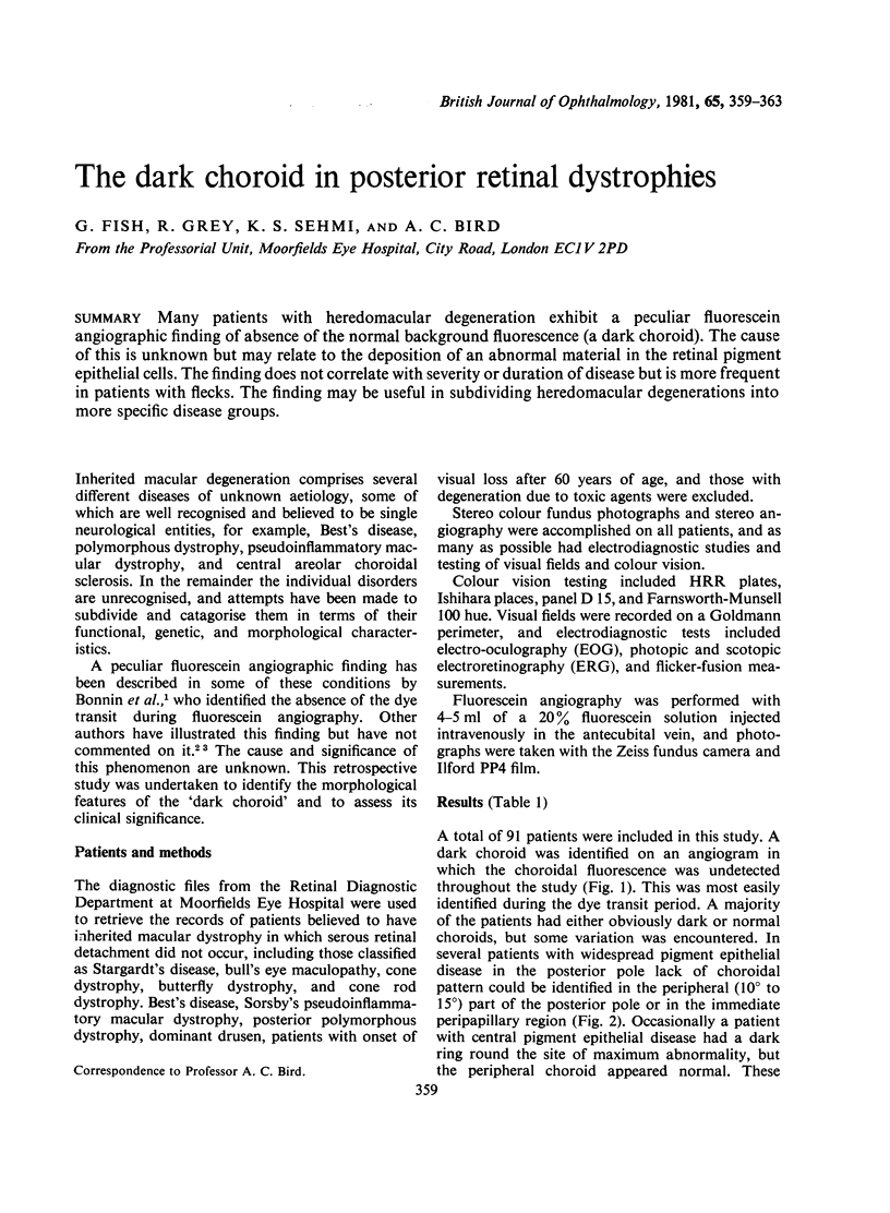


Images in this article


Selected References
These references are in PubMed. This may not be the complete list of references from this article.
- DOWLING J. E., SIDMAN R. L. Inherited retinal dystrophy in the rat. J Cell Biol. 1962 Jul;14:73–109. doi: 10.1083/jcb.14.1.73. [DOI] [PMC free article] [PubMed] [Google Scholar]
- Eagle R. C., Jr, Lucier A. C., Bernardino V. B., Jr, Yanoff M. Retinal pigment epithelial abnormalities in fundus flavimaculatus: a light and electron microscopic study. Ophthalmology. 1980 Dec;87(12):1189–1200. doi: 10.1016/s0161-6420(80)35106-3. [DOI] [PubMed] [Google Scholar]
- Gabel V. P., Birngruber R., Hillenkamp F. Individuelle Unterschiede der Lichtabsorption am Augenhintergrund im sichtbaren und infrarotin Spektralbereich. Ber Zusammenkunft Dtsch Ophthalmol Ges. 1977;74:418–421. [PubMed] [Google Scholar]
- Klien B. A., Krill A. E. Fundus flavimaculatus. Clinical, functional and histopathologic observations. Am J Ophthalmol. 1967 Jul;64(1):3–23. [PubMed] [Google Scholar]